. 2020 Feb;27(2):540-555.
doi: 10.1038/s41418-019-0372-z. Epub 2019 Jun 17.

FNDC5 alleviates oxidative stress and cardiomyocyte apoptosis in doxorubicin-induced cardiotoxicity via activating AKT

Affiliations

FNDC5 alleviates oxidative stress and cardiomyocyte apoptosis in doxorubicin-induced cardiotoxicity via activating AKT

Xin Zhang et al. Cell Death Differ. 2020 Feb.

Abstract

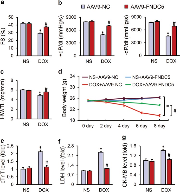
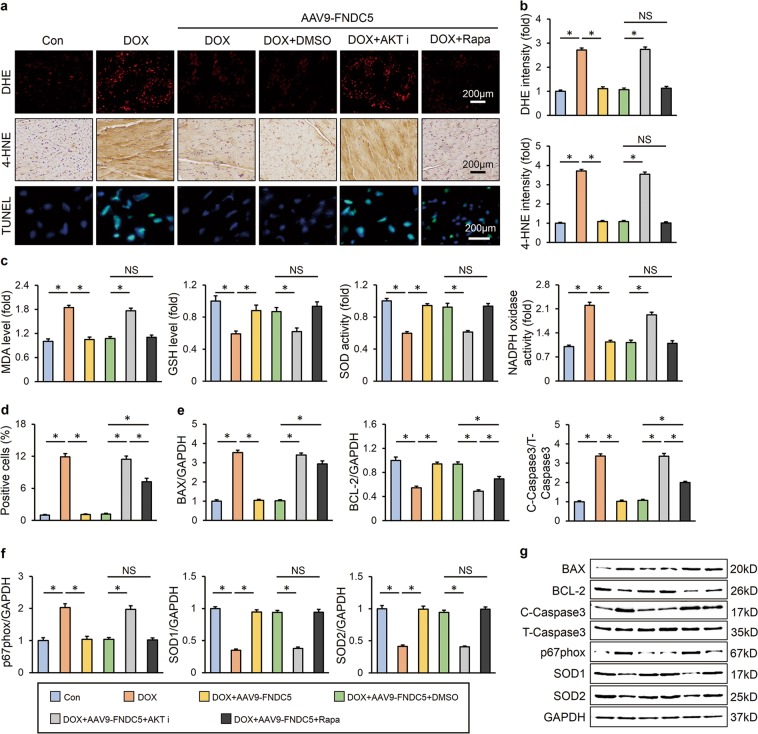
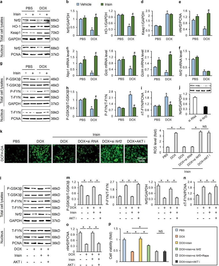
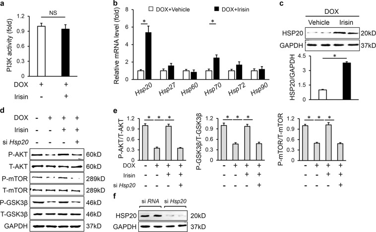
Oxidative stress and cardiomyocyte apoptosis play critical roles in doxorubicin (DOX)-induced cardiotoxicity. Previous studies indicated that fibronectin type III domain-containing 5 (FNDC5) and its cleaved form, irisin, could preserve mitochondrial function and attenuate oxidative damage as well as cell apoptosis, however, its role in DOX-induced cardiotoxicity remains unknown. Our present study aimed to investigate the role and underlying mechanism of FNDC5 on oxidative stress and cardiomyocyte apoptosis in DOX-induced cardiotoxicity. Cardiomyocyte-specific FNDC5 overexpression was achieved using an adeno-associated virus system, and then the mice were exposed to a single intraperitoneal injection of DOX (15 mg/kg) to generate DOX-induced cardiotoxicity. Herein, we found that FNDC5 expression was downregulated in DOX-treated murine hearts and cardiomyocytes. Fndc5 deficiency resulted in increased oxidative damage and apoptosis in H9C2 cells under basal conditions, imitating the phenotype of DOX-induced cardiomyopathy in vitro, conversely, FNDC5 overexpression or irisin treatment alleviated DOX-induced oxidative stress and cardiomyocyte apoptosis in vivo and in vitro. Mechanistically, we identified that FNDC5/Irisin activated AKT/mTOR signaling and decreased DOX-induced cardiomyocyte apoptosis, and moreover, we provided direct evidence that the anti-oxidant effect of FNDC5/Irisin was mediated by the AKT/GSK3β/FYN/Nrf2 axis in an mTOR-independent manner. And we also demonstrated that heat shock protein 20 was responsible for the activation of AKT caused by FNDC5/Irisin. In line with the data in acute model, we also found that FNDC5/Irisin exerted beneficial effects in chronic model of DOX-induced cardiotoxicity (5 mg/kg, i.p., once a week for three times, the total cumulative dose is 15 mg/kg) in mice. Based on these findings, we supposed that FNDC5/Irisin was a potential therapeutic agent against DOX-induced cardiotoxicity.


Conflict of interest statement

The authors declare that they have no conflict of interest.

Figures

Fig. 1
Fig. 1
FNDC5 attenuated doxorubicin (DOX)-induced cardiac dysfunction in mice. a Fractional shortening (FS) of mice as determined via echocardiography 8 days after DOX injection (n = 8). b Hemodynamic parameter of mice with or without FNDC5 overexpression (n = 8). c Statistical results of the heart weight/tibia length ((HW/TL) (n = 8). (d) Body weight alterations (n = 8). eg Biochemical determination of cTnT, LDH, CK-MB serum levels (n = 10). Values represent the mean ± SEM. *P < 0.05 versus the corresponding normal saline (NS) group mice injected with negative control (NC) adeno-associated virus 9, #P < 0.05 versus DOX-treated mice injected with AAV9-NC
Fig. 2
Fig. 2
FNDC5 suppressed oxidative stress and cardiomyocyte apoptosis in DOX-treated mice. a, b Representative DHE, 4-HNE staining images and the quantitative results (n = 6). c, d Western blots and statistical results (n = 6). e Quantitative results of myocardial MDA, GSH levels and SOD, NADPH oxidase activities (n = 6). f, g TUNEL staining and the quantitative results (n = 8). h, i Western blots and the statistical results of apoptosis-related proteins (n = 6). Values represent the mean ± SEM. *P < 0.05 versus the corresponding normal saline (NS) group mice injected with negative control (NC) adeno-associated virus 9, #P < 0.05 versus DOX-treated mice injected with AAV9-NC
Fig. 3
Fig. 3
FNDC5 alleviated DOX-induced oxidative stress and cardiomyocyte apoptosis in vitro. a, b Western blots and statistical results (n = 6). c, d DCFH-DA staining, TUNEL staining and the quantitative results of ROS level (n = 6). e Cell viability detected by CCK-8 assay (n = 6). f Quantitative results of MDA, GSH levels and SOD, NADPH oxidase activities in cultured H9C2 cells (n = 6). Values represent the mean ± SEM. *P < 0.05 versus PBS group treated with control vehicle, #P < 0.05 versus DOX-treated H9C2 cells with irisin protection
Fig. 4
Fig. 4
FNDC5 activated AKT/mTOR signaling pathway in vivo and in vitro. a, b Western blots and quantitative results in murine hearts (n = 6). c, d Western blots and statistical results in cultured H9C2 cells (n = 6). Values represent the mean ± SEM. In figure (a, b), *P < 0.05 versus the corresponding normal saline (NS) group mice injected with negative control (NC) adeno-associated virus 9, #P < 0.05 versus DOX-treated mice injected with AAV9-NC. In figure (c, d), *P < 0.05 versus PBS group treated with control vehicle, #P < 0.05 versus DOX-treated H9C2 cells with irisin protection
Fig. 5
Fig. 5
AKT/mTOR signaling was responsible for FNDC5-mediated protective role on cardiomyocyte apoptosis. a Representative DHE, 4-HNE, TUNEL staining images (n = 6). b Quantitative results of DHE and 4-HNE intensity (n = 6). c Quantitative results of myocardial MDA, GSH levels and SOD, NADPH oxidase activities (n = 6). d Quantitative results of TUNEL-positive cells (n = 6). eg Western blots and statistical results (n = 6). Values represent the mean ± SEM. *P < 0.05 versus the matched group. NS means no significance
Fig. 6
Fig. 6
AKT/GSK3β/FYN/Nrf2 signaling was responsible for FNDC5-mediated protective role on oxidative damage. a, b Western blots and statistical results (n = 6). c Relative mRNA levels of Nqo1, Gclc, and Gclm in cultured H9C2 cells (n = 6). d, e Keap1 protein levels in total cell lysates and Nrf2 protein levels in nuclear lysates (n = 6). f Relative Nrf2 mRNA level (n = 6). g, h Representative western blots (n = 6). i FYN protein levels in nuclear lysates (n = 6). j Efficiency of si Nrf2 determined by western blots (n = 6). k DCFH-DA staining and the statistical data (n = 6). lo Western blots and quantitative results (n = 6). p Cell viability detected by CCK-8 assay (n = 6). Values represent the mean ± SEM. *P < 0.05 versus the matched group. NS means no significance
Fig. 7
Fig. 7
HSP20 was involved in the activation of AKT caused by FNDC5. a Quantitative results of PI3K activity (n = 5). b Relative mRNA levels (n = 6). c HSP20 protein levels and the statistical results (n = 6). d, e Western blots and quantitative data (n = 6). f Efficiency of si Hsp20 detected by western blots (n = 6). Values represent the mean ± SEM. *P < 0.05 versus the matched group. NS means no significance
Fig. 8
Fig. 8
FNDC5/Irisin was a potential therapeutic agent against DOX-induced cardiotoxicity. a, b Representative DHE, 4-HNE, TUNEL staining images and statistical results (n = 6). c Quantitative results of myocardial MDA levels and SOD, NADPH oxidase activities (n = 6). df Western blots and statistical results (n = 6). g Statistical results of the heart weight/tibia length ((HW/TL) (n = 8). (h) Serum cTnT levels (n = 6). i Echocardiographic and hemodynamic parameters (n = 8). j Serum liver enzymes levels (n = 8). k Fractional shortening (FS) of mice as determined via echocardiography (n = 8). Values represent the mean ± SEM. *P < 0.05 versus the corresponding normal saline (NS) group mice treated with saline, #P < 0.05 versus DOX-treated mice treated with irisin. In figure (k), *P < 0.05 versus the matched group

References

    1. Carvalho C, Santos RX, Cardoso S, Correia S, Oliveira PJ, Santos MS, et al. Doxorubicin: the good, the bad and the ugly effect. Curr Med Chem. 2009;16:3267–85. - PubMed
    1. Silber JH, Barber G. Doxorubicin-induced cardiotoxicity. N Engl J Med. 1995;333:1359–60. - PubMed
    1. Zhang S, Liu X, Bawa-Khalfe T, Lu LS, Lyu YL, Liu LF, et al. Identification of the molecular basis of doxorubicin-induced cardiotoxicity. Nat Med. 2012;18:1639–42. - PubMed
    1. Takemura G, Fujiwara H. Doxorubicin-induced cardiomyopathy from the cardiotoxic mechanisms to management. Prog Cardiovasc Dis. 2007;49:330–52. - PubMed
    1. Mukhopadhyay P, Rajesh M, Batkai S, Patel V, Kashiwaya Y, Liaudet L, et al. CB1 cannabinoid receptors promote oxidative stress and cell death in murine models of doxorubicin-induced cardiomyopathy and in human cardiomyocytes. Cardiovasc Res. 2010;85:773–84. - PMC - PubMed

Publication types