. 2019 Jul;51(7):1113-1122.
doi: 10.1038/s41588-019-0423-x. Epub 2019 Jun 17.

Quantitative evidence for early metastatic seeding in colorectal cancer

Affiliations

Quantitative evidence for early metastatic seeding in colorectal cancer

Zheng Hu et al. Nat Genet. 2019 Jul.

Abstract

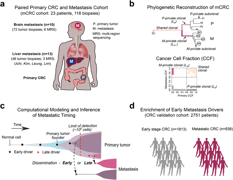
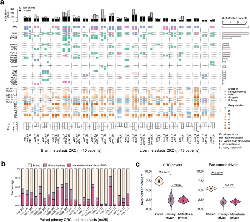
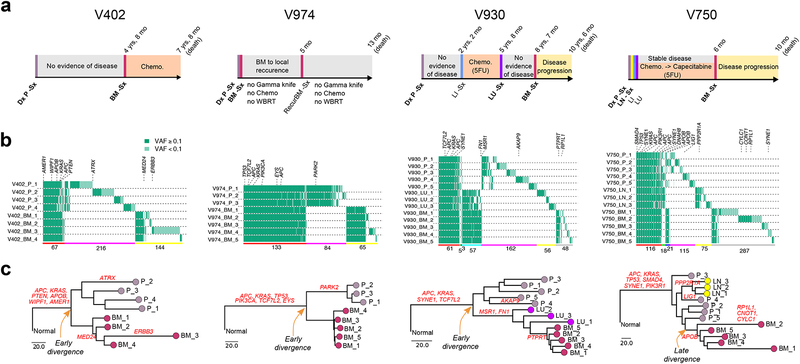
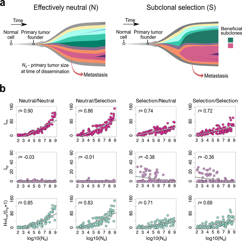
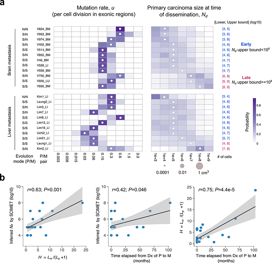
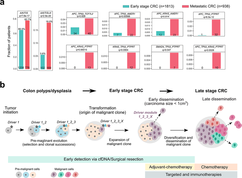
Both the timing and molecular determinants of metastasis are unknown, hindering treatment and prevention efforts. Here we characterize the evolutionary dynamics of this lethal process by analyzing exome-sequencing data from 118 biopsies from 23 patients with colorectal cancer with metastases to the liver or brain. The data show that the genomic divergence between the primary tumor and metastasis is low and that canonical driver genes were acquired early. Analysis within a spatial tumor growth model and statistical inference framework indicates that early disseminated cells commonly (81%, 17 out of 21 evaluable patients) seed metastases while the carcinoma is clinically undetectable (typically, less than 0.01 cm3). We validated the association between early drivers and metastasis in an independent cohort of 2,751 colorectal cancers, demonstrating their utility as biomarkers of metastasis. This conceptual and analytical framework provides quantitative in vivo evidence that systemic spread can occur early in colorectal cancer and illuminates strategies for patient stratification and therapeutic targeting of the canonical drivers of tumorigenesis.


Figures

Figure 1.
Figure 1.. Study overview.
(a) The metastatic colorectal cancer (mCRC) patient cohort includes 118 tumor biopsies from 23 patients. Paired CRCs with metastases to the brain and other sites (liver, lung, lymph nodes) from 10 patients and 72 tumor biopsies were whole-exome sequenced, including 6 cases with multi-region sequencing (MRS) of 3–5 regions each from the primary CRC and metastasis. Additionally, four publicly available cohorts with paired CRCs and liver metastases from 13 patients and 46 tumor biopsies were reanalyzed within the same bioinformatics framework, including 3 cases with MRS. (b) Tumor phylogenies were reconstructed from somatic alterations (sSNVs+indels). The mutational cancer cell fraction (CCF) was computed and compared for each primary CRCs and metastasis pair. (c) Schematic illustration of tumor evolution starting from a normal cell that acquires mutations leading to malignant transformation, growth of the primary tumor, metastatic dissemination, seeding and outgrowth. It is unknown whether dissemination occurs early from a dominant subclone when the size of the primary tumor is below the limits of clinical detection (108 cells or 1 cm3) (early dissemination) or later from a minor subclone after the acquisition of additional driver alterations (late dissemination). To address this question, we developed a 3-D model of tumor growth and statistical inference framework to time metastasis from patient genomic data. (d) We further leveraged a large collection of metastatic (n=938) and non-metastatic (n=1,813) CRCs with targeted sequencing data to evaluate the association between specific combinations of early driver genes (modules) identified in the mCRC cohort.
Figure 2.
Figure 2.. The mutational landscape and patterns of genetic divergence in paired primary CRCs and metastases.
(a) Concordance amongst somatic alterations (sSNVs, indels and CNAs) in known CRC ‘driver’ genes between paired primary CRCs and metastases. Stacked barplots illustrate the total number of sSNVs and indels in exonic regions with a lower cutoff of variant allele frequency (VAF)=0.1 in the corresponding site (primary or metastasis). (b) The percentage of clonal sSNVs that are shared, primary-private, or metastasis-private out of all clonal sSNVs with CCF>60% in any of paired primaries and distant metastases. (c) Violin plots illustrate the probability density of driver gene fold enrichment amongst shared, primary-private, and metastasis-private clonal non-silent sSNVs based on known CRC or pan-cancer ‘drivers’. The inset box corresponds to the 25th to 75th percentile (interquartile range, IQR); the horizontal line indicates the median; and the vertical line includes data within 1.5 times the IQR. A test statistic was computed based on n=100 down-samplings amongst patients (Methods). P-value, Wilcoxon Rank-Sum Test (two-sided).
Figure 3.
Figure 3.. Within and between lesion heterogeneity in paired primary CRCs and metastases.
(a) Clinical and treatment history for four representative CRC patients with brain metastases. Dx: diagnosis; Sx: surgical resection. (b) Patterns of within and between lesion heterogeneity amongst sSNVs and indels based on multi-region sequencing of paired primary CRCs and metastases, where canonical CRC driver genes are labeled. The number of mutations shared or private amongst different lesions is indicated below the corresponding colored horizontal bars: ubiquitously P-M shared (red), partially P-M shared (green-M1 or blue-M2), P-private (pink) or M-private (yellow-M1 or gray-M2 or cyan-M1 and M2). P corresponds to primary. M1 and M2 correspond to different metastatic sites in the same patient. (c) Phylogeny reconstruction via maximum parsimony (PHYLIP) based on mutational presence/absence, where canonical CRC drivers genes are labeled. VAF, variant allele frequency.
Figure 4.
Figure 4.. Correlation between the Lp, Lm and H and primary carcinoma size at the time of dissemination (Nd).
(a) Schematic illustration of effectively neutral (N) evolution and stringent subclonal selection (S), two distinct evolutionary modes that can occur during growth of the primary tumor or metastasis. It is assumed that metastatic dissemination occurs during expansion of the primary CRC where Nd corresponds to the size of the primary carcinoma at the time of dissemination. (b) The correlation between the timing of dissemination, Nd, and Lm, Lp or H, based on the spatial simulation of tumor growth (n=100 tumors for each scenario; Pearson’s r is reported). Lp and Lm correspond to the number of private clonal sSNVs (CCF>60% in one site and CCF<1% on the other site) in the whole primary carcinoma and metastasis, respectively and H=Lm/(Lp+1).
Figure 5.
Figure 5.. Patient-specific inference of the timing of metastasis in CRC.
(a) Heatmap of the posterior probability distributions inferred by SCIMET for the mutation rate u (per cell division in exonic regions) and Nd (timing of metastatic dissemination relative to primary carcinoma size) in individual P/M pairs (n=23) from 21 mCRC patients. The median of the inferred posterior distribution (referred to as the inferred Nd orNd˜) is indicated by a white circle at the corresponding value. For patients with more than one distant metastasis, each was analyzed independently. The mode of tumor evolution in each P/M pair was determined based on model selection within the statistical inference framework (Methods). We define early dissemination as Nd (upper bound) <108 cells (~1 cm3 in volume) and use the 3rd quartile of the posterior distribution as the upper bound to be conservative. Late dissemination is defined as Nd (upper bound) 108. P/M pairs where dissemination and seeding are inferred to have occurred early are denoted in blue, whereas those inferred to have disseminated late are denoted in magenta. (b) Correlations between the inferred timing of dissemination (Nd˜) based on SCIMET and the H metric as well as the time elapsed from diagnosis (Dx) of the primary to diagnosis of the metastasis (n=23). The Pearson’s r and P values are reported. Shading corresponds to 95% confidence interval of the linear regression.
Figure 6.
Figure 6.. Enrichment of early driver gene modules in mCRC and clinical implications of early dissemination.
(a) The enrichment of canonical ‘core’ CRC driver genes (APC, KRAS, TP53 or SMAD4; A, K, T or S) plus recurrent mutations in candidate drivers (AMER1, ATM, BRAF, PIK3CA, PTPRT or TCF7L2) identified in the mCRC cohort was evaluated in an independent cohort of 2,751 CRC patients. The combined barplots (left) illustrate the overall frequency of the ‘core’ module alone or with an additional candidate driver (‘X’) in early stage versus metastatic CRCs. Individual barplots indicate the frequency of specific ‘modules’. Q-values are based on two-sided Fisher’s exact tests with Benjamini–Hochberg adjustment. (b) Three stages of CRC progression are outlined: pre-malignancy (between initiation and transformation), early-stage (between transformation and dissemination) and late-stage (after dissemination). A set of potential interventions to prevent cancer mortality targets each stage: for pre-malignant lesions, resection (after detection via colonoscopy or possibly cell free DNA; cfDNA); for early-stage CRC, surgical resection and possibly adjuvant chemotherapy; and for late-stage CRC, chemotherapy and/or targeted/immune therapies. Given the high rate (80% here) of early dissemination, prior to clinical detectability of the early-stage CRC, detection and resection of pre-malignant lesions will have the greatest impact on preventing cancer mortality. For tumors that undergo dissemination prior to clinical detectability, surgical resection alone, even of a small tumor, cannot prevent metastasis. Once the early-stage tumor is discovered, newly defined metastatic modules (panel a) may inform patient stratification to aid the directed use of adjuvant chemotherapy.

Comment in

References

    1. Vanharanta S & Massague J Origins of metastatic traits. Cancer Cell 24, 410–21 (2013). - PMC - PubMed
    1. Turajlic S & Swanton C Metastasis as an evolutionary process. Science 352, 169–75 (2016). - PubMed
    1. Lambert AW, Pattabiraman DR & Weinberg RA Emerging Biological Principles of Metastasis. Cell 168, 670–691 (2017). - PMC - PubMed
    1. Jones S et al. Comparative lesion sequencing provides insights into tumor evolution. Proc Natl Acad Sci U S A 105, 4283–8 (2008). - PMC - PubMed
    1. Campbell PJ et al. The patterns and dynamics of genomic instability in metastatic pancreatic cancer. Nature 467, 1109–13 (2010). - PMC - PubMed

Methods-only References

    1. Berghoff AS et al. Differential role of angiogenesis and tumour cell proliferation in brain metastases according to primary tumour type: analysis of 639 cases. Neuropathol Appl Neurobiol 41, e41–55 (2015). - PubMed
    1. Berghoff AS et al. Invasion patterns in brain metastases of solid cancers. Neuro Oncol 15, 1664–72 (2013). - PMC - PubMed
    1. Li H & Durbin R Fast and accurate short read alignment with Burrows-Wheeler transform. Bioinformatics 25, 1754–60 (2009). - PMC - PubMed
    1. Cibulskis K et al. Sensitive detection of somatic point mutations in impure and heterogeneous cancer samples. Nat Biotechnol 31, 213–9 (2013). - PMC - PubMed
    1. Koboldt DC et al. VarScan 2: somatic mutation and copy number alteration discovery in cancer by exome sequencing. Genome Res 22, 568–76 (2012). - PMC - PubMed

Publication types

Substances