Review
. 2019 Jul 1;142(7):1858-1875.
doi: 10.1093/brain/awz144.

Assessment of lesions on magnetic resonance imaging in multiple sclerosis: practical guidelines

Affiliations
Review

Assessment of lesions on magnetic resonance imaging in multiple sclerosis: practical guidelines

Massimo Filippi et al. Brain. .

Abstract

MRI has improved the diagnostic work-up of multiple sclerosis, but inappropriate image interpretation and application of MRI diagnostic criteria contribute to misdiagnosis. Some diseases, now recognized as conditions distinct from multiple sclerosis, may satisfy the MRI criteria for multiple sclerosis (e.g. neuromyelitis optica spectrum disorders, Susac syndrome), thus making the diagnosis of multiple sclerosis more challenging, especially if biomarker testing (such as serum anti-AQP4 antibodies) is not informative. Improvements in MRI technology contribute and promise to better define the typical features of multiple sclerosis lesions (e.g. juxtacortical and periventricular location, cortical involvement). Greater understanding of some key aspects of multiple sclerosis pathobiology has allowed the identification of characteristics more specific to multiple sclerosis (e.g. central vein sign, subpial demyelination and lesional rims), which are not included in the current multiple sclerosis diagnostic criteria. In this review, we provide the clinicians and researchers with a practical guide to enhance the proper recognition of multiple sclerosis lesions, including a thorough definition and illustration of typical MRI features, as well as a discussion of red flags suggestive of alternative diagnoses. We also discuss the possible place of emerging qualitative features of lesions which may become important in the near future.

Keywords: diagnostic criteria; guidelines; lesions; magnetic resonance imaging; multiple sclerosis.


Figures

Figure 1
Figure 1
Characteristics of periventricular multiple sclerosis lesions that are typical (‘green flags’), atypical (‘red flags’), and those that should not be included in lesion count. Left column: Green flags: (A) examples of periventricular lesions suggestive of multiple sclerosis; (B) periventricular lesions perpendicular to the corpus callosum (‘Dawson’s fingers’). Middle column: Red flags: (C) multiple white matter lesions involving paraventricular and deep grey matter regions, suggestive of ischaemic small-vessel disease; (D) extensive posterior corpus callosum involvement and bilateral diencephalic hyperintense lesions in neuromyelitis optica spectrum disorders; (E) multiple lesions affecting deep white matter, external capsule, and temporal lobes in cerebral autosomal dominant arteriopathy with subcortical infarcts and leukoencephalopathy; (F) intra-callosal ‘snowball’ lesions in Susac syndrome; (G) diffuse and extensive lesions affecting both white matter and deep grey matter in systemic lupus erythematosus. Right column: Lesions that should not be considered periventricular: (H) lesion not touching the lateral ventricles; (I) anterior and posterior symmetric periventricular ‘capping’; (J) lesion smaller than 3 mm in longest axis; (K) symmetric linear hyperintensities abutting the lateral ventricles. PV = periventricular.
Figure 2
Figure 2
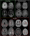
Characteristics of cortical/juxtacortical multiple sclerosis that are typical (‘green flags’) and atypical (‘red flags’), as well as those that should not be included. Top left: Green flags: examples of (A) juxtacortical lesions and (B) cortical lesions suggestive of multiple sclerosis. Top right: (C) white matter lesions not touching the cortex or within the cortex (subcortical). Bottom: Red flags: (D) multiple white matter lesions involving subcortical and deep white matter, suggestive of small-vessel disease; (E) lesions involving the grey matter-white matter border of different brain lobes with ill-defined borders in progressive multifocal leukoencephalopathy; (F) multiple well-defined CSF-like abnormalities that appear as dots or stripes in enlarged Virchow-Robin space; (G) hypointensity on T2-weighted sequence suggesting haemosiderin deposit due to a microbleed; (H) multiple leptomeningeal/cortical hyperintensities on T1-weighted imaging with associated hypointensity on gradient-echo sequence in CNS vasculitis. JC/CL = juxtacortical/cortical.
Figure 3
Figure 3
Characteristics of infratentorial multiple sclerosis lesions that are typical (‘green flags’) and atypical (‘red flags’). Left column: Green flags: (A) examples of infratentorial lesions suggestive of multiple sclerosis. Right column: Red flags: (B) symmetric central pontine lesions in small-vessel disease; (C) periaqueductal lesion in neuromyelitis optica spectrum disorder; (D) area postrema lesions in neuromyelitis optica spectrum disorder; (E) mesencephalic-diencecephalic lesion in anti-MOG syndrome; (F) large ovoid lesion close to the floor of the fourth ventricle in neuro-Behçet disease.
Figure 4
Figure 4
Characteristics of spinal cord multiple sclerosis lesions that are typical (‘green flags’) and atypical (‘red flags’), and those that should not be included. Top left: Green flags: examples of (A) spinal cord lesions in the cervical and thoracic cord on sagittal short-tau inversion recovery sequence; (B) cervical cord lesions showing hypointensity on T1-weighted sequences at 3 T (green arrowheads); (C) a cervical cord lesion showing involvement of the lateral column and central grey matter (green arrows) on T2-weighted and phase sensitive inversion recovery sequences. Top right: (D) ‘Diffuse’ spinal cord lesions with ill-defined borders not included for the definition of spinal cord involvement. Bottom: Red flags: (E) longitudinally extensive transverse myelitis affecting more than three vertebral segments in neuromyelitis optica spectrum disorder; (F) longitudinally extensive spinal cord lesion affecting more than three vertebral segments associated with leptomeningeal and peripheral spinal cord contrast-enhancement in neuro-sarcoidosis; (G) extensive and selective involvement of lateral and posterior columns in subacute combined neurodegeneration; (H) spinal cord cavities in syringomyelia; (I) extensive T2-hyperintense lesion extending rostrally from the conus, with spotted and tortuous regions of contrast enhancement in an arteriovenous fistula; (J) hyperintense lesion in the anterior portion of the thoracic spinal cord extending for more than two vertebral segments in a case of subacute ischaemic myelopathy; (K) T2-hyperintense lesion of the cervical cord showing ‘pancake-like’ gadolinium enhancement in a case of spondylotic myelopathy. SC = spinal cord.
Figure 5
Figure 5
Characteristics of gadolinium-enhancing multiple sclerosis lesions that are typical (‘green flags’) and atypical (‘red flags’), and those that should not be included. Top left: Green flags: examples of contrast enhancement suggestive of multiple sclerosis: (A) nodular; (B) open-ring; (C) closed-ring; (D) spinal cord nodular enhancement. Top right: (E) Capillary telangiectasia (not to be counted for the diagnostic criteria). Bottom: Red flags: (F) inhomogeneous enhancement of a large (>2 cm) tumefactive lesion suggestive of an atypical idiopathic inflammatory demyelinating lesion; (G) band-like enhancement in Balò disease; (H) enhancement of the diencephalon, the corpus callosum (in a ‘cloud-like’ pattern), and a longitudinally extensive spinal cord lesion in neuromyelitis optica spectrum disorder; (I) irregular leptomeningeal, cortical, and subcortical enhancement in a vasculitis of the CNS; (J) leptomeningeal and pial enhancement and the ‘trident sign’ on axial images in neurosarcoidosis; (K) homogeneous diencephalic enhancement in anti-Ma2 encephalitis; (L) irregular and inhomogeneous enhancement in glioblastoma.

References

    1. Absinta M, Rocca MA, Colombo B, Copetti M, De Feo D, Falini A, et al.Patients with migraine do not have MRI-visible cortical lesions. J Neurol 2012; 259: 2695–8. - PubMed
    1. Absinta M, Sati P, Reich DS. Advanced MRI and staging of multiple sclerosis lesions. Nat Rev Neurol 2016a; 12: 358–68. - PMC - PubMed
    1. Absinta M, Sati P, Schindler M, Leibovitch EC, Ohayon J, Wu T, et al.Persistent 7-tesla phase rim predicts poor outcome in new multiple sclerosis patient lesions. J Clin Invest 2016b; 126: 2597–609. - PMC - PubMed
    1. Absinta M, Vuolo L, Rao A, Nair G, Sati P, Cortese IC, et al.Gadolinium-based MRI characterization of leptomeningeal inflammation in multiple sclerosis. Neurology 2015; 85: 18–28. - PMC - PubMed
    1. Aiken AH, Mukherjee P, Green AJ, Glastonbury CM. MR imaging of optic neuropathy with extended echo-train acquisition fluid-attenuated inversion recovery. AJNR Am J Neuroradiol 2011; 32: 301–5. - PMC - PubMed

Publication types