

Prospective acceleration of parallel RF transmission-based 3D chemical exchange saturation transfer imaging with compressed sensing
- PMID: 31209938
- PMCID: PMC6660350
- DOI: 10.1002/mrm.27875
Prospective acceleration of parallel RF transmission-based 3D chemical exchange saturation transfer imaging with compressed sensing
Abstract
Purpose: To develop prospectively accelerated 3D CEST imaging using compressed sensing (CS), combined with a saturation scheme based on time-interleaved parallel transmission.
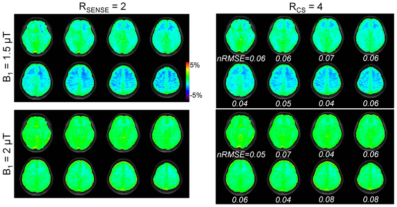
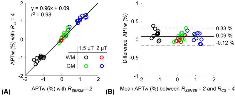
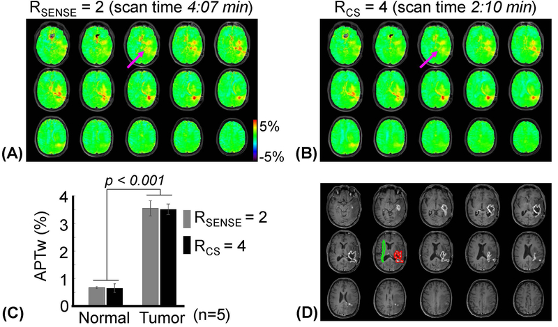
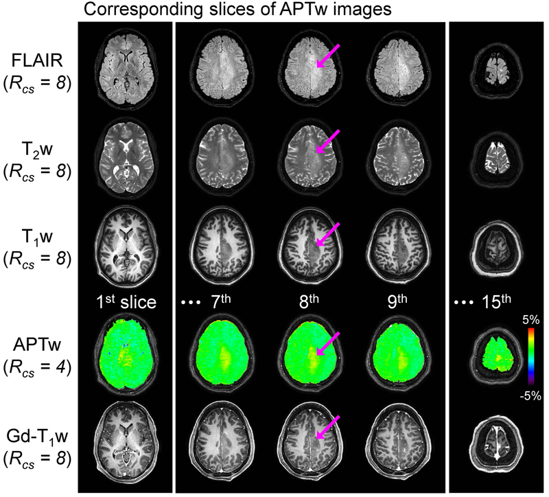
Methods: A variable density pseudo-random sampling pattern with a centric elliptical k-space ordering was used for CS acceleration in 3D. Retrospective CS studies were performed with CEST phantoms to test the reconstruction scheme. Prospectively CS-accelerated 3D-CEST images were acquired in 10 healthy volunteers and 6 brain tumor patients with an acceleration factor (RCS ) of 4 and compared with conventional SENSE reconstructed images. Amide proton transfer weighted (APTw) signals under varied RF saturation powers were compared with varied acceleration factors.
Results: The APTw signals obtained from the CS with acceleration factor of 4 were well-preserved as compared with the reference image (SENSE R = 2) both in retrospective phantom and prospective healthy volunteer studies. In the patient study, the APTw signals were significantly higher in the tumor region (gadolinium [Gd]-enhancing tumor core) than in the normal tissue (p < .001). There was no significant APTw difference between the CS-accelerated images and the reference image. The scan time of CS-accelerated 3D APTw imaging was dramatically reduced to 2:10 minutes (in-plane spatial resolution of 1.8 1.8 mm2 ; 15 slices with 4-mm slice thickness) as compared with SENSE (4:07 minutes).
Conclusion: Compressed sensing acceleration was successfully extended to 3D-CEST imaging without compromising CEST image quality and quantification. The CS-based CEST imaging can easily be integrated into clinical protocols and would be beneficial for a wide range of applications.
Keywords: APT; CEST; brain tumor; compressed sensing; parallel RF transmission.
© 2019 International Society for Magnetic Resonance in Medicine.
Figures

Similar articles
-
Accelerating chemical exchange saturation transfer (CEST) MRI by combining compressed sensing and sensitivity encoding techniques.Magn Reson Med. 2017 Feb;77(2):779-786. doi: 10.1002/mrm.26141. Epub 2016 Feb 17. Magn Reson Med. 2017. PMID: 26888295 Free PMC article.
-
Fast 3D chemical exchange saturation transfer imaging with variably-accelerated sensitivity encoding (vSENSE).Magn Reson Med. 2019 Dec;82(6):2046-2061. doi: 10.1002/mrm.27881. Epub 2019 Jul 1. Magn Reson Med. 2019. PMID: 31264278
-
Accelerating chemical exchange saturation transfer MRI with parallel blind compressed sensing.Magn Reson Med. 2019 Jan;81(1):504-513. doi: 10.1002/mrm.27400. Epub 2018 Aug 26. Magn Reson Med. 2019. PMID: 30146714 Free PMC article.
-
Three-dimensional chemical exchange saturation transfer imaging using compressed SENSE for full z-spectrum acquisition.Magn Reson Imaging. 2022 Oct;92:58-66. doi: 10.1016/j.mri.2022.05.014. Epub 2022 May 28. Magn Reson Imaging. 2022. PMID: 35640858
-
Clinical applications of chemical exchange saturation transfer (CEST) MRI.J Magn Reson Imaging. 2018 Jan;47(1):11-27. doi: 10.1002/jmri.25838. Epub 2017 Aug 9. J Magn Reson Imaging. 2018. PMID: 28792646 Free PMC article. Review.
Cited by
-
CEST-MRI for body oncologic imaging: are we there yet?NMR Biomed. 2023 Jun;36(6):e4906. doi: 10.1002/nbm.4906. Epub 2023 Feb 15. NMR Biomed. 2023. PMID: 36640112 Free PMC article. Review.
-
Bloch simulator-driven deep recurrent neural network for magnetization transfer contrast MR fingerprinting and CEST imaging.Magn Reson Med. 2023 Oct;90(4):1518-1536. doi: 10.1002/mrm.29748. Epub 2023 Jun 15. Magn Reson Med. 2023. PMID: 37317675 Free PMC article.
-
CEST and nuclear Overhauser enhancement imaging with deep learning-extrapolated semisolid magnetization transfer reference: Scan-rescan reproducibility and reliability studies.Magn Reson Med. 2024 Mar;91(3):1002-1015. doi: 10.1002/mrm.29937. Epub 2023 Nov 27. Magn Reson Med. 2024. PMID: 38009996 Free PMC article.
-
Learning-based optimization of acquisition schedule for magnetization transfer contrast MR fingerprinting.NMR Biomed. 2022 May;35(5):e4662. doi: 10.1002/nbm.4662. Epub 2021 Dec 22. NMR Biomed. 2022. PMID: 34939236 Free PMC article.
-
Accelerated multi-target chemical exchange saturation transfer magnetic resonance imaging of the mouse heart.Phys Med Biol. 2021 Jul 16;66(14):10.1088/1361-6560/ac0e78. doi: 10.1088/1361-6560/ac0e78. Phys Med Biol. 2021. PMID: 34167100 Free PMC article.
References
-
- Ward KM, Aletras AH, Balaban RS. A new class of contrast agents for MRI based on proton chemical exchange dependent saturation transfer (CEST). J Magn Reson 2000;143:79–87. - PubMed
-
- Zhou J, Payen J, Wilson DA, Traystman RJ, van Zijl PCM. Using the amide proton signals of intracellular proteins and peptides to detect pH effects in MRI. Nature Med 2003;9:1085–1090. - PubMed
Publication types
MeSH terms
Substances
Grants and funding
LinkOut - more resources
Full Text Sources
Medical
