

The Modulation of Pain by Metabotropic Glutamate Receptors 7 and 8 in the Dorsal Striatum
- PMID: 31210112
- PMCID: PMC7327935
- DOI: 10.2174/1570159X17666190618121859
The Modulation of Pain by Metabotropic Glutamate Receptors 7 and 8 in the Dorsal Striatum
Abstract
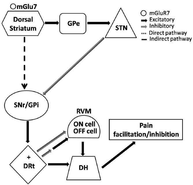
The dorsal striatum, apart from controlling voluntary movement, displays a recently demonstrated pain inhibition. It is connected to the descending pain modulatory system and in particular to the rostral ventromedial medulla through the medullary dorsal reticular nucleus. Diseases of the basal ganglia, such as Parkinson's disease, in addition to being characterized by motor disorders, are associated with pain and hyperactivation of the excitatory transmission. A way to counteract glutamatergic hyperactivation is through the activation of group III metabotropic glutamate receptors (mGluRs), which are located on presynaptic terminals inhibiting neurotransmitter release. So far the mGluRs of group III have been the least investigated, owing to a lack of selective tools. More recently, selective ligands for each mGluR of group III, in particular positive and negative allosteric modulators, have been developed and the role of each subtype is starting to emerge. The neuroprotective potential of group III mGluRs in pathological conditions, such as those characterized by elevate glutamate, has been recently shown. In the dorsal striatum, mGluR7 and mGluR8 are located at glutamatergic corticostriatal terminals and their stimulation inhibits pain in pathological conditions such as neuropathic pain. The two receptors in the dorsal striatum have instead a different role in pain control in normal conditions. This review will discuss recent results focusing on the contribution of mGluR7 and mGluR8 in the dorsal striatal control of pain. The role of mGluR4, whose antiparkinsonian activity is widely reported, will also be addressed.
Keywords: chronic pain; descending pain modulatory system; dorsal striatum; hyperglutamatergism; mGluR7; mGluR8..
Copyright© Bentham Science Publishers; For any queries, please email at epub@benthamscience.net.
Figures


Similar articles
-
Nociception modulation by supraspinal group III metabotropic glutamate receptors.J Neurochem. 2017 May;141(4):507-519. doi: 10.1111/jnc.13725. Epub 2017 Feb 22. J Neurochem. 2017. PMID: 27363363 Review.
-
Metabotropic glutamate receptor subtype 7 in the dorsal striatum oppositely modulates pain in sham and neuropathic rats.Neuropharmacology. 2018 Jun;135:86-99. doi: 10.1016/j.neuropharm.2018.03.003. Epub 2018 Mar 2. Neuropharmacology. 2018. PMID: 29505788
-
Supraspinal metabotropic glutamate receptor subtype 8: a switch to turn off pain.Amino Acids. 2014 Jun;46(6):1441-8. doi: 10.1007/s00726-014-1703-5. Epub 2014 Mar 13. Amino Acids. 2014. PMID: 24623118 Review.
-
Group III human metabotropic glutamate receptors 4, 7 and 8: molecular cloning, functional expression, and comparison of pharmacological properties in RGT cells.Brain Res Mol Brain Res. 1998 Jan;53(1-2):88-97. doi: 10.1016/s0169-328x(97)00277-5. Brain Res Mol Brain Res. 1998. PMID: 9473604
-
Supraspinal metabotropic glutamate receptors: a target for pain relief and beyond.Eur J Neurosci. 2014 Feb;39(3):444-54. doi: 10.1111/ejn.12398. Eur J Neurosci. 2014. PMID: 24494684 Review.
Cited by
-
The Role of the Subnucleus Reticularis Dorsalis (SRD) in Pain Modulation: A Literature Review.Curr Med Sci. 2025 Aug;45(4):745-754. doi: 10.1007/s11596-025-00082-8. Epub 2025 Jul 7. Curr Med Sci. 2025. PMID: 40622437 Review.
-
GRM7 deficiency, from excitotoxicity and neuroinflammation to neurodegeneration: Systematic review of GRM7 deficient patients.Brain Behav Immun Health. 2024 Jun 17;39:100808. doi: 10.1016/j.bbih.2024.100808. eCollection 2024 Aug. Brain Behav Immun Health. 2024. PMID: 38983774 Free PMC article. Review.
-
Novel Molecular Hallmarks of Group 3 Medulloblastoma by Single-Cell Transcriptomics.Front Oncol. 2021 Mar 18;11:622430. doi: 10.3389/fonc.2021.622430. eCollection 2021. Front Oncol. 2021. PMID: 33816256 Free PMC article.
-
Translational Value of the Transdermal Administration of Bergamot Essential Oil and of Its Fractions.Pharmaceutics. 2022 May 7;14(5):1006. doi: 10.3390/pharmaceutics14051006. Pharmaceutics. 2022. PMID: 35631592 Free PMC article.
-
L-Acetylcarnitine causes analgesia in mice modeling Fabry disease by up-regulating type-2 metabotropic glutamate receptors.Mol Pain. 2022 Jan-Dec;18:17448069221087033. doi: 10.1177/17448069221087033. Mol Pain. 2022. PMID: 35255745 Free PMC article.
References
Publication types
MeSH terms
Substances
LinkOut - more resources
Full Text Sources
Medical
