

Relationship Study of The Verified H uman Epidermal Growth Factor Receptor 2 Amplification with Other Tumor Markers and Clinicohistopathological Characteristics in Patients with Invasive Breast Cancer, Using Chromogenic In Situ Hybridization
- PMID: 31210439
- PMCID: PMC6582419
- DOI: 10.22074/cellj.2019.6219
Relationship Study of The Verified H uman Epidermal Growth Factor Receptor 2 Amplification with Other Tumor Markers and Clinicohistopathological Characteristics in Patients with Invasive Breast Cancer, Using Chromogenic In Situ Hybridization
Abstract
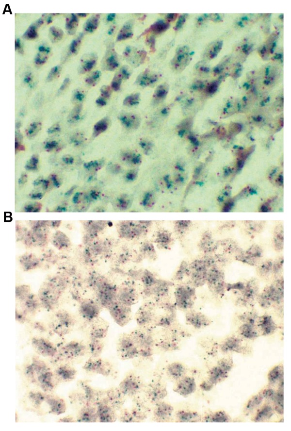
Objective: Human epidermal growth factor receptor 2 (HER-2), as a crucial factor involved in about 20% of breast cancer cases, is one of the most reliable tumor markers to determine prognosis and therapeutic trend of this disease. This marker is generally assessed by immunohistochemistry (IHC) technique. In the cases that result of IHC test cast doubt (+2), the test should be repeated or validated by applying in situ hybridization techniques, like chromogenic in situ hybridization (CISH). In this regard, the goal of current study was to figure out the link between different clinicopathological characteristics of patients suffering from invasive breast cancer, using tumor markers, hormone receptor (HR) and HER-2. Comparing IHC and CISH techniques for evaluating diagnostic value and usefulness of HER-2 were also the other objective of this study.
Materials and methods: Based on this retrospective study, histological markers of 113 individuals suffering from invasive breast cancer -such as estrogen receptor (ER), progesterone receptor, HER-2 receptor, E-cadherin, CK5/6, vimentin and Ki67 were examined by IHC technique. HER-2 amplification of all patients was also evaluated by CISH. Clinicopathological information of the patients was also extracted from medical documents and their associations with tumor markers were statistically evaluated.
Results: There is a significant relationship between tumor size, CK5/6 and tumor grade with HR status. Similar relationship was observed between HER-2 status and HR status, as well as vascular invasion (P<0.05). The comparison of HER-2 amplification showed no complete concordance of the result obtained from these two techniques, with score +3.
Conclusion: Since the status of HER-2 is very important in decision making of the treatment process, CISH technique is recommended in the malignant conditions as the primary test, instead of IHC. In this study, we also determined that HER-2 expression is greatly correlated with ER- and PR- status. This might propose a better prognosis for HER-2+ patients.
Keywords: Breast Cancer; Chromogenic In Situ Hybridization; HER-2; Tumor Markers.
Copyright© by Royan Institute. All rights reserved.
Conflict of interest statement
There is no conflict of interest in this study.
Figures

References
-
- Nosrati H, Mozdarani H, Hadad P, Omranipour R, Mozdarani S, Bakhshandeh M, et al. Inherent radiation sensitivity of lymphocytes of triple negative (TN) and luminal a: a comparison between patients with breast cancer and normal individuals as assayed by the micronucleus test. Arch Breast Cancer. 2017;4(1):10–15.
LinkOut - more resources
Full Text Sources
Research Materials
Miscellaneous