

Ultrasonographic evaluation of the fetal central nervous system: review of guidelines
- PMID: 31210692
- PMCID: PMC6561375
- DOI: 10.1590/0100-3984.2018.0056
Ultrasonographic evaluation of the fetal central nervous system: review of guidelines
Abstract
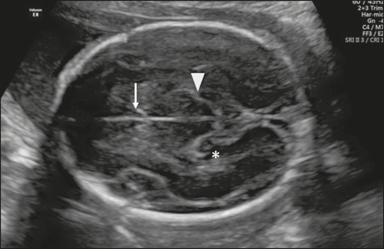
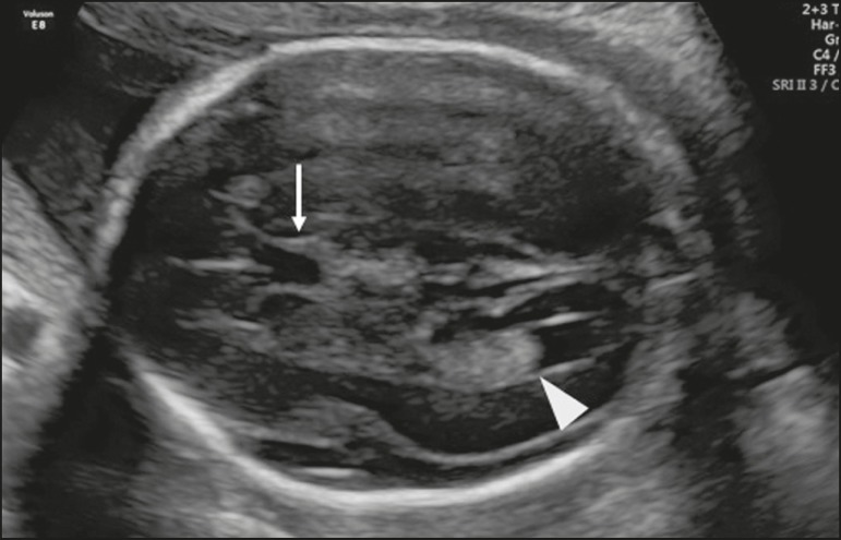
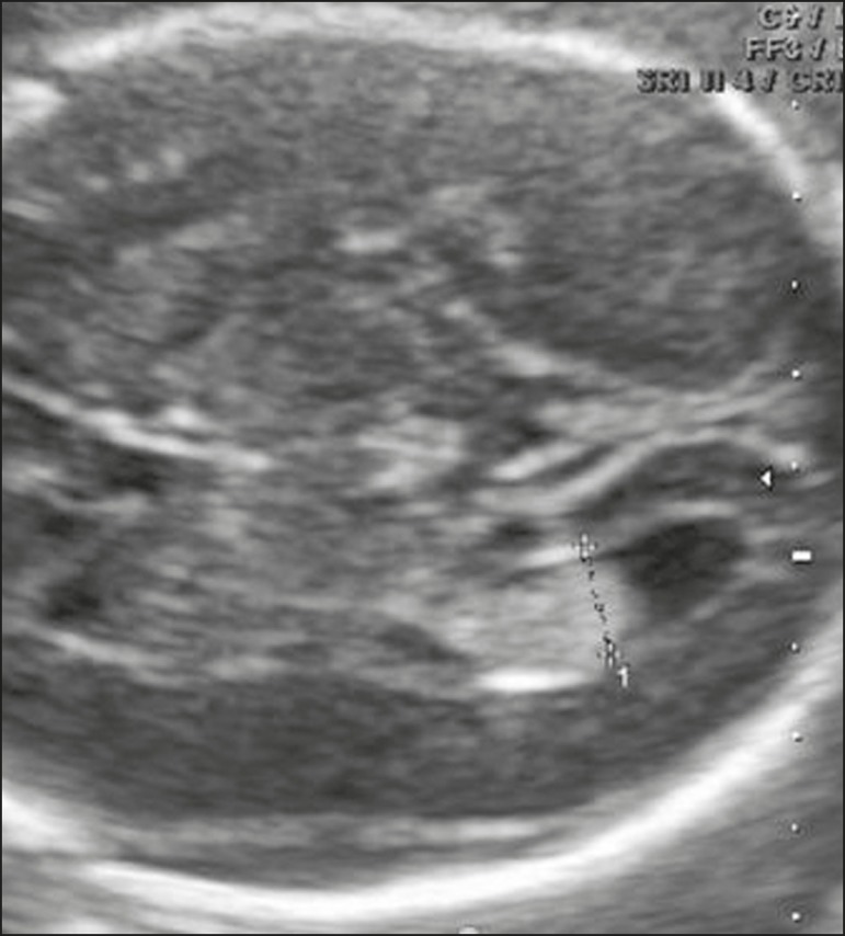
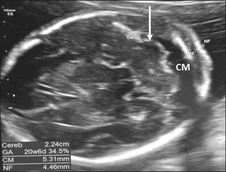
Central nervous system malformations constitute the second most common group of anomalies in fetuses. Such malformations have assumed clinical importance because of their association with high rates of perinatal morbidity and mortality. Therefore, it is extremely important to assess the fetal central nervous system during the prenatal period, in order to identify any changes in its development and thereby gain sufficient information to advise parents about pregnancy follow-up, options for fetal therapy, and the timing/type of delivery, as well as the postnatal treatment and prognosis. The objective of this review was to describe the ultrasonographic evaluation of the fetal central nervous system as per the guidelines of the International Society of Ultrasound in Obstetrics and Gynecology.
Malformações do sistema nervoso central são o segundo mais frequente grupo de anomalias que afetam o feto. Elas têm adquirido grande importância em razão da sua associação com altas taxas de morbidade e mortalidade perinatal. Além do mais, é extremamente importante avaliar o sistema nervoso central fetal durante o período pré-natal, de modo a se diagnosticar possíveis mudanças no desenvolvimento e aconselhar os casais sobre o seguimento da gestação, possibilidades terapêuticas fetais, tempo e tipo de parto, tratamento pós-natal e prognóstico. O objetivo desta revisão foi descrever a avaliação do sistema nervoso central fetal por meio das recomendações da International Society of Ultrasound in Obstetrics and Gynecology.
Keywords: Central nervous system; Fetus; Practice guidelines as topic; Ultrasonography.
Figures
References
-
- Wticzak M, Ferenc T, Wilczyński J. Pathogenesis and genetics of neural tube defects. Ginekol Pol. 2007;78:981–985. - PubMed
-
- Malinger G, Lerman-Sagie T. Normal two- and three-dimensional neurosonography of the prenatal brain. In: Timor-Tritsch IE, Monteagudo A, Pilu G, et al., editors. Ultrasonography of the prenatal brain. 3rd ed. New York, NY: McGrawHill Medical; 2012. pp. 15–102.
-
- International Society of Ultrasound in Obstetrics & Gynecology Education Committee, editor. Sonographic examination of the fetal central nervous system: guidelines for performing the 'basic examination' and the 'fetal neurosonogram'. Ultrasound Obstet Gynecol. 2007;29:109–116. - PubMed
-
- Malinger G, Lev D, Lerman-Sagie T. Normal and abnormal fetal brain development during the third trimester as demonstrated by neurosonography. Eur J Radiol. 2006;57:226–232. - PubMed
LinkOut - more resources
Full Text Sources