. 2019 May 9:2019:3182627.
doi: 10.1155/2019/3182627. eCollection 2019.

Lupeol, a Dietary Triterpene, Enhances Wound Healing in Streptozotocin-Induced Hyperglycemic Rats with Modulatory Effects on Inflammation, Oxidative Stress, and Angiogenesis

Affiliations

Lupeol, a Dietary Triterpene, Enhances Wound Healing in Streptozotocin-Induced Hyperglycemic Rats with Modulatory Effects on Inflammation, Oxidative Stress, and Angiogenesis

Fernando Pereira Beserra et al. Oxid Med Cell Longev. .

Erratum in

Abstract

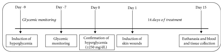
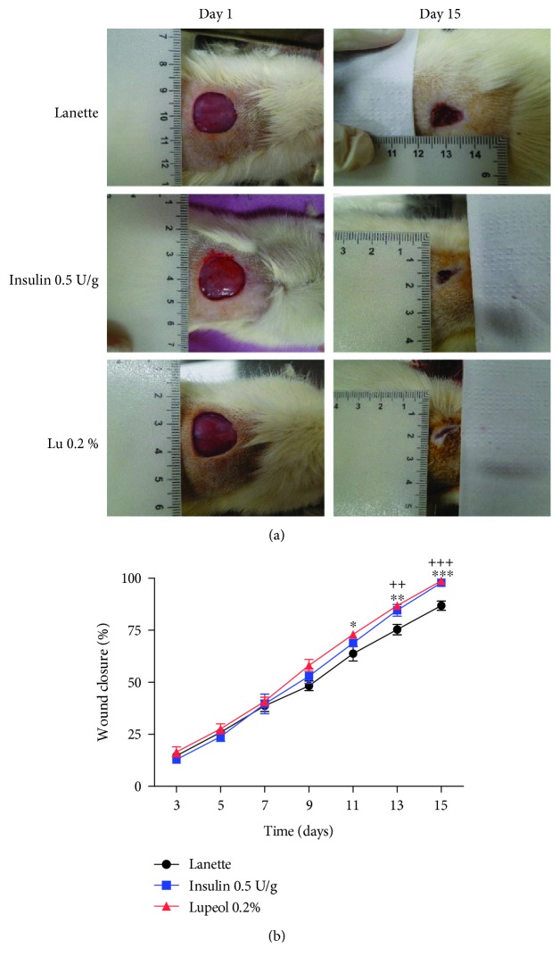
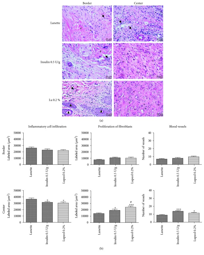
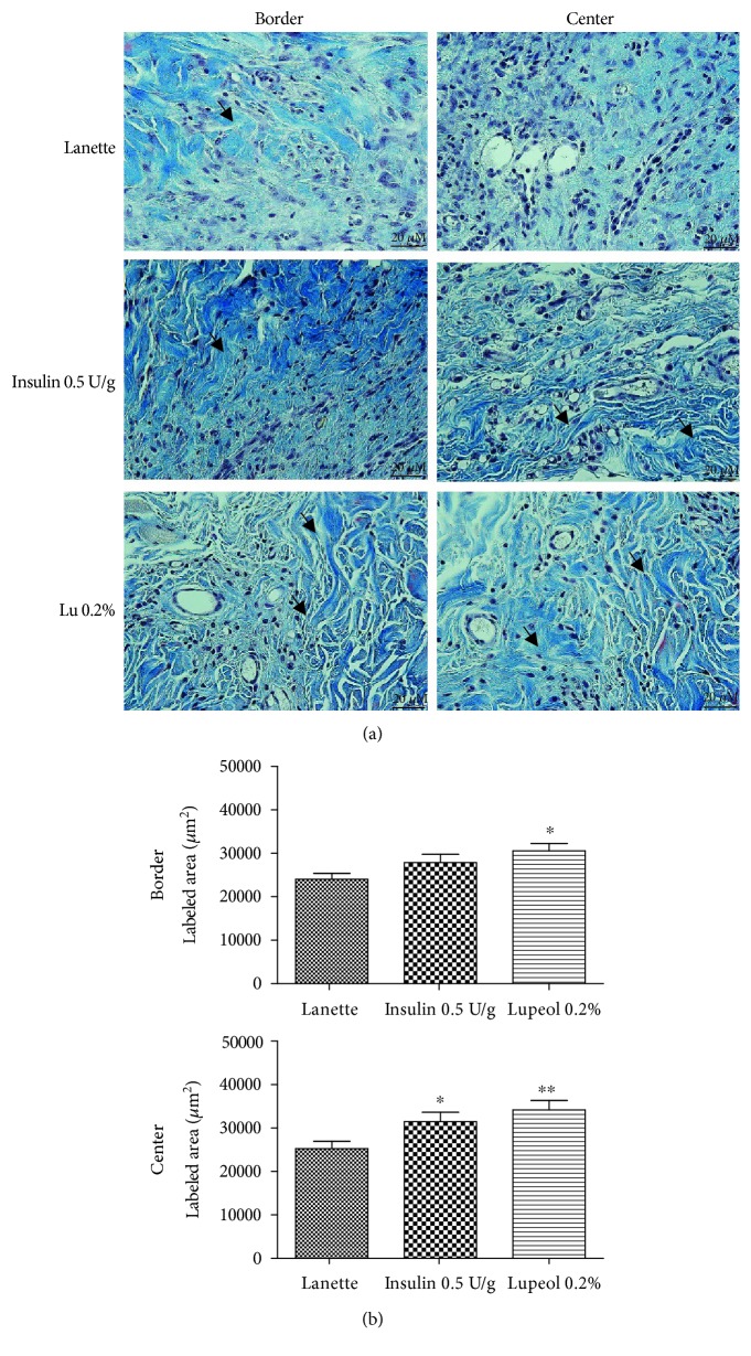
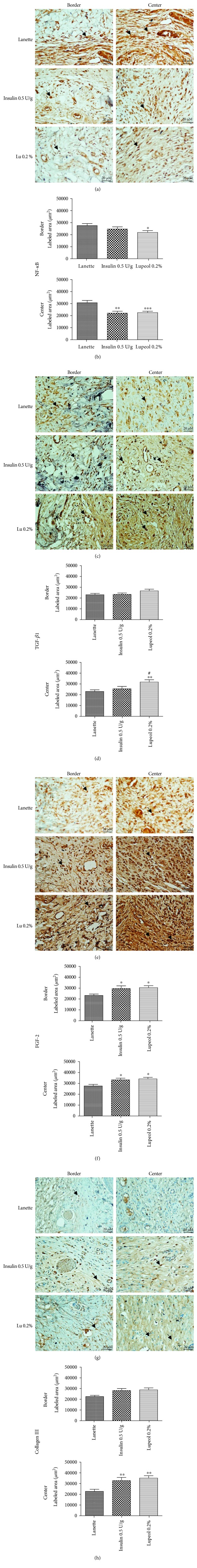
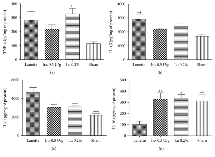
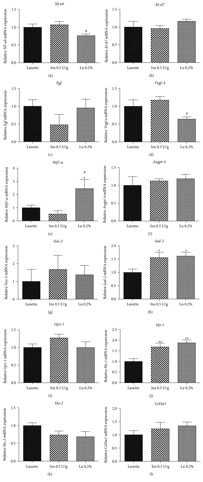
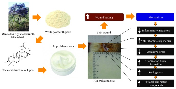
Impaired wound healing is a debilitating complication of diabetes that leads to significant morbidity, particularly foot ulcers. Natural products have shown to be effective in treating skin wounds. Lupeol is known to stimulate angiogenesis, fibroblast proliferation, and expressions of cytokines and growth factors involved in wound healing. The study is performed to evaluate the wound healing activity of lupeol in streptozotocin-induced hyperglycemic rats by macroscopical, histological, immunohistochemical, immunoenzymatic, and molecular methods. Percentage of wound closure and contraction was increased in the lupeol-treated group when compared to the Lanette group. Histopathological observation revealed decreased inflammatory cell infiltration and increased proliferation of fibroblasts, vascularization, and deposition of collagen fibers after lupeol treatment. Immunohistochemical analyses showed decreased intensity of NF-κB and increased intensity of FGF-2, TGF-β1, and collagen III. ELISA results revealed downregulated IL-6 levels and upregulated IL-10 levels in response to lupeol. The mRNA expression levels of Hif-1α, Sod-2, and Ho-1 were significantly increased in response to lupeol as compared to Lanette whereas Nf-κb and Vegf-A levels were decreased in relation to insulin and lupeol treatment. These findings indicate that lupeol possesses wound healing potential in hyperglycemic conditions and may be useful as a treatment for chronic wounds in diabetic patients.


Figures

Figure 1
Figure 1
Overview of the experimental protocol for wound healing in streptozotocin-induced hyperglycemic rats.
Figure 2
Figure 2
Effect of lupeol on skin wound healing in streptozotocin-induced hyperglycemic rats. Representative images of the lesion area (initial and from the last day of experimentation) of animals treated with Lanette, insulin 0.5 U/g, and lupeol 0.2% (a) and wound closure (%) on days 3, 5, 7, 9, 11, 13, and 15 postwounding (b). p < 0.05, ∗∗ p < 0.01, and ∗∗∗ p < 0.001 represent lupeol vs. Lanette group. ++ p < 0.01 and +++ p < 0.001 represent insulin vs. Lanette group, using ANOVA followed by the Newman-Keuls test. Lu 0.2% = lupeol 0.2%.
Figure 3
Figure 3
HE-stained skin tissue sections on day 14 postwound induction in streptozotocin-induced hyperglycemic rats (a). Inflammatory cell infiltration, proliferation of fibroblasts, and number of blood vessels (b) in HE staining of the border and central region of rats' hyperglycemic wounds treated with Lanette, insulin 0.5 U/g, or lupeol 0.2% for 14 days. p < 0.05 and ∗∗∗ p < 0.001 vs. Lanette group. # p < 0.05 vs. insulin group, using ANOVA followed by the Newman-Keuls test. Bar represents 20 μm. Black arrows indicate the presence of inflammatory cells, # indicates collagen fibers, indicates blood vessels, + indicates fibroblasts, and ▲ indicates sebaceous glands. Lu 0.2% = lupeol 0.2%.
Figure 4
Figure 4
Masson's trichrome-stained skin tissue sections on day 14 postwound induction in streptozotocin-induced hyperglycemic rats (a). Labeled area of total collagen fibers (b) (μm2) in the border and central region of rats' hyperglycemic wounds treated with Lanette, insulin 0.5 U/g, or lupeol 0.2% for 14 days. p < 0.05 and ∗∗ p < 0.01 vs. Lanette group, using ANOVA followed by the Newman-Keuls test. Bar represents 20 μm. Black arrows indicate the presence of total collagen fibers. Lu 0.2% = lupeol 0.2%.
Figure 5
Figure 5
Photomicrography of the immunostaining and immunolabeled area (μm2) for NF-κB (a, b), TGF-β1 (c, d), FGF-2 (e, f), and collagen III (g, h) in the border and central region of rats' hyperglycemic wounds treated with Lanette, insulin 0.5 U/g, or lupeol 0.2% for 14 days. p < 0.05, ∗∗ p < 0.01, and ∗∗∗ p < 0.001 vs. Lanette group. # p < 0.05 vs. insulin group, using ANOVA followed by the Newman-Keuls test. Bar represents 20 μm. Black arrows indicate antibody staining against NF-κB, TGF-β1, FGF-2, and collagen III. Lu 0.2% = lupeol 0.2%.
Figure 6
Figure 6
Quantification of TNF-α (a), IL-1β (b), IL-6 (c), and IL-10 (d) levels (pg/mg protein) in rats' hyperglycemic wounds treated with Lanette, insulin 0.5 U/g, or lupeol 0.2% for 14 days. p < 0.05, ∗∗ p < 0.01, and ∗∗∗ p < 0.001 vs. Lanette group. + p < 0.05 and ++ p < 0.01 vs. sham group, using ANOVA followed by the Newman-Keuls test. Lu 0.2% = lupeol 0.2%.
Figure 7
Figure 7
Gene expression (RT-qPCR) of Nf-κb (a), Ki-67 (b), Egf (c), Vegf-A (d), Hif-1α (e), Angiopoietin-4 (f), Nos-2 (g), Sod-2 (h), Gpx-1 (i), Ho-1 (j), Ho-2 (k), and Col3α1 (l) in rats' hyperglycemic wounds treated with Lanette, insulin 0.5 U/g, or lupeol 0.2% for 14 days. p < 0.05 and ∗∗ p < 0.01 vs. Lanette group. # p < 0.05 vs. insulin group, using ANOVA followed by the Newman-Keuls test. Lu 0.2% = lupeol 0.2%.
Figure 8
Figure 8
Schematic representation of the regulatory mechanisms of lupeol-based cream in wound healing of hyperglycemic rats.

References

    1. Tripathi B. K., Srivastava A. K. Diabetes mellitus: complications and therapeutics. Medical Science Monitor. 2006;12(7):RA130–RA147. - PubMed
    1. World Health Organization. Global report on diabetes. 2006, January 2019, https://www.who.int/news-room/fact-sheets/detail/diabetes/
    1. Tan W. S., Arulselvan P., Ng S. F., Mat Taib C. N., Sarian M. N., Fakurazi S. Improvement of diabetic wound healing by topical application of Vicenin-2 hydrocolloid film on Sprague Dawley rats. BMC Complementary and Alternative Medicine. 2019;19(1):p. 20. doi: 10.1186/s12906-018-2427-y. - DOI - PMC - PubMed
    1. Zhang X. N., Ma Z. J., Wang Y., et al. Angelica dahurica ethanolic extract improves impaired wound healing by activating angiogenesis in diabetes. PLoS One. 2017;12(5, article e0177862) doi: 10.1371/journal.pone.0177862. - DOI - PMC - PubMed
    1. Kido D., Mizutani K., Takeda K., et al. Impact of diabetes on gingival wound healing via oxidative stress. PLoS One. 2017;12(12, article e0189601) doi: 10.1371/journal.pone.0189601. - DOI - PMC - PubMed

MeSH terms