. 2019 Jun 18;321(23):2316-2325.
doi: 10.1001/jama.2019.7437.

Associations of Amyloid, Tau, and Neurodegeneration Biomarker Profiles With Rates of Memory Decline Among Individuals Without Dementia

Affiliations

Associations of Amyloid, Tau, and Neurodegeneration Biomarker Profiles With Rates of Memory Decline Among Individuals Without Dementia

Clifford R Jack Jr et al. JAMA. .

Abstract

Importance: A National Institute on Aging and Alzheimer's Association workgroup proposed a research framework for Alzheimer disease in which biomarker classification of research participants is labeled AT(N) for amyloid, tau, and neurodegeneration biomarkers.

Objective: To determine the associations between AT(N) biomarker profiles and memory decline in a population-based cohort of individuals without dementia age 60 years or older, and to determine whether biomarkers provide incremental prognostic value beyond more readily available clinical and genetic information.

Design, setting, and participants: Population-based cohort study of cognitive aging in Olmsted County, Minnesota, that included 480 nondemented Mayo Clinic Study of Aging participants who had a clinical evaluation and amyloid positron emission tomography (PET) (A), tau PET (T), and magnetic resonance imaging (MRI) cortical thickness (N) measures between April 16, 2015, and November 1, 2017, and at least 1 clinical evaluation follow-up by November 12, 2018.

Exposures: Age, sex, education, cardiovascular and metabolic conditions score, APOE genotype, and AT(N) biomarker profiles. Each of A, T, or (N) can be abnormal (+) or normal (-), resulting in 8 AT(N) profiles.

Main outcomes and measures: Primary outcome was a composite memory score measured longitudinally at 15-month intervals. Analyses measured the associations between predictor variables and the memory score, and whether AT(N) biomarker profiles significantly improved prediction of memory z score rates of change beyond a model with clinical and genetic variables only.

Results: Participants were followed up for a median of 4.8 years (interquartile range [IQR], 3.8-5.1) and 44% were women (211/480). Median (IQR) ages ranged from 67 years (65-73) in the A-T-(N)- group to 83 years (76-87) in the A+T+(N)+ group. Of the participants, 92% (441/480) were cognitively unimpaired but the A+T+(N)+ group had the largest proportion of mild cognitive impairment (30%). AT(N) biomarkers improved the prediction of memory performance over a clinical model from an R2 of 0.26 to 0.31 (P < .001). Memory declined fastest in the A+T+(N)+, A+T+(N)-, and A+T-(N)+ groups compared with the other 5 AT(N) groups (P = .002). Estimated rates of decline in the 3 fastest declining groups were -0.13 (95% CI, -0.17 to -0.09), -0.10 (95% CI, -0.16 to -0.05), and -0.10 (95% CI, -0.13 to -0.06) z score units per year, respectively, for an 85-year-old APOE ε4 noncarrier.

Conclusions and relevance: Among older persons without baseline dementia followed for a median of 4.8 years, a prediction model that included amyloid PET, tau PET, and MRI cortical thickness resulted in a small but statistically significant improvement in predicting memory decline over a model with more readily available clinical and genetic variables. The clinical importance of this difference is uncertain.


Conflict of interest statement

Conflict of Interest Disclosures: Dr Jack reported receiving grants from the National Institutes of Health (NIH) and Alexander Family Professorship of Alzheimer’s Disease Research during the conduct of the study, consulting for Eli Lily, and serving on an independent data monitoring board for Roche outside the submitted work but receives no personal compensation from any commercial entity. Drs Therneau, Vemuri, Machulda, and Schwarz reported receiving grants from the NIH. Dr Knopman reported receiving personal fees from the Dominantly Inherited Alzheimer Network–Trials Unit data and safety monitoring board; serving as an investigator in clinical trials sponsored by Biogen, Lilly Pharmaceuticals, and the University of Southern California; and receiving research support from the NIH/National Institute on Aging (NIA) outside the submitted work. Dr Mielke reported receiving grants from the NIH/NIA during the conduct of the study and grants from Biogen, Lundbeck, and Roche and personal fees from Eli Lilly outside the submitted work. Dr Lowe reported consulting for Bayer Schering Pharma, Piramal Life Science, and Merck Research and receiving research support from GE Healthcare, Siemens Molecular Imaging, AVID Radiopharmaceuticals, and the NIH. Dr Senjem reported owning shares of the following medical-related stocks, unrelated to the current work, at the time of manuscript submission: Align Technology Inc, LHC Group Inc, Mesa Laboratories Inc, Natus Medical Inc, and Varex Imaging Corp. Within the past 3 years, he reported owning the following medical-related stocks, unrelated to the current work: CRISPR Therapeutics, Gilead Sciences Inc, Globus Medical Inc, Inovio Biomedical Corp, Ionis Pharmaceuticals, Johnson & Johnson, Medtronic Inc, and Parexel International Corp. Dr Graff-Radford reported receiving grants from the NIA during the conduct of the study. Dr Jones reported receiving grants from the NIH and State of Minnesota during the conduct of the study. Dr Petersen reported receiving personal fees from Hoffman-La Roche Inc, Merck Inc, Genentech Inc, Biogen Inc, GE Healthcare, and Eisai Inc and royalties from Oxford University Press for “Mild Cognitive Impairment: Aging to Alzheimer’s Disease” outside the submitted work. No other disclosures were reported.

Figures

Figure 1.
Figure 1.. Amyloid, Tau, and Neurodegeneration Biomarker (AT[N]) Examples
The amyloid positron emission tomography (PET) images show axial views and the tau PET and magnetic resonance imaing (MRI) images show coronal views. Amyloidosis characteristically appears diffusely throughout the cortex, whereas in the typical aging to Alzheimer disease continuum, both tauopathy and atrophy are most prominent in the medial-basal-lateral temporal lobes and thus are best displayed with coronal views. Five different participants are illustrated down the rows: A−T−(N)−, a 60-year old cognitively unimpaired man with nonspecific uptake of the amyloid PET ligand present in the white matter but no uptake present in the cortex; A−T+(N)−, an 81-year-old cognitively unimpaired woman; A+T−(N)−, a 70-year-old cognitively unimpaired woman; A+T+(N)−, a 75-year-old cognitively unimpaired man; and A+T+(N)+, an 84-year-old man with mild cognitive impairment.
Figure 2.
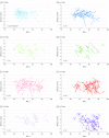
Figure 2.. Plots of Individual Memory z Score Trajectories by Age and Amyloid, Tau, and Neurodegeneration Biomarker (AT[N]) Group
Each line represents 1 participant’s trajectory, with the thinner portion of the line indicating memory z scores prior to the index (baseline) AT(N) imaging visit and the thicker portion of the line indicating memory z scores after the index AT(N) imaging visit. The z scores have been adjusted to account for the number of exposures to the cognitive battery by subtracting the estimated mean learning effect. The individual trajectories illustrate that there is substantial intraindividual variation in memory z scores over time. aFor the A−T−(N)− panel, a random subset of 50% of the data are shown to reduce overlap in the lines.
Figure 3.
Figure 3.. Estimated Rates of Annual Change in Memory z Score for 3 Exemplar Ages and APOE ε4 Carriers and Noncarriers
Rate estimates (95% CIs) are from 2 models: a clinical model in which rates depended on age and APOE status and an AT(N) model in which rates depended on age, APOE status, and AT(N) group. As illustrated in each age panel and for both APOE ε4 carriers and ε4 noncarriers, the rates of memory decline vary by AT(N), indicating better prediction of mean memory decline by age and APOE status (ie, the clinical model).
Figure 4.
Figure 4.. Estimates of Annual Rate of Change in Memory z Score at Each Age
The solid blue line shows the expected annual rate of decline in memory z score at each age in the cohort. This reflects both aging alone and increasing fractions who will have a more abnormal amyloid, tau, and neurodegeneration biomarker (AT[N]) profile. The solid orange line shows the estimated portion of the rate of decline that is partitioned to aging alone, ie, if individuals of different ages but the same AT(N) profile were compared. The shaded blue region represents the portion of the rate that is partitioned to the change in AT(N) biomarker prevalence with age. Vertical bars show 95% confidence limits at 3 different ages for each line.

Comment in

Similar articles

Cited by

  • Exploring the ATN classification system using brain morphology.
    Heinzinger N, Maass A, Berron D, Yakupov R, Peters O, Fiebach J, Villringer K, Preis L, Priller J, Spruth EJ, Altenstein S, Schneider A, Fliessbach K, Wiltfang J, Bartels C, Jessen F, Maier F, Glanz W, Buerger K, Janowitz D, Perneczky R, Rauchmann BS, Teipel S, Killimann I, Göerß D, Laske C, Munk MH, Spottke A, Roy N, Heneka MT, Brosseron F, Dobisch L, Ewers M, Dechent P, Haynes JD, Scheffler K, Wolfsgruber S, Kleineidam L, Schmid M, Berger M, Düzel E, Ziegler G; Alzheimer’s Disease Neuroimaging Initiative. Heinzinger N, et al. Alzheimers Res Ther. 2023 Mar 13;15(1):50. doi: 10.1186/s13195-023-01185-x. Alzheimers Res Ther. 2023. PMID: 36915139 Free PMC article.
  • Contribution of Alzheimer's biomarkers and risk factors to cognitive impairment and decline across the Alzheimer's disease continuum.
    Tosun D, Demir Z, Veitch DP, Weintraub D, Aisen P, Jack CR Jr, Jagust WJ, Petersen RC, Saykin AJ, Shaw LM, Trojanowski JQ, Weiner MW; Alzheimer's Disease Neuroimaging Initiative. Tosun D, et al. Alzheimers Dement. 2022 Jul;18(7):1370-1382. doi: 10.1002/alz.12480. Epub 2021 Oct 14. Alzheimers Dement. 2022. PMID: 34647694 Free PMC article.
  • Brain amyloid, cortical thickness, and changes in activities of daily living.
    Vassilaki M, Aakre JA, Kremers WK, Lesnick TG, Mielke MM, Geda YE, Machulda MM, Knopman DS, Butler L, Traber M, Vemuri P, Lowe VJ, Jack CR Jr, Roberts RO, Petersen RC. Vassilaki M, et al. Ann Clin Transl Neurol. 2020 Apr;7(4):474-485. doi: 10.1002/acn3.51010. Epub 2020 Apr 21. Ann Clin Transl Neurol. 2020. PMID: 32314554 Free PMC article.
  • Editorial: Hippocampal mechanisms in aging and clinical memory decline.
    Ginsberg SD, Tarantini S. Ginsberg SD, et al. Front Aging Neurosci. 2023 May 5;15:1204954. doi: 10.3389/fnagi.2023.1204954. eCollection 2023. Front Aging Neurosci. 2023. PMID: 37213539 Free PMC article. No abstract available.
  • Distinct cerebral small vessel disease impairment in early- and late-onset Alzheimer's disease.
    Luo X, Hong H, Li K, Zeng Q, Wang S, Li Z, Fu Y, Liu X, Hong L, Li J, Zhang X, Zhong S, Jiaerken Y, Liu Z, Chen Y, Huang P, Zhang M; Alzheimer's Disease Neuroimaging Initiative (ADNI). Luo X, et al. Ann Clin Transl Neurol. 2023 Aug;10(8):1326-1337. doi: 10.1002/acn3.51824. Epub 2023 Jun 22. Ann Clin Transl Neurol. 2023. PMID: 37345812 Free PMC article.

References

    1. Jack CR Jr, Bennett DA, Blennow K, et al. ; Contributors . NIA-AA research framework: toward a biological definition of Alzheimer’s disease. Alzheimers Dement. 2018;14(4):535-562. doi:10.1016/j.jalz.2018.02.018 - DOI - PMC - PubMed
    1. Hyman BT, Phelps CH, Beach TG, et al. . National Institute on Aging-Alzheimer’s Association guidelines for the neuropathologic assessment of Alzheimer’s disease. Alzheimers Dement. 2012;8(1):1-13. doi:10.1016/j.jalz.2011.10.007 - DOI - PMC - PubMed
    1. Jack CR Jr, Wiste HJ, Weigand SD, et al. . Defining imaging biomarker cut points for brain aging and Alzheimer’s disease. Alzheimers Dement. 2017;13(3):205-216. doi:10.1016/j.jalz.2016.08.005 - DOI - PMC - PubMed
    1. Caselli RJ, Dueck AC, Osborne D, et al. . Longitudinal modeling of age-related memory decline and the APOE epsilon4 effect. N Engl J Med. 2009;361(3):255-263. doi:10.1056/NEJMoa0809437 - DOI - PMC - PubMed
    1. Roberts RO, Geda YE, Knopman DS, et al. . The Mayo Clinic Study of Aging: design and sampling, participation, baseline measures and sample characteristics. Neuroepidemiology. 2008;30(1):58-69. doi:10.1159/000115751 - DOI - PMC - PubMed

Publication types