. 2019 Sep;33(9):10300-10314.
doi: 10.1096/fj.201900322R. Epub 2019 Jun 18.

Infection-induced endothelial amyloids impair memory

Affiliations

Infection-induced endothelial amyloids impair memory

Ron Balczon et al. FASEB J. 2019 Sep.

Abstract

Patients with nosocomial pneumonia exhibit elevated levels of neurotoxic amyloid and tau proteins in the cerebrospinal fluid (CSF). In vitro studies indicate that pulmonary endothelium infected with clinical isolates of either Pseudomonas aeruginosa, Klebsiella pneumoniae, or Staphylococcus aureus produces and releases cytotoxic amyloid and tau proteins. However, the effects of the pulmonary endothelium-derived amyloid and tau proteins on brain function have not been elucidated. Here, we show that P. aeruginosa infection elicits accumulation of detergent insoluble tau protein in the mouse brain and inhibits synaptic plasticity. Mice receiving endothelium-derived amyloid and tau proteins via intracerebroventricular injection exhibit a learning and memory deficit in object recognition, fear conditioning, and Morris water maze studies. We compared endothelial supernatants obtained after the endothelia were infected with P. aeruginosa possessing an intact [P. aeruginosa isolated from patient 103 (PA103) supernatant] or defective [mutant strain of P. aeruginosa lacking a functional type 3 secretion system needle tip complex (ΔPcrV) supernatant] type 3 secretion system. Whereas the PA103 supernatant impaired working memory, the ΔPcrV supernatant had no effect. Immunodepleting amyloid or tau proteins from the PA103 supernatant with the A11 or T22 antibodies, respectively, overtly rescued working memory. Recordings from hippocampal slices treated with endothelial supernatants or CSF from patients with or without nosocomial pneumonia indicated that endothelium-derived neurotoxins disrupted the postsynaptic synaptic response. Taken together, these results establish a plausible mechanism for the neurologic sequelae consequent to nosocomial bacterial pneumonia.-Balczon, R., Pittet, J.-F., Wagener, B. M., Moser, S. A., Voth, S., Vorhees, C. V., Williams, M. T., Bridges, J. P., Alvarez, D. F., Koloteva, A., Xu, Y., Zha, X.-M., Audia, J. P., Stevens, T., Lin, M. T. Infection-induced endothelial amyloids impair memory.

Keywords: cerebrospinal fluid; dementia; depression; learning; nosocomial pneumonia.


Conflict of interest statement

The authors thank the members of the Department of Comparative Medicine and the University of South Alabama for their efforts in animal husbandry, and the members of the Intensive Care Unit team and Clinical Microbiology Laboratory at the University of Alabama at Birmingham for their care in handling and processing clinical samples. The authors also thank Dr. James Maylie (Oregon Health and Science University) for generous help with Igor Pro. This work was funded by a College of Medicine Intramural Award and U.S. National Institutes of Health (NIH) National Heart, Lung, and Blood Institute Grants HL140182 (to M.T.L.), HL118334 (to J.P.A. and D.F.A.), HL66299 and HL135003 (to T.S. and R.B.), HL60024 (to T.S.), and NIH National Institute of Neurological Disorders and Stroke Grant NS102495 (to X.-M.Z.). S.V. is a predoctoral fellow (T32 HL076125). The authors declare no conflicts of interest.

Figures

Figure 1
Figure 1
PA103 lung infection elicits mouse brain tau protein accumulation and inhibits synaptic plasticity. A) Immunoblot of sarkosyl-precipitated brain homogenate shows an increased oligomeric tau protein accumulation 1 wk after mice were inoculated with PA103 (105 CFU) or saline. Sarkosyl precipitation concentrates tau protein (T22) but not Aβ (MOAB-2). An arrow highlights the tau protein band. B) Time course of normalized field excitatory postsynaptic potential (fEPSP) (means ± sem) obtained 1 wk after PA103 or saline inoculation. Representative mean traces shown on the right were obtained from −5 to 0 min (black), 7 to 12 min (blue), and 55 to 60 min (red). An arrow at time 0 indicates LTP induction. C) Representative voltage traces evoked by synaptic stimuli of increasing intensity (left). The amplitude of FV and initial slope of fEPSP were obtained from the vertical dotted line and shaded area, respectively. Recordings were performed on brains collected from saline- or PA103-inoculated mice (middle). Plots of input-output relations from traces show FV vs. stimulus intensity (presynaptic component) and fEPSP slope vs. FV (postsynaptic response). Data were fitted with corresponding color lines (right). Bar graphs summarizing the slopes of fitted lines for presynaptic component and postsynaptic response are presented. *P < 0.05 compared with saline (Student’s t test).
Figure 2
Figure 2
PA103 supernatant induces subtle behavioral changes in open-field studies. Mice received sham surgery or were injected into the right lateral ventricle ICV with saline, ΔPcrV supernatant, and PA103 supernatant neutralized with A11 or T22 antibody (A11 or T22, respectively), or amyloids eluted from A11 (A11-eluate) or T22 (T22-eluate). Results show open-field studies conducted 1 wk after the surgery. Each mouse was placed in an 18-inch cube box for a 5-min trial per day for 3 consecutive d. A) Raw 2-dimensional movement tracks from an individual mouse (top panel) or overlay of 4 mice tracks (middle panel) on the first trial. Bottom panel shows analyses of 4 mice traces as heat maps. Dark regions represent areas where mice spent longer dwelling time at the box locations. Notice that the PA103 mice leaped numerous times (loops). B, C) Bar graphs show travel speed (B) and distance (C) analyzed from the 5-min trials and averaged over 3 d. D, E) Traveling distance was further separated into either center region (D) or along the edges (E). F, G) Number of times animals crossed into the center region (F) and time (%) mice dwelled in the center region over 5 min (G). Bars show means ± sem. Numbers on bars indicate number of mice per group.
Figure 3
Figure 3
Amyloids in PA103 supernatant decrease animal working memory and dampen interest in exploring a novel object. On the training day, mice were placed in an 18-inch cube box containing 2 identical objects placed at opposite corners. They were allowed to explore the objects for a total combination of 18 s. A) After 24 h, mice were returned to the same box, except one of the objects was replaced with a novel object. Mice were allowed to explore freely for 90 s, and their movement and activity inside the box were tracked. Offline analysis subdivided the box into 4 regions, in which Q1 contained the novel object (N) and quadrant 4 contained the old object (O). B) Novel object dwell time: percent time mouse spent exploring Q1. Dashed line indicates chance exploration (25%). C) Novel object preference: normalized time mouse spent exploring the novel vs. old object. Dashed line shows 50%, denoting no preference toward either object. Bars show means ± sem. *P < 0.05 vs. saline (ANOVA with Tukey’s post hoc tests).
Figure 4
Figure 4
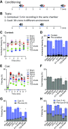
PA103 supernatant impairs fear response. A, B) Context-cue fear experimental paradigm. Mice received a context preexposure session in which mice were allowed to explore an activity-tracking chamber for 5 min (d 0). Mice were returned 24 h for conditioning. A total of 3 30-s tones (blue bars) were presented with a coterminating foot shock (red thunder) (A). To examine context fear memory, mice were returned after a 24 h retention interval. No shock or tone was presented during 5 min context test (top) (B). To examine cued fear, mice were placed into a novel chamber, and the 30-s tone was presented twice (bottom). C, D) Results of context fear as freezing response. Percent freezing was calculated per minute; 50% = 30 s freezing per min. Scatter plots show freezing (%) time course (C). Bar graph shows freezing (%) averaged over 5 min, before (black) and after conditioning (context; gray bars) (D). EH) Results of cued fear response. Scatter plots show freezing (%) time course (E). Blue bars on the x axis indicate the 30-s cue tone induced at 1 and 4 min. Bar graphs show total freezing response (%) averaged over 7 min (F), precue (i.e., during 0–1 min) vs. first cue-induced freezing increase (i.e., during 1–2 min) (G), and freezing response associated with (cue; i.e., during 1–2 and 4–5 min; blue bars) or before (Precue; i.e., during 0–1 and 3–4 min; gray bars) the tone (H). Numbers in parentheses indicate the time during which freezing responses were measured. Bars show means ± sem *P < 0.05 vs. saline (ANOVA with Tukey’s post hoc test).
Figure 5
Figure 5
PA103 supernatant–induced working memory impairment is amyloid dependent. A) Morris water maze protocol. ICV-mice were trained for 3 consecutive d with a hidden platform located in the top-left (Q1) or bottom-right corner (quadrant 4) prior to probe 1 and 2 tests performed on d 4 and 8, respectively. The hidden platform in d 1–3 and 5–7 were directly opposite and furthest away from each other. B) Summary of escape latency from daily swim trials. Trial 1 runs (upper panel) were performed 24 h apart, and trial 2 runs (lower panel) were performed 15 min after trial 1. C, D) Raw probe swim traces from 4 mice per group obtained from probe 1 (C) and probe 2 (D) analyzed into 2-dimensional location heat maps. E, F) Percent time mice spent in the correct quadrant for probe 1 (i.e., Q1) (E) and probe 2 (i.e., quadrant 4) (F). G) Mean time mice spent in the respective correct quadrants during both probe tests. H) Maximum (blue) and mean swim speed during probe tests. Bars show means ± sem *P < 0.05 vs. saline (ANOVA with Tukey’s post hoc test).
Figure 6
Figure 6
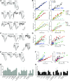
Amyloid proteins in human CSF obtained from infected patients disrupt neural information transfer. A) Representative voltage traces evoked by synaptic stimuli of increasing intensity. The amplitude of FV and initial slope of field excitatory postsynaptic potential (fEPSP) were obtained from the vertical dotted line and shaded areas, respectively. Recordings were performed on control, PA103-bathed, or ΔPcrV-bathed slices (top panel), on slices bathed in A11-, T22-, IgG- or both A11- and T22-immunodepleted PA103 supernatant (middle panel) or bathed in amyloids eluted from the respective antibody complex (bottom panel). B) Plots of input-output relations from traces (A) show FV vs. stimulus intensity (presynaptic component; left panel) and fEPSP slope vs. FV (postsynaptic response; right panel). Data were fitted with corresponding color lines. C, D) Representative recordings and analyses from slices treated with human CSF obtained from a noninfected control or a K. pneumoniae patient without or with A11, T22, or control Ig-immunodepletion or amyloids eluted from A11 or IgG antibody complex. Dotted lines show extrapolated line fit for K. pneumonia (dark blue) and IgG-eluate (light blue) (D). E, F) Representative recordings and analyses from slices treated with human CSF obtained from a noninfected control or an S. aureus patient without or with A11 or T22 immunodepletion. G, H) Bar graphs summarizing the slopes of fitted lines for the presynaptic component (obtained from FV vs. stimulus) and postsynaptic response (from fEPSP vs. FV). *P < 0.05 vs. controls (ANOVA with Tukey’s post hoc test), #P < 0.05 vs. infected (Student’s t test).
Figure 7
Figure 7
Schematic illustrating how nosocomial pneumonia impairs neuronal communication in an amyloid-dependent manner. A) The presynaptic action potential in which the action potential stimulates neurotransmitter release from presynaptic terminals. B) The postsynaptic response in which glutamate binding to postsynaptic AMPA and NMDA receptors activates excitatory postsynaptic responses. C) Early-phase LTP (E-LTP; blue arrows) in which stimulation of synaptic NMDA receptors promotes Ca2+ influx and kinase activation, both of which are required for the induction of LTP. Phosphorylated AMPA receptor insertion into extrasynaptic sites and lateral trafficking into the electron-dense zone enhance synaptic response. D) Late-phase LTP (L-LTP; red arrows) in which activated spine Ca2+ and kinases further increase protein translation at the base of the spine and in the soma. Newly synthesized proteins and receptors are transported to the potentiated spines, resulting in spine reorganization and synaptic growth.

References

    1. Patel M. B., Morandi A., Pandharipande P. P. (2015) What’s new in post-ICU cognitive impairment Intensive Care Med. 41, 708–711 - PubMed
    1. Iwashyna T. J., Ely E. W., Smith D. M., Langa K. M. (2010) Long-term cognitive impairment and functional disability among survivors of severe sepsis. JAMA 304, 1787–1794 - PMC - PubMed
    1. Needham D. M., Dinglas V. D., Morris P. E., Jackson J. C., Hough C. L., Mendez-Tellez P. A., Wozniak A. W., Colantuoni E., Ely E. W., Rice T. W., Hopkins R. O., NIH NHLBI ARDS Network (2013) Physical and cognitive performance of patients with acute lung injury 1 year after initial trophic versus full enteral feeding. EDEN trial follow-up. Am. J. Respir. Crit. Care Med. 188, 567–576 - PMC - PubMed
    1. Annane D., Sharshar T. (2015) Cognitive decline after sepsis. Lancet Respir. Med. 3, 61–69 - PubMed
    1. Ehlenbach W. J., Hough C. L., Crane P. K., Haneuse S. J., Carson S. S., Curtis J. R., Larson E. B. (2010) Association between acute care and critical illness hospitalization and cognitive function in older adults. JAMA 303, 763–770 - PMC - PubMed

Publication types

MeSH terms