. 2019 Oct:130:104508.
doi: 10.1016/j.nbd.2019.104508. Epub 2019 Jun 15.

MicroRNA inhibition upregulates hippocampal A-type potassium current and reduces seizure frequency in a mouse model of epilepsy

Affiliations

MicroRNA inhibition upregulates hippocampal A-type potassium current and reduces seizure frequency in a mouse model of epilepsy

Durgesh Tiwari et al. Neurobiol Dis. 2019 Oct.

Abstract

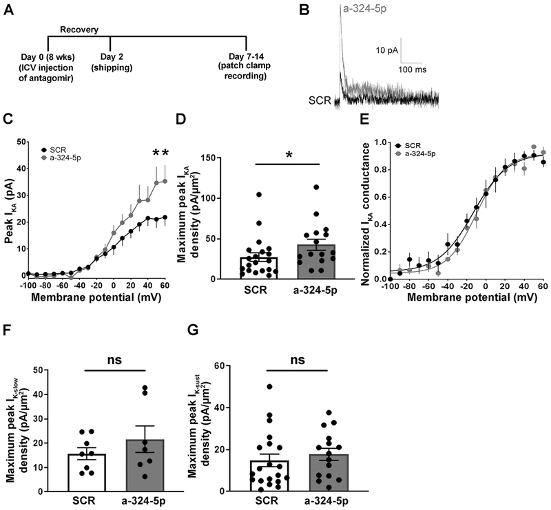
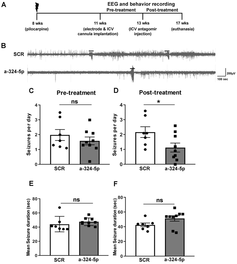
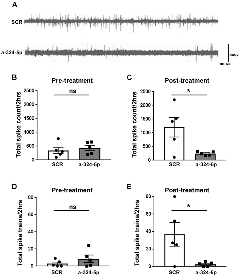
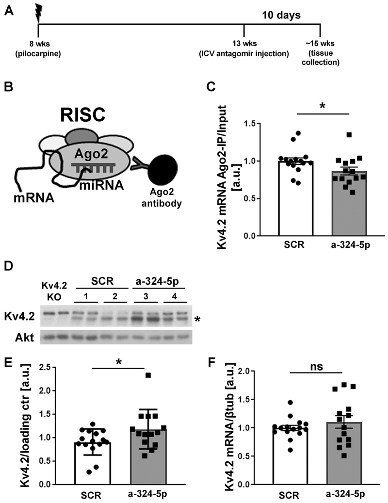
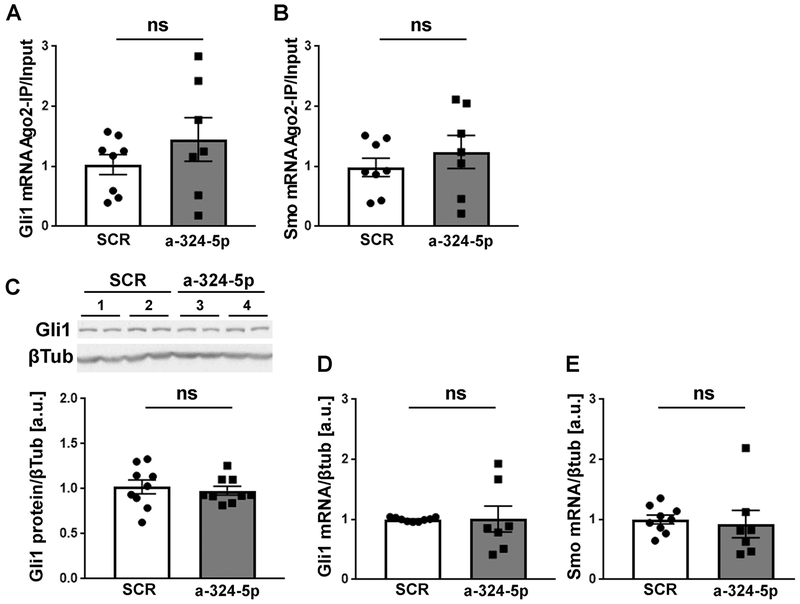
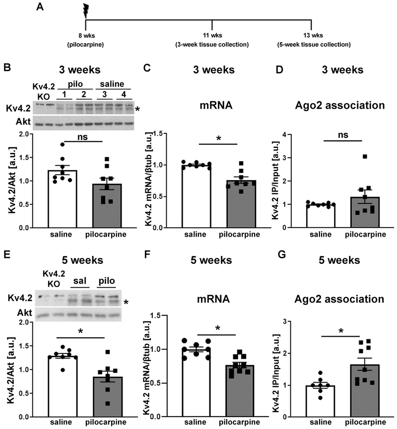
Epilepsy is often associated with altered expression or function of ion channels. One example of such a channelopathy is the reduction of A-type potassium currents in the hippocampal CA1 region. The underlying mechanisms of reduced A-type channel function in epilepsy are unclear. Here, we show that inhibiting a single microRNA, miR-324-5p, which targets the pore-forming A-type potassium channel subunit Kv4.2, selectively increased A-type potassium currents in hippocampal CA1 pyramidal neurons in mice. Resting membrane potential, input resistance and other potassium currents were not altered. In a mouse model of acquired chronic epilepsy, inhibition of miR-324-5p reduced the frequency of spontaneous seizures and interictal epileptiform spikes supporting the physiological relevance of miR-324-5p-mediated control of A-type currents in regulating neuronal excitability. Mechanistic analyses demonstrated that microRNA-induced silencing of Kv4.2 mRNA is increased in epileptic mice leading to reduced Kv4.2 protein levels, which is mitigated by miR-324-5p inhibition. By contrast, other targets of miR-324-5p were unchanged. These results suggest a selective miR-324-5p-dependent mechanism in epilepsy regulating potassium channel function, hyperexcitability and seizures.

Keywords: A-type potassium currents; Antagomir; Epilepsy; Epileptiform spikes; Kv4.2; RISC; RNA-induced silencing complex; Seizures; miR-324-5p; microRNA.


Conflict of interest statement

Declaration of Interest

C.G. is co-inventor on US patent 9,932,585 B2. All other authors declare no competing financial interests.

Figures

Figure 1:
Figure 1:. Antagonizing miR-324-5p increases hippocampal A-type potassium currents in mice.
(A) Timeline depicting ICV antagomir injection, shipping and patch clamp recording for potassium current measurement. (B) Representative traces showing A-type potassium current recorded from a CA1 pyramidal neuron from mice injected with either scrambled or miR-324-5p antagomir. (C) Summary plot showing that the peak IKA was significantly greater at the most depolarized test voltages in mice treated with the antagomir compared to scrambled (SCR: n=9 from 3 mice; a-324-5p: n=7 from 3 mice; 2-way RM ANOVA with Sidak’s multiple comparison post hoc testing, interaction antagomir X membrane potential: F(16,224)=3.194, p<0.0001; *p=0.004 at VM = 50, and *p=0.005 at VM = 60 mV). (D) The maximum IKA current density was significantly greater in mice treated with antagomir compared to scrambled (SCR: n=20 from 9 mice; a-324-5p: n=16 from 9 mice; unpaired two-tailed Mann-Whitney test, *p=0.036). (E) There was no difference in the voltage-dependence of activation of IKA between scrambled and miR-324-5p antagomir-injected mice (n=8 from 3 mice each). (F,G) There was no significant difference in either the slowly inactivating (F: SCR: n=8 from 9 mice; a-324-5p: n=7 from 9 mice; unpaired two-tailed t-test, t(13)=1.032, p=0.321) or sustained (G: SCR: n=20 from 9 mice; a-324-5p: n=15 from 9 mice; two-tailed Mann-Whitney test, p=0.382) potassium currents between neurons from miR-324-5p antagomir- and scrambled antagomir-injected mice. Error bars represent SEM. Additional analyses shown in Fig. S1.
Figure 2:
Figure 2:. Antagonizing miR-324-5p in vivo reduces seizure frequency in a pilocarpine mouse model of epilepsy.
(A) Timeline depicting age of mice during pilocarpine injection and antagomir treatment. (B) Representative EEG traces of spontaneous seizures in the pilocarpine model following scrambled or miR-324-5p antagomir treatment. Seizures are indicated with lines above the EEG. (C) No significant difference was observed in seizure frequency in mice before injection of scrambled or miR-324-5p-specific antagomirs (unpaired two-tailed t-test, t(14)=0.898, p=0.384). (D) ICV injection of a miR-324-5p-specific antagomir reduces seizure frequency on average by half compared to the scrambled control (unpaired two-tailed t-test, t(14)=2.221, *p=0.043). (E) No significant effect between the two treatment groups was observed on the mean seizure duration between scrambled or miR-324-5p antagomir-injected mice during the pre-treatment period (two-tailed Mann-Whitney test, p=0.142). (F) Similarly, no difference in seizure duration after miR-324-5p antagomir treatment was observed (two-tailed Mann-Whitney test, p=0.142). N was 7 for scrambled, and 9 for miR-324-5p antagomir-treated mice. Error bars represent SEM.
Figure 3:
Figure 3:. Antagonizing miR-324-5p in vivo reduces the number of interictal spikes and spike trains in epileptic mice.
(A) Representative EEG traces of interictal spikes in scrambled or miR-324-5p antagomir-injected mice. (B,D) Before treatment, no significant differences were observed in total spike count (B, unpaired two-tailed t-test, t(8)=0.590, p=0.572) and the number of spike trains (D, unpaired two-tailed t-test, t(8)=1.195, p=0.266) between scrambled and miR-324-5p mice (2-hour period over 3 days). (C,E) After treatment, total spike number (C, unpaired two-tailed t-test, t(8)=2.676, *p=0.028) and total spike trains (E, unpaired two-tailed t-test, t(8)=2.51, *p=0.036) were significantly lower in miR-324-5p-specific antagomir-injected mice compared to the scrambled control (2-hour period over 3 days). Error bars represent SEM, n=5 for both conditions. Also see extended EEG trace in Fig. S6.
Figure 4:
Figure 4:. miR-324-5p antagomir reduces microRNA-induced silencing of Kv4.2 in the pilocarpine model.
(A) Timeline depicting age of the mice during pilocarpine injection, antagomir treatment and tissue collection. (B) Schematic of Ago2-specific immunoprecipitation to assess Kv4.2 mRNA levels in the RISC. (C) Reduced association of Kv4.2 mRNA with Ago2 was observed after miR-324-5p antagomir treatment compared to scrambled control (C: SCR: n=15, a-324-5p: n=14, unpaired one-tailed t-test, t(27)=1.940, *p=0.031). mRNA levels in Ago2-IPs were quantified by qRT-PCR and normalized to input levels. (D,E) Western blot analyses of hippocampal lysates show significantly increased Kv4.2 expression in chronically seizing mice 10 days after miR-324-5p antagomir treatment compared to scrambled control (SCR: n=15, a-324-5p: n=14, unpaired one-tailed Mann-Whitney test, *p=0.047). Example blot of 2 different mice per condition (#1-4, loaded in duplicates) shown in D, cumulative quantification shown in E. Kv4.2-specific signal was normalized to loading control (Akt or βTubulin) on the same blot. Asterisk indicates Kv4.2-specific band that is absent in lysates from Kv4.2 KO mice. (F) Kv4.2 mRNA levels (normalized to βtubulin) were not significantly changed after antagomir treatment (SCR: n=15, a-324-5p: n=14, unpaired one-tailed t-test, t(27)=0.916, p=0.184). Error bars represent SEM.
Figure 5:
Figure 5:. Gli1 and Smo expression are not affected by miR-324-5p inhibition in epileptic mice.
(A,B) In contrast to Kv4.2 mRNA, association of Gli1 (A) and Smo (B) mRNA with Ago2 is not significantly changed in epileptic mice after miR-324-5p inhibition (SCR: n=8, a-324-5p: n=7, unpaired one-tailed t-test, A: t(13)=1.087, p=0.148; B: t(13)=0.836, p=0.209). (C-E) Likewise, neither Gli1 protein (C) nor Gli1 (D) or Smo (E) mRNA are affected by the in vivo antagomir treatment (C: n=9, unpaired one-tailed t-test, t(16)=0.450, p=0.330; D: SCR: n=9, a-324-5p: n=7, unpaired one-tailed t-test, t(14)=0.039, p=0.485; E: SCR: n=9, a-324-5p: n=7, one-tailed Mann-Whitney test, p=0.105). Example western blot of two mice per condition (#1-4, loaded in duplicates) for C shown at top. Ago-IPs were normalized to input. Error bars represent SEM.
Figure 6:
Figure 6:. MicroRNA-induced silencing of Kv4.2 is increased in epileptic mice.
(A) Timeline depicting age of mice during pilocarpine injection and tissue collection. (B-D) Three weeks after pilocarpine injection, when mice start to develop spontaneous seizures, no significant reduction in Kv4.2 protein levels (B: n=8, unpaired two-tailed t-test, t(14)=1.899, p=0.078) or Kv4.2 mRNA association with Ago2 (i.e. the RISC) (D: n=8, unpaired two-tailed t-test, t(14)=1.168, p=0.262), but significantly reduced Kv4.2 mRNA (C: n=8, unpaired two-tailed t-test, t(14)=4.470, *p<0.001) was observed. (E-G) Five weeks following pilocarpine treatment, when mice have frequent spontaneous seizures, significantly reduced Kv4.2 protein (E: n=8, unpaired two-tailed t-test, t(14)=3.513, *p=0.003) and mRNA levels (F: saline: n=8, pilo: n=9; unpaired two-tailed t-test, t(15)=4.218, *p<0.001), and significantly increased association of Kv4.2 mRNA with Ago2 (G: saline: n=7, pilocarpine: n=9, two-tailed Mann-Whitney test, *p=0.008) compared to saline control was observed. Example western blots for B and E are shown at top (samples loaded in duplicates), Kv4.2-specific signal was normalized to Akt on the same blot. Asterisk indicates Kv4.2-specific band that is absent in lysates from Kv4.2 KO mice. Total mRNA levels in C and F were quantified by qRT-PCR and normalized to βtubulin. mRNA levels in Ago2-IPs in D and G were quantified by qRT-PCR and normalized to input levels. Error bars represent SEM.

Similar articles

Cited by

References

    1. Arida RM, Scorza FA, de Araujo Peres C, Cavalheiro EA, 1999. The course of untreated seizures in the pilocarpine model of epilepsy. Epilepsy Res 34, 99–107. 10.1016/S0920-1211(98)00092-8 - DOI - PubMed
    1. Banerjee SB, Rajendran R, Dias BG, Ladiwala U, Tole S, Vaidya VA, 2005. Recruitment of the Sonic hedgehog signalling cascade in electroconvulsive seizure-mediated regulation of adult rat hippocampal neurogenesis. Eur J Neurosci 22, 1570–1580. 10.1111/j.1460-9568.2005.04317.x - DOI - PMC - PubMed
    1. Bar-Klein G, Klee R, Brandt C, Bankstahl M, Bascunana P, Tollner K, Dalipaj H, Bankstahl JP, Friedman A, Loscher W, 2016. Isoflurane prevents acquired epilepsy in rat models of temporal lobe epilepsy. Ann Neurol 80, 896–908. 10.1002/ana.24804 - DOI - PubMed
    1. Barnwell LFS, Lugo JN, Lee WL, Willis SE, Gertz SJ, Hrachovy RA, Anderson AE, 2009. Kv4.2 knockout mice demonstrate increased susceptibility to convulsant stimulation. Epilepsia. 50, 1741–1751. 10.1111/j.1528-1167.2009.02086.x - DOI - PMC - PubMed
    1. Bartel DP, 2018. Metazoan MicroRNAs. Cell. 173, 20–51. 10.1016/j.cell.2018.03.006 - DOI - PMC - PubMed

Publication types