. 2019 Jun 12;20(12):2854.
doi: 10.3390/ijms20122854.

WC-1 and the Proximal GATA Sequence Mediate a Cis-/Trans-Acting Repressive Regulation of Light-Dependent Gene Transcription in the Dark

Affiliations

WC-1 and the Proximal GATA Sequence Mediate a Cis-/Trans-Acting Repressive Regulation of Light-Dependent Gene Transcription in the Dark

Andrea Brenna et al. Int J Mol Sci. .

Abstract

Light influences a wide range of physiological processes from prokaryotes to mammals. Neurospora crassa represents an important model system used for studying this signal pathway. At molecular levels, the WHITE COLLAR Complex (WCC), a heterodimer formed by WC-1 (the blue light photo-sensor) and WC-2 (the transcriptional activator), is the critical positive regulator of light-dependent gene expression. GATN (N indicates any other nucleotide) repeats are consensus sequences within the promoters of light-dependent genes recognized by the WCC. The distal GATN is also known as C-box since it is involved in the circadian clock. However, we know very little about the role of the proximal GATN, and the molecular mechanism that controls the transcription of light-induced genes during the dark/light transition it is still unclear. Here we showed a first indication that mutagenesis of the proximal GATA sequence within the target promoter of the albino-3 gene or deletion of the WC-1 zinc finger domain led to a rise in expression of light-dependent genes already in the dark, effectively decoupling light stimuli and transcriptional activation. This is the first observation of cis-/trans-acting repressive machinery, which is not consistent with the light-dependent regulatory mechanism observed in the eukaryotic world so far.

Keywords: GATA; WC-1; light responses; zinc finger.


Conflict of interest statement

The authors declare that they have no conflict of interest.

Figures
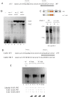
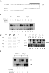
Figure 1
Figure 1
Dark/Light binding of WHITE COLLAR Complex (WCC) to the light-responsive elements (LREs) of al-3. (A) The DNA sequence of LRE in the promoter of al-3 used as a radioactive probe for the Electrophoresis Mobility Shift Assay (EMSA) assay. The sequence was based on previously published data [26]. The arrow indicates the direction towards the transcriptional start site. (B) EMSA assay was performed incubating a double-stranded oligonucleotide probe containing the LREs of al-3 promoter and a nuclear extract from Neurospora crassa. Lane 1 does not contain extract. In lanes 2–3 the probe was incubated with the nuclear extract obtained from wc-1 null mutants grown either in the dark (lane 2) or light-treated for 20 min (lane 3). Same conditions were used in lanes 4–5 (wc-2 mutant, 234 w) and 6–7 (wt). (C) Interaction of WC proteins is dependent on WC-1 C-Term region. WC-2 protein was immunoprecipitated with an anti-WC-2 antiserum from wt or wmn [23] (grown in the dark or 20 min after the light pulse. The immunoprecipitated complexes were resuspended in Laemmli buffer and loaded on SDS-PAGE. Western blot was performed using an anti-WC-1 affinity purified antibody. (D) DNA sequence used as cold probes for the EMSA competitive assay (D) based on (A). The upper sequence is a wt LRE region of the al-3 promoter. In the lower sequence, the GATA is mutagenized. (E) EMSA assay was performed by incubating the labelled double-stranded oligonucleotide with no nuclear extract (lane1), an equal amount of nuclear extract (10 μg) derived from dark growth mycelia (lane 2), or mycelia induced to light for 20 min. (lane 3). The specificity of the binding was verified by co-incubating the labelled oligonucleotide and nuclear extracts obtained from dark growth mycelia or light-treated for 20 min as following. Samples were incubated in the presence of a 10- or 50-fold excess of cold-specific competitor (lanes 4–5, 8–9), or 10- or 50-fold excess of unlabeled specific competitor harboring mutations in both the GATA motifs (lanes 6–7, 10–11).
Figure 2
Figure 2
Role of proximal and distal GATA sequences in dark/light shift. (A) Schematic representation of al-3 promoter GATA region that was used for the EMSA. The arrow indicates the direction towards the transcriptional start site. (B) EMSA was performed on N. crassa nuclear extracts of mycelia grown in the dark or after light pulse and incubated with the al-3 promoter GATA regions as the probe (lanes 1–2). Competition assay was performed by co-incubating labelled GATA with increasing concentrations (+ = 10-fold, ++ = 50-fold) of each indicated cold competitor sequence (lanes 3–4, 9–10: D/P GATA; lanes 4–5, 11–12: D-GATA; lanes 6–7, 13–14: D-GATA). (C) A diagrammatic representation of the exogenous al-3 promoter bearing mutations of the GATA motifs. (D) Representative Northern blot analyses of Neurospora transformants containing a shortened version of the al-3 gene. Asterisk: endogenous al-3 expression. Arrow: Δ 675. (E) EMSA was performed using the nuclear extract of wt N. crassa mycelia grown in the dark (lanes 1–3) or light-treated for 20 min (4–6). Competition experiment was performed by adding cold probes as indicated in the figure.
Figure 3
Figure 3
WC-1 zinc finger domain is required for repressing in the dark-/light-dependent phenotypes. (A) Schematic representation of functional domains of WC-1 Myc and WC-1 Myc zinc finger deleted proteins. (B) Wt WC-1 Myc and WC-1 Myc ZnF ∆ strains were grown in slant for 4 d in the dark, and carotenoid accumulation was checked right after, without giving illumination (left) or after the light pulse (right). (C) An increasing amount of conidia resuspended in water (5, 10, 20, and 25 µL) was spotted on a nitrocellulose filter to observe the increment of the orange color corresponding to carotenoid content. (D) The carotenoid content was analyzed by densitometry quantification and graphically represented. (E) Conidia count in wt and zinc finger deleted strains were measured using a Burker cell counter. The data are shown as the mean ± S.E.M. from three different biological replicas. t-test: ** p-value < 0.0023.
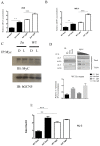
Figure 4
Figure 4
Zinc finger deletion influences increased RNA accumulation and histone acetylation of light-dependent genes in the dark. RNA abundances of al-3 (A) and wc-1 (B) were measured using real-time PCR in the littermate wt, and zinc finger deleted mutant strains, kept either in the dark or after illumination (light 5 min, dark 20 min for a total of 25 min after the light pulse). Data showed a different biological replica that was plotted on a graph with the mean ± S.E.M. from three different experiments. Two-way Anova, Bonferroni: ** p-value < 0.05. (C) WC-1 was immunoprecipitated using a commercial anti-Myc antibody, and the coresolution of NGF-1 was detected using hGCN5 antibody either in wt WC-1 Myc or WC-1 Myc ZnF ∆ mutant. Immunoprecipitation was performed starting from 400 µg of mycelia grown for 3 d in the dark before harvesting or exposed to saturated light (as in (A) and (B)). (D) A ChIP assay was performed on extract obtained from mycelia grown for 3 d in the dark before harvesting or exposed to saturated light. Anti-H3 and anti-H3-acetylated were used for immunoprecipitating the chromatin. PCR was performed on the free DNA after decrosslinking and further purification. Input: three different dilutions of total input were used for PCR amplification (1/4, 1/8, and 1/16). PCRs were finally quantified by densitometry and plotted on a graph. The data are the mean ± S.E.M. from three independent biological experiments (lower panel). (E) H3ac/H3 ratio was measured in wt and zinc finger strains, and data were plotted on a graph to compare accumulation of acetylated form of H3 in both strains in both environmental conditions (dark, light). The plotted data were obtained from three different biological replicas. Two-way Anova, Bonferroni: ** p-value < 0.05; *** p < 0.001.
Figure 5
Figure 5
Proposed molecular model. Schematic representation that summarizes the data discussed in the paper. White collar complex (WCC), slow mobility complex, and fast mobility complex form a preassembled molecular scaffold, present already in the dark. Moreover, the WCC binds the acetyltransferase NGF-1. The presence of a putative repressive complex is suggested even if it is not clear what proteins are involved. After light pulse, the WC-2:FMC complex binds the promoter, and the repression is abolished. The acetyl transferase NGF-1 is placed onto the promoter in order to modify the light-dependent chromatin acetylation, and gene expression is promoted.

References

    1. Dodd A.N., Salathia N., Hall A., Kevei E., Toth R., Nagy F., Hibberd J.M., Millar A.J., Webb A.A. Plant circadian clocks increase photosynthesis, growth, survival, and competitive advantage. Science. 2005;309:630–633. doi: 10.1126/science.1115581. - DOI - PubMed
    1. Michael T.P., Salome P.A., McClung C.R. Two Arabidopsis circadian oscillators can be distinguished by differential temperature sensitivity. Proc. Natl. Acad. Sci. USA. 2003;100:6878–6883. doi: 10.1073/pnas.1131995100. - DOI - PMC - PubMed
    1. Woelfle M.A., Ouyang Y., Phanvijhitsiri K., Johnson C.H. The adaptive value of circadian clocks: An experimental assessment in cyanobacteria. Curr. Biol. 2004;14:1481–1486. doi: 10.1016/j.cub.2004.08.023. - DOI - PubMed
    1. Corrochano L.M. Fungal photoreceptors: Sensory molecules for fungal development and behaviour. Photochem. Photobiol. Sci. 2007;6:725–736. doi: 10.1039/b702155k. - DOI - PubMed
    1. Chen C.H., Loros J.J. Neurospora sees the light: Light signaling components in a model system. Commun. Integr. Biol. 2009;2:448–451. doi: 10.4161/cib.2.5.8835. - DOI - PMC - PubMed

MeSH terms

LinkOut - more resources