

Atypical Histiocytoid Cells and Multinucleated Giant Cells in Fine-Needle Aspiration Cytology of the Thyroid Predict Lymph Node Metastasis of Papillary Thyroid Carcinoma
- PMID: 31212879
- PMCID: PMC6627749
- DOI: 10.3390/cancers11060816
Atypical Histiocytoid Cells and Multinucleated Giant Cells in Fine-Needle Aspiration Cytology of the Thyroid Predict Lymph Node Metastasis of Papillary Thyroid Carcinoma
Abstract
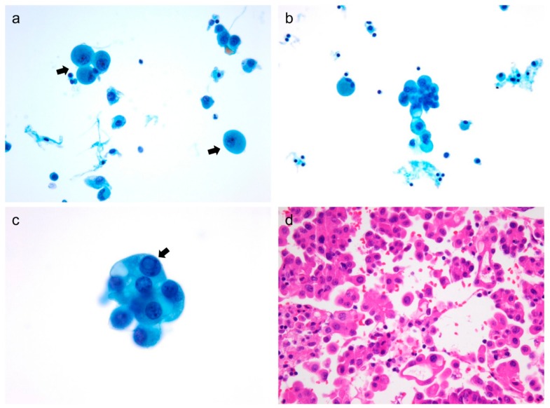
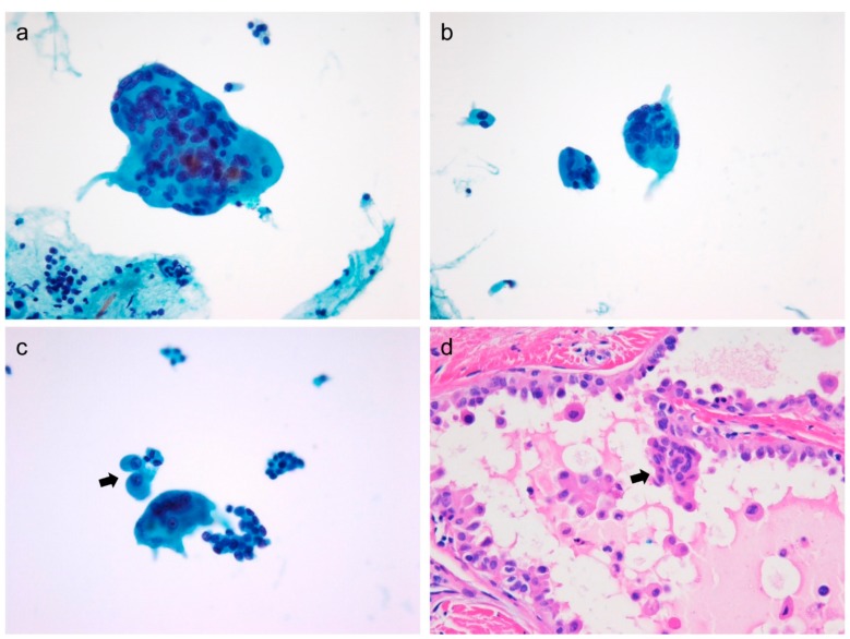
Preoperative detection of cervical lymph node metastasis in papillary thyroid carcinoma (PTC) is crucial for determining the surgical strategy to prevent locoregional recurrence of the disease. We identified the cytological predictors of lymph node metastasis in 222 consecutive patients with PTC using fine-needle aspiration cytology (FNAC) of the thyroid. Cervical lymph node metastases occurred in 99 (44.6%) of 222 PTC patients. Lymph node metastasis was significantly associated with tumor multifocality (p = 0.003), and high cellularity (p = 0.021), atypical histiocytoid cells (p < 0.001), and multinucleated giant cells (p < 0.001) in thyroid FNAC. The BRAF V600E mutation was marginally associated with lymph node metastasis (p = 0.054). Multivariate analysis revealed that atypical histiocytoid cells (odds ratio = 2.717; p = 0.001) and multinucleated giant cells (odds ratio = 3.070; p = 0.031) were independent predictors of lymph node metastasis in patients with PTC. In a subgroup analysis of 164 patients with microcarcinomas, atypical histiocytoid cells (odds ratio = 2.761; p = 0.005) was an independent predictor of lymph node metastasis. Cytological detection of atypical histiocytoid cells and multinucleated giant cells on thyroid FNAC can be used to preoperatively predict cervical lymph node metastasis in patients with PTC.
Keywords: fine needle aspiration; liquid-based preparation; lymph node metastasis; papillary carcinoma; thyroid cytopathology.
Conflict of interest statement
The authors declare no conflict of interest.
Figures
Similar articles
-
[Thyroglobulin concentration measurement in fine-needle aspiration fluid from cystic cervical lymph node metastases of papillary thyroid carcinoma].Nihon Jibiinkoka Gakkai Kaiho. 2011 Dec;114(12):912-6. doi: 10.3950/jibiinkoka.114.912. Nihon Jibiinkoka Gakkai Kaiho. 2011. PMID: 22352010 Japanese.
-
Atypical histiocytoid cells in fine-needle aspiration cytology of metastatic papillary thyroid carcinoma: A potential diagnostic pitfall.Diagn Cytopathol. 2022 Mar;50(3):133-140. doi: 10.1002/dc.24932. Epub 2022 Jan 18. Diagn Cytopathol. 2022. PMID: 35040599
-
Clinical implication of highly sensitive detection of the BRAF V600E mutation in fine-needle aspirations of thyroid nodules: a comparative analysis of three molecular assays in 4585 consecutive cases in a BRAF V600E mutation-prevalent area.J Clin Endocrinol Metab. 2012 Jul;97(7):2299-306. doi: 10.1210/jc.2011-3135. Epub 2012 Apr 12. J Clin Endocrinol Metab. 2012. PMID: 22500044
-
Pancreatic metastasis of papillary thyroid carcinoma preoperatively diagnosed by endoscopic ultrasound-guided fine-needle aspiration biopsy: a case report with review of literatures.Clin J Gastroenterol. 2018 Dec;11(6):521-529. doi: 10.1007/s12328-018-0875-z. Epub 2018 Jun 8. Clin J Gastroenterol. 2018. PMID: 29948817 Review.
-
Intraglandular dissemination: a special pathological feature.Front Oncol. 2024 Jul 29;14:1428274. doi: 10.3389/fonc.2024.1428274. eCollection 2024. Front Oncol. 2024. PMID: 39135992 Free PMC article. Review.
Cited by
-
What Is New in Thyroid Cancer: The Special Issue of the Journal Cancers.Cancers (Basel). 2020 Oct 19;12(10):3036. doi: 10.3390/cancers12103036. Cancers (Basel). 2020. PMID: 33086491 Free PMC article.
-
Cytologic hallmarks and differential diagnosis of papillary thyroid carcinoma subtypes.J Pathol Transl Med. 2024 Nov;58(6):265-282. doi: 10.4132/jptm.2024.10.11. Epub 2024 Nov 7. J Pathol Transl Med. 2024. PMID: 39557408 Free PMC article. Review.
-
A clinical and molecular pathology prediction model for central lymph node metastasis in cN0 papillary thyroid microcarcinoma.Front Endocrinol (Lausanne). 2023 Feb 2;14:1075598. doi: 10.3389/fendo.2023.1075598. eCollection 2023. Front Endocrinol (Lausanne). 2023. PMID: 36817603 Free PMC article.
-
Immune-related key gene CLDN10 correlates with lymph node metastasis but predicts favorable prognosis in papillary thyroid carcinoma.Aging (Albany NY). 2020 Feb 11;12(3):2825-2839. doi: 10.18632/aging.102780. Epub 2020 Feb 11. Aging (Albany NY). 2020. PMID: 32045884 Free PMC article.
-
TPO as an indicator of lymph node metastasis and recurrence in papillary thyroid carcinoma.Sci Rep. 2023 Jul 5;13(1):10848. doi: 10.1038/s41598-023-37932-1. Sci Rep. 2023. PMID: 37407700 Free PMC article.
References
-
- Haugen B.R., Alexander E.K., Bible K.C., Doherty G.M., Mandel S.J., Nikiforov Y.E., Pacini F., Randolph G.W., Sawka A.M., Schlumberger M., et al. 2015 American Thyroid Association Management Guidelines for Adult Patients with Thyroid Nodules and Differentiated Thyroid Cancer: The American Thyroid Association Guidelines Task Force on Thyroid Nodules and Differentiated Thyroid Cancer. Thyroid. 2016;26:1–133. doi: 10.1089/thy.2015.0020. - DOI - PMC - PubMed
Grants and funding
LinkOut - more resources
Full Text Sources
Research Materials
