. 2019 Jun 17;11(6):346.
doi: 10.3390/toxins11060346.

Recombinant Antibodies against Mycolactone

Affiliations

Recombinant Antibodies against Mycolactone

Leslie Naranjo et al. Toxins (Basel). .

Abstract

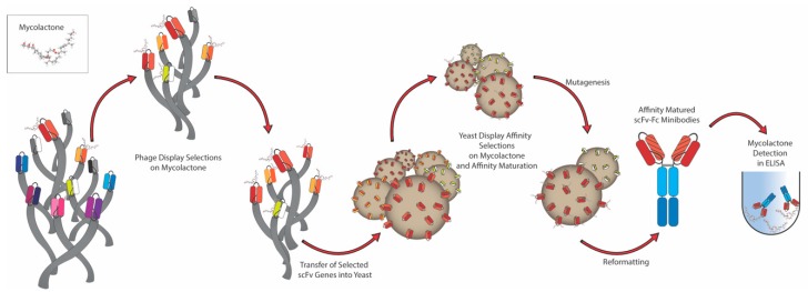
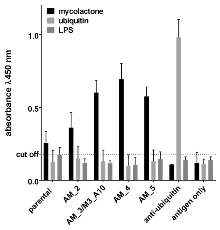
In the past, it has proved challenging to generate antibodies against mycolactone, the primary lipidic toxin A of Mycobacterium ulcerans causing Buruli ulcer, due to its immunosuppressive properties. Here we show that in vitro display, comprising both phage and yeast display, can be used to select antibodies recognizing mycolactone from a large human naïve phage antibody library. Ten different antibodies were isolated, and hundreds more identified by next generation sequencing. These results indicate the value of in vitro display methods to generate antibodies against difficult antigenic targets such as toxins, which cannot be used for immunization unless inactivated by structural modification. The possibility to easily generate anti-mycolactone antibodies is an exciting prospect for the development of rapid and simple diagnostic/detection methods.

Keywords: Buruli ulcer; mycolactone; phage display; recombinant antibody; single chain Fv; yeast display.


Conflict of interest statement

The authors declare no conflict of interest, and the funders had no role in the design of the study; in the collection, analyses, or interpretation of data; in the writing of the manuscript, or in the decision to publish the results.

Figures

Figure 1
Figure 1
Strategy used to select antibodies using both phage and yeast display. Antibodies are first selected against mycolactone using two rounds of phage display, after which the whole selection output is cloned into a yeast display vector. A further one or two rounds of sorting by flow cytometry allow the subsequent isolation and testing of single clones, followed by affinity maturation by mutagenesis to select for higher affinity binders. The final selected antibodies are finally expressed as ‘scFv-Fc fusions’—where the variable domain of the antibody (scFv) is fused with the CH2-CH3 constant region (Fc) of human immunoglobulin IgG1—and further validated in an enzymatic assay (ELISA) for its specific binding to the toxin of interest.
Figure 2
Figure 2
Analysis of the flow cytometric output of scFv selections displayed on the surface of yeast. Selection 1 was carried out using plastic containers, while Selection 2 was carried out using glass containers. For Selection 2 the enrichment progress during the sorting steps is shown, including the gates used for the sorting of binding cells. The populations labeled as “no mycolactone” show the background binding for the fluorescently conjugated streptavidin.
Figure 3
Figure 3
Binding profile analysis by Flow cytometry of individual selected scFvs tested against 240 and 80 nM mycolactone and a negative control. M3_B11 was the only clone identified in Selection 1 and among the 10 identified after Selection 2.
Figure 4
Figure 4
Detection activity of the scFv-Fc derived from individual affinity matured scFvs tested by ELISA against 300 nM biotinylated mycolactone. Ubiquitin (300 nM) was used as negative control antigen and an anti-ubiquitin scFv-Fc derived from the same library was used as an assay positive control. * Indicate a p value < 0.05 compared to the parental clone.
Figure 5
Figure 5
Detection activity of the scFv-Fc derived from individual affinity matured scFvs tested by ELISA against 300 nM of non-biotinylated mycolactone. Ubiquitin and LPS (300 nM) were used as negative control antigens and an anti-ubiquitin scFv-Fc derived from the same library was used as an assay positive control.
Figure 6
Figure 6
(A) Plot of anti-mycolactone HCDR3 antibody sequences plotted against the percentage abundance (y-axis). The bubble sizes are proportional to the number of different HCD3 sequences present in each cluster. (B) Plot of the first two components of the principal component analysis (PCA) performed using the Levenshtein distance of the HCDR3 amino acid sequences of sorted antibodies recognizing mycolactone. Different colors represent different clusters of HCDR3 identified. Clones represented as crosses did not cluster with any other sequence. (C) Clonotypic analysis of individual clusters showing the variability in each position of the HCDR3 sequence. The consensus sequences are generated from alignment of all HCDR3 sequences belonging to a given cluster. In some cases, there are a few HCDR3 sequences that contains additional amino-acids not present in most other sequences resulting in apparent ‘gaps’ in the consensus sequences.

References

    1. Connor D.H., Lunn H.F. Mycobacterium ulcerans infection (with comments on pathogenesis) Int. J. Lepr. 1965;33:698–709. - PubMed
    1. Connor D.H., Lunn H.F. Buruli Ulceration: A clincopathologic study of 38 Ugandans with Mycobacterium ulcerans ulceration. Arch. Pathol. 1966;81:183–199.
    1. Dobos K.M., Small P.L., Deslauriers M., Quinn F.D., King C.H. Mycobacterium ulcerans cytotoxicity in an adipose cell model. Infect. Immun. 2001;69:7182–7186. doi: 10.1128/IAI.69.11.7182-7186.2001. - DOI - PMC - PubMed
    1. George K.M., Pascopella L., Welty D.M., Small P.L. A Mycobacterium ulcerans toxin, mycolactone, causes apoptosis in guinea pig ulcers and tissue culture cells. Infect. Immun. 2000;68:877–883. doi: 10.1128/IAI.68.2.877-883.2000. - DOI - PMC - PubMed
    1. Sarfo F.S., Converse P.J., Almeida D.V., Zhang J., Robinson C., Wansbrough-Jones M., Grosset J.H. Microbiological, Histological, Immunological, and Toxin Response to Antibiotic Treatment in the Mouse Model of Mycobacterium ulcerans Disease. PLoS Negl. Trop. Dis. 2013;7:e2101. doi: 10.1371/journal.pntd.0002101. - DOI - PMC - PubMed

Publication types

LinkOut - more resources