. 2019 Jun 17;20(12):2955.
doi: 10.3390/ijms20122955.

Distinct Localization of Mature HGF from its Precursor Form in Developing and Repairing the Stomach

Affiliations

Distinct Localization of Mature HGF from its Precursor Form in Developing and Repairing the Stomach

Nawaphat Jangphattananont et al. Int J Mol Sci. .

Abstract

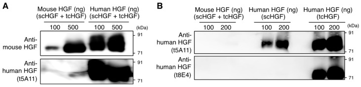
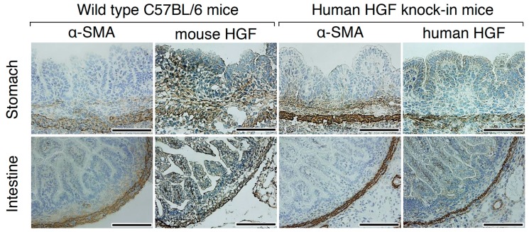
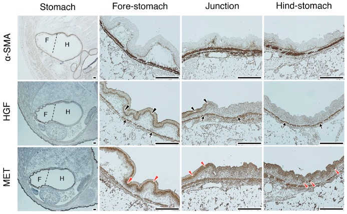
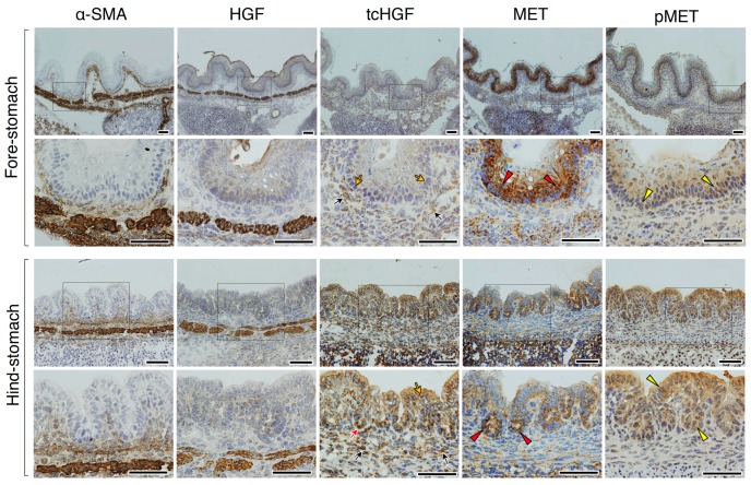
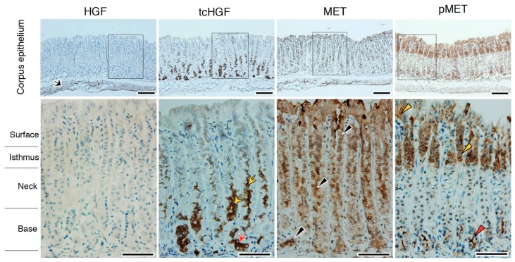
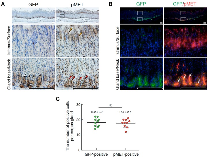
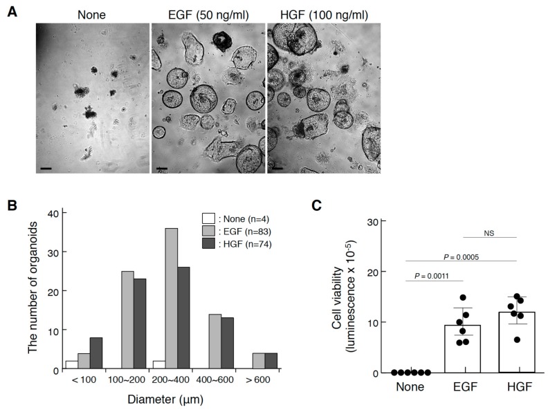
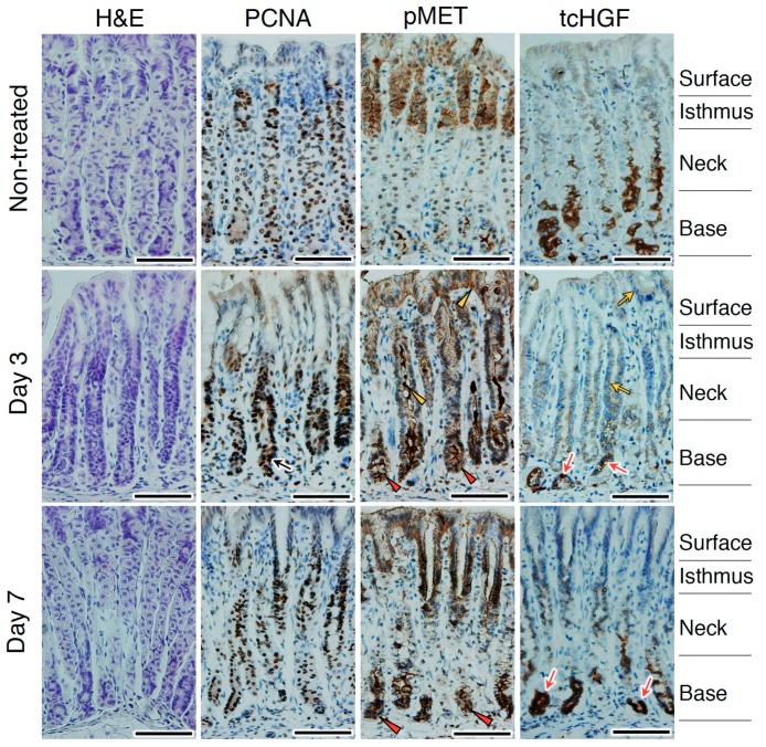
Hepatocyte growth factor (HGF) is secreted as an inactive single-chain HGF (scHGF); however, only proteolytically processed two-chain HGF (tcHGF) can activate the MET receptor. We investigated the localization of tcHGF and activated/phosphorylated MET (pMET) using a tcHGF-specific antibody. In day 16.5 mouse embryos, total HGF (scHGF + tcHGF) was mainly localized in smooth muscle cells close to, but separate from, MET-positive epithelial cells in endodermal organs, including the stomach. In the adult stomach, total HGF was localized in smooth muscle cells, and tcHGF was mainly localized in the glandular base region. Immunostaining for pMET and Lgr5-driven green fluorescent protein (GFP) indicated that pMET localization overlapped with Lgr5+ gastric stem cells. HGF promoted organoid formation similar to EGF, indicating the potential for HGF to promote the survival and growth of gastric stem cells. pMET and tcHGF localizations changed during regeneration following gastric injury. These results indicate that MET is constantly activated in gastric stem cells and that the localization of pMET differs from the primary localization of precursor HGF but has a close relationship to tcHGF. Our results suggest the importance of the microenvironmental generation of tcHGF in the regulation of development, regeneration, and stem cell behavior.

Keywords: HGF; MET receptor; regeneration; smooth muscle cell; stem cell.


Conflict of interest statement

The authors declare no conflicts of interest.

Figures

Figure 1
Figure 1
Specificity of anti-human HGF antibodies. (A) Western blot analysis for the cross-reactivity of anti-mouse HGF polyclonal antibody and t5A11 anti-human HGF monoclonal antibody between mouse and human HGF. (B) Specificity of anti-HGF monoclonal antibodies (t5A11 and t8E4) to mouse HGF, scHGF, and tcHGF. Mouse HGF and human HGF were subjected to SDS-PAGE under non-reducing conditions. Human HGF (composed of scHGF and tcHGF), scHGF, and tcHGF were expressed and purified as described in the Materials and Methods section.
Figure 2
Figure 2
Localization of HGF in the developing stomach and intestine of wild-type C57BL/6 and hHGF-ki mice. Immunohistochemistry was performed using anti-mouse HGF polyclonal antibodies and t5A11 anti-human HGF monoclonal antibodies, respectively, in C57BL/6 and hHGF-ki mice. Similar expression and localization patterns were obtained in sections from two different mice. Tissues were obtained from day 16.5 embryos. Scale bars represent 200 µm.
Figure 3
Figure 3
Localizations of HGF and MET receptors in the developing stomach. Immunohistochemical staining was performed using t5A11 anti-human HGF monoclonal antibody or anti-MET antibody. Stomachs in day 16.5 embryos were divided into the anterior/fore-stomach and posterior/hind-stomach, distinguished by dotted lines. Black arrows indicate HGF localization in smooth muscle cells. Black arrowheads indicate HGF localized in the epithelial cells. Red arrowheads indicate MET expression in epithelial cells. Similar localization patterns were obtained in sections from two different mice. Tissues were obtained from day 16.5 embryos of hHGF-ki mice. Scale bars represent 200 µm.
Figure 4
Figure 4
Localizations of HGF, tcHGF, MET, and phosphorylated MET (pMET) in the developing stomach. Immunohistochemical staining was performed using t5A11 (for scHGF and tcHGF), t8E4 (for tcHGF), anti-MET, or anti-phospho-MET antibody. The fore-stomach and hind-stomach were respectively characterized by multilayered squamous epithelia and developing glandular epithelium. Black and yellow arrows indicate tcHGF localized in the sub-epithelial and epithelial cells, respectively. The red arrow indicates strong tcHGF localization in developing glandular regions. Red arrowheads indicate strongly MET-positive cells in the epithelial cells. Yellow arrowheads indicate pMET in epithelial cells. The images in the lower panel are magnified images of the boxed areas in the upper panel. Similar immunohistochemical localization patterns were obtained in sections from two different mice. Tissues were obtained from day 16.5 embryos of hHGF-ki mice. Scale bars represent 200 µm.
Figure 5
Figure 5
Localizations of HGF, tcHGF, MET, and pMET in the corpus epithelium of the adult stomach. Immunohistochemical staining was performed using t5A11 (for scHGF and tcHGF), t8E4 (for tcHGF), anti-MET, or anti-phospho-MET antibody. Black arrows, HGF in smooth muscle cells; red arrow, tcHGF in the glandular base region; yellow arrows, tcHGF in the neck region; black arrowheads, MET in glandular epithelial structure; red arrowhead, pMET in the glandular base region; yellow arrowhead, pMET in the isthmus region and the surface region. The images in the lower panel are magnified images of the boxed areas in the upper panel. Similar immunohistochemical localization patterns were obtained in sections from two different mice. Tissues were obtained from hHGF-ki mice. Scale bars represent 200 µm.
Figure 6
Figure 6
Localizations of Lgr5+ stem cells and pMET in the corpus epithelium of the adult stomach. (A) Immunohistochemical staining for GFP and pMET. (B) Double immunofluorescence staining for GFP and pMET. In (A), black arrows, Lgr5-driven GFP; red arrowheads, pMET in the glandular base region; yellow arrowheads, pMET in the isthmus and/or surface regions. In (B), white arrows indicate the co-localization of pMET and GFP in Lgr5+ stem cells. The images in the lower panel are magnified images of the boxed areas in the upper panel. Tissues were obtained from Lgr5-DTR-EGFP mice. Scale bars represent 200 µm. (C) The numbers of GFP- and pMET-positive cells per corpus gland. These values were obtained by counting in 10 glands (n = 10). Data were tested for significance using an unpaired two-tailed t-test. NS, not significant.
Figure 7
Figure 7
Promotion of gastric corpus organoids by HGF. (A) The appearance of organoids cultured in the absence or presence of EGF or HGF. Scale bars represent 200 µm. (B) The size distribution of organoids. (C) The viability of organoids. In (A), corpus gland stem cells isolated from a C57BL/6 mouse were cultured in Matrigel in the absence or presence of EGF (50 ng/mL) or recombinant HGF (100 ng/mL) for 21 days. In (B), the size distribution was analyzed by using Image-J. In (C), the viability was measured using the CellTiter-Glo® 3D cell viability assay. Values are presented as mean ± S.D. (n = 6). Data were tested for significance using an unpaired two-tailed t-test. NS, not significant. Organoid culture was performed twice, from which similar results were obtained.
Figure 8
Figure 8
Changes in the localizations of tcHGF and pMET following stomach injury. hHGF-ki mice were subjected to a tamoxifen treatment. The black arrow indicates PCNA-positive proliferating cells. Red arrowheads and red arrows indicate pMET and tcHGF localizations, respectively, in the glandular base region. Yellow arrowheads and yellow arrows indicate pMET and tcHGF localizations, respectively, in the neck and/or surface regions. H&E, hematoxylin and eosin. Similar results were obtained in sections from two different mice independently subjected to tamoxifen treatment. Scale bars represent 200 µm.

Similar articles

Cited by

References

    1. Birchmeier C., Gherardi E. Developmental roles of HGF/SF and its receptor, the c-met tyrosine kinase. Trends Cell Biol. 1998;8:404–410. doi: 10.1016/S0962-8924(98)01359-2. - DOI - PubMed
    1. Trusolino L., Bertotti A., Comoglio P.M. MET signalling: Principles and functions in development, organ regeneration and cancer. Nat. Rev. Mol. Cell Biol. 2010;11:834–848. doi: 10.1038/nrm3012. - DOI - PubMed
    1. Sakai K., Aoki S., Matsumoto K. Hepatocyte growth factor and met in drug discovery. J. Biochem. 2015;157:271–284. doi: 10.1093/jb/mvv027. - DOI - PubMed
    1. Montesano R., Matsumoto K., Nakamura T., Orci L. Identification of a fibroblast-derived epithelial morphogen as hepatocyte growth factor. Cell. 1991;67:901–908. doi: 10.1016/0092-8674(91)90363-4. - DOI - PubMed
    1. Soriano J.V., Pepper M.S., Nakamura T., Orci L., Montesano R. Hepatocyte growth factor stimulates extensive development of branching duct-like structures by cloned mammary gland epithelial cells. J. Cell Sci. 1995;108:413–430. - PubMed

LinkOut - more resources