

Quercetin Inhibits the Production of IL-1β-Induced Inflammatory Cytokines and Chemokines in ARPE-19 Cells via the MAPK and NF-κB Signaling Pathways
- PMID: 31212975
- PMCID: PMC6628093
- DOI: 10.3390/ijms20122957
Quercetin Inhibits the Production of IL-1β-Induced Inflammatory Cytokines and Chemokines in ARPE-19 Cells via the MAPK and NF-κB Signaling Pathways
Abstract
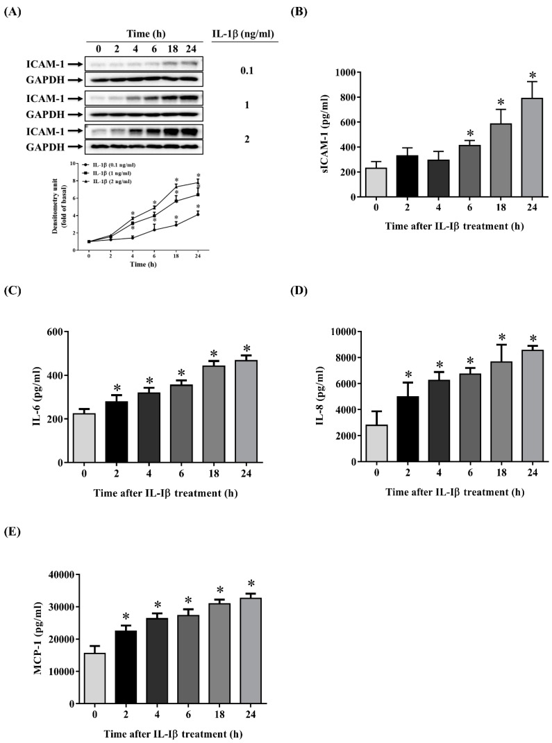
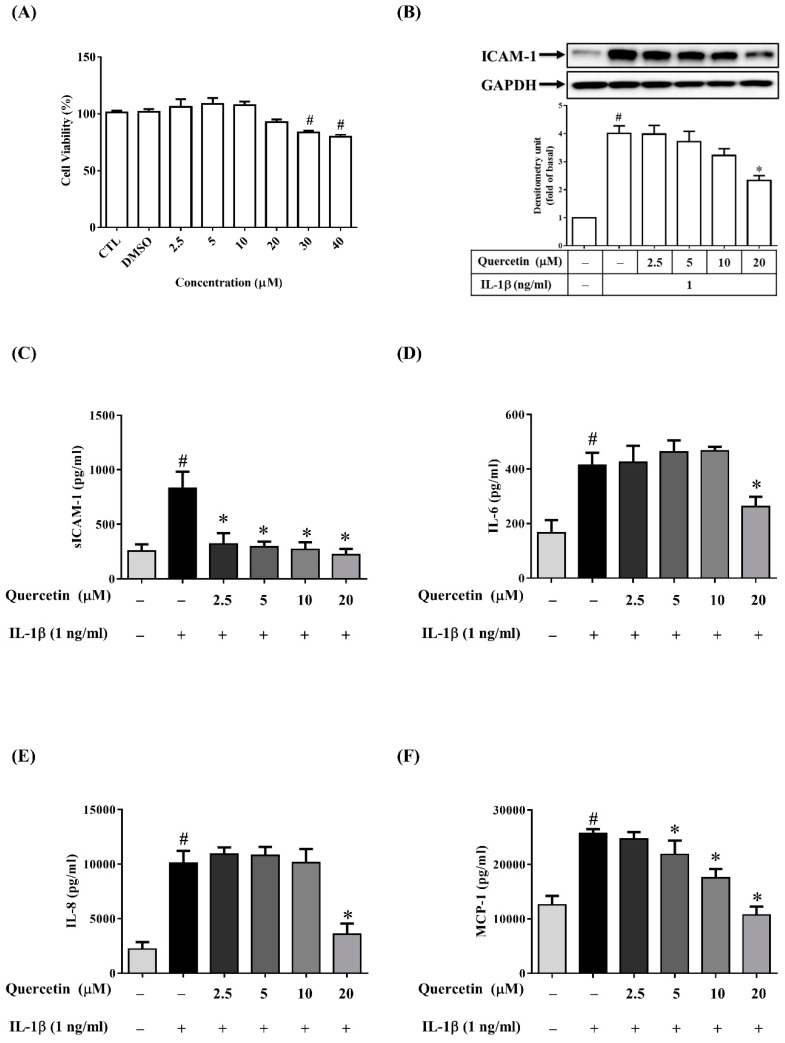
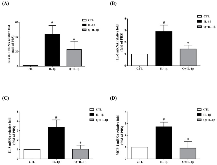
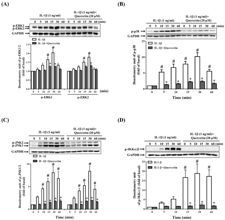
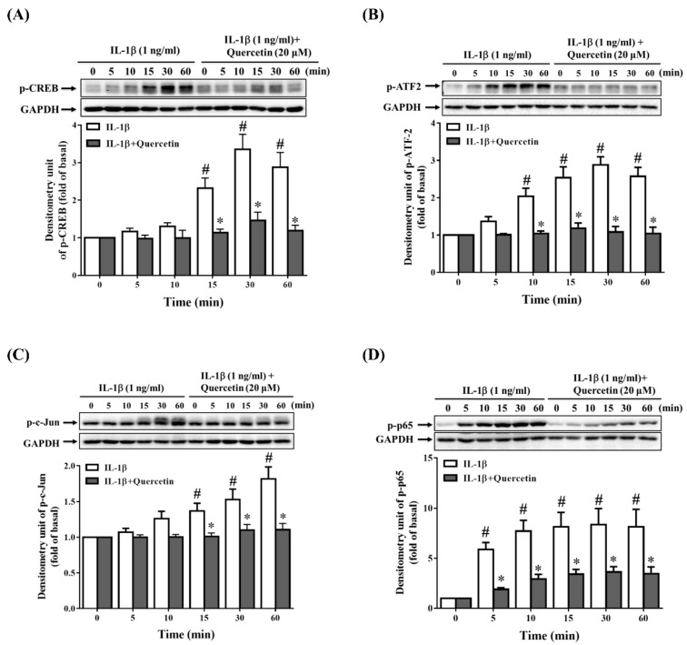
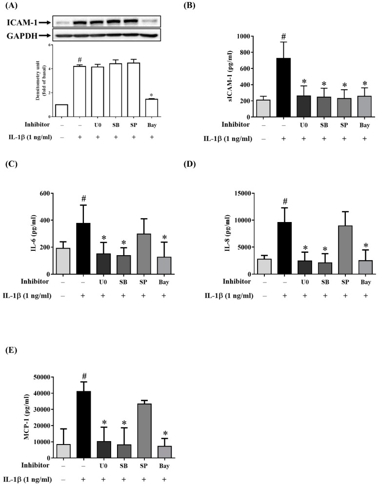
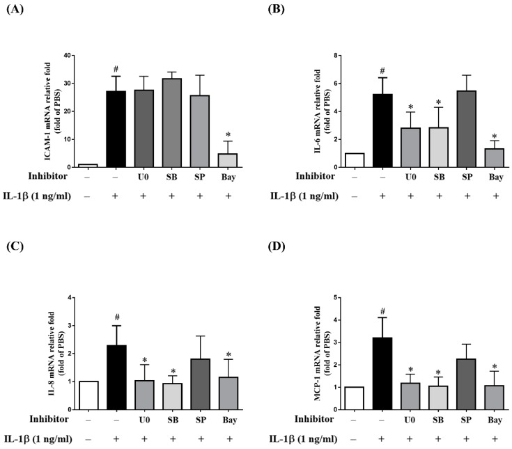
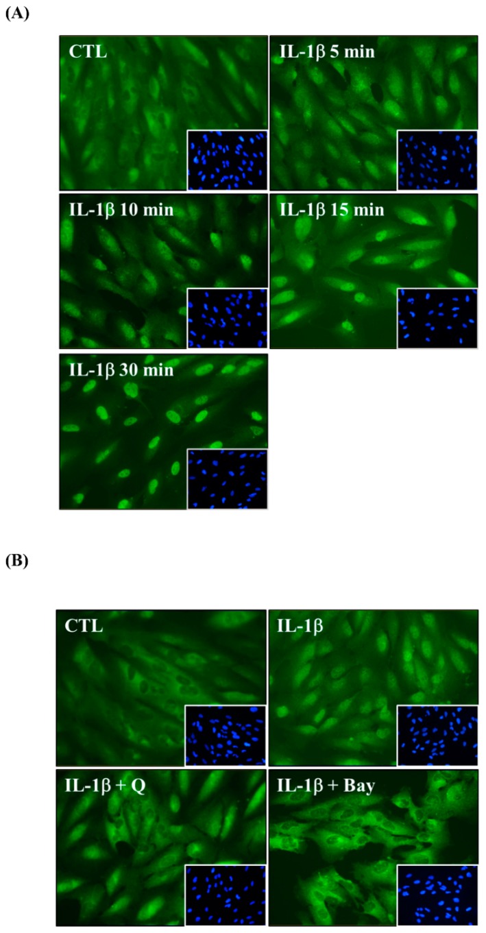
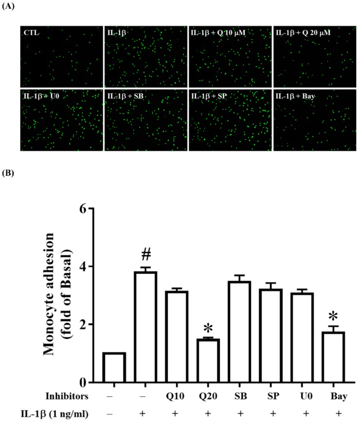
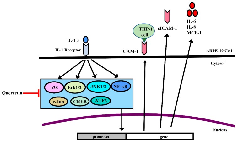
Quercetin, a bioflavonoid derived from vegetables and fruits, exerts anti-inflammatory effects in various diseases. Our previous study revealed that quercetin could suppress the expression of matrix metalloprotease-9 (MMP-9) and intercellular adhesion molecule-1 (ICAM-1) to achieve anti-inflammatory effects in tumor necrosis factor-α (TNF-α)-stimulated human retinal pigment epithelial (ARPE-19) cells. The present study explored whether quercetin can inhibit the interleukin-1β (IL-1β)-induced production of inflammatory cytokines and chemokines in ARPE-19 cells. Prior to stimulation by IL-1β, ARPE-19 cells were pretreated with quercetin at various concentrations (2.5-20 µM). The results showed that quercetin could dose-dependently decrease the mRNA and protein levels of ICAM-1, IL-6, IL-8 and monocyte chemoattractant protein-1 (MCP-1). It also attenuated the adherence of the human monocytic leukemia cell line THP-1 to IL-1β-stimulated ARPE-19 cells. We also demonstrated that quercetin inhibited signaling pathways related to the inflammatory process, including phosphorylation of mitogen-activated protein kinases (MAPKs), inhibitor of nuclear factor κ-B kinase (IKK)α/β, c-Jun, cAMP response element-binding protein (CREB), activating transcription factor 2 (ATF2) and nuclear factor (NF)-κB p65, and blocked the translocation of NF-κB p65 into the nucleus. Furthermore, MAPK inhibitors including an extracellular signal-regulated kinase (ERK) 1/2 inhibitor (U0126), a p38 inhibitor (SB202190) and a c-Jun N-terminal kinase (JNK) inhibitor (SP600125) decreased the expression of soluble ICAM-1 (sICAM-1), but not ICAM-1. U0126 and SB202190 could inhibit the expression of IL-6, IL-8 and MCP-1, but SP600125 could not. An NF-κB inhibitor (Bay 11-7082) also reduced the expression of ICAM-1, sICAM-1, IL-6, IL-8 and MCP-1. Taken together, these results provide evidence that quercetin protects ARPE-19 cells from the IL-1β-stimulated increase in ICAM-1, sICAM-1, IL-6, IL-8 and MCP-1 production by blocking the activation of MAPK and NF-κB signaling pathways to ameliorate the inflammatory response.
Keywords: anti-inflammatory; chemokines; cytokines; quercetin; retinal pigment epithelial cells.
Conflict of interest statement
The authors declare that there are no conflicts of interest.
Figures
Similar articles
-
Quercetin disrupts tyrosine-phosphorylated phosphatidylinositol 3-kinase and myeloid differentiation factor-88 association, and inhibits MAPK/AP-1 and IKK/NF-κB-induced inflammatory mediators production in RAW 264.7 cells.Immunobiology. 2013 Dec;218(12):1452-67. doi: 10.1016/j.imbio.2013.04.019. Epub 2013 May 9. Immunobiology. 2013. PMID: 23735482
-
Nepetin inhibits IL-1β induced inflammation via NF-κB and MAPKs signaling pathways in ARPE-19 cells.Biomed Pharmacother. 2018 May;101:87-93. doi: 10.1016/j.biopha.2018.02.054. Epub 2018 Feb 23. Biomed Pharmacother. 2018. PMID: 29477475
-
Oleuropein Protects Human Retinal Pigment Epithelium Cells from IL-1β-Induced Inflammation by Blocking MAPK/NF-κB Signaling Pathways.Inflammation. 2022 Feb;45(1):297-307. doi: 10.1007/s10753-021-01546-4. Epub 2021 Oct 6. Inflammation. 2022. PMID: 34613549
-
The immunosuppressive activity of artemisinin-type drugs towards inflammatory and autoimmune diseases.Med Res Rev. 2021 Nov;41(6):3023-3061. doi: 10.1002/med.21842. Epub 2021 Jul 21. Med Res Rev. 2021. PMID: 34288018 Review.
-
Exploring the Role of Licorice and Its Derivatives in Cell Signaling Pathway NF-κB and MAPK.J Nutr Metab. 2024 Oct 23;2024:9988167. doi: 10.1155/2024/9988167. eCollection 2024. J Nutr Metab. 2024. PMID: 39479405 Free PMC article. Review.
Cited by
-
Exhaustion of Protective Heat Shock Response Induces Significant Tumor Damage by Apoptosis after Modulated Electro-Hyperthermia Treatment of Triple Negative Breast Cancer Isografts in Mice.Cancers (Basel). 2020 Sep 10;12(9):2581. doi: 10.3390/cancers12092581. Cancers (Basel). 2020. PMID: 32927720 Free PMC article.
-
Long-Chain Polyunsaturated Fatty Acids and Their Metabolites Regulate Inflammation in Age-Related Macular Degeneration.J Inflamm Res. 2022 Feb 9;15:865-880. doi: 10.2147/JIR.S347231. eCollection 2022. J Inflamm Res. 2022. PMID: 35173457 Free PMC article. Review.
-
Yindan Jiedu granules exhibit anti-inflammatory effect in patients with novel Coronavirus disease (COVID-19) by suppressing the NF-κB signaling pathway.Phytomedicine. 2022 Jan;95:153784. doi: 10.1016/j.phymed.2021.153784. Epub 2021 Oct 1. Phytomedicine. 2022. PMID: 34785108 Free PMC article. Clinical Trial.
-
Plausibility of natural immunomodulators in the treatment of COVID-19-A comprehensive analysis and future recommendations.Heliyon. 2023 Jun;9(6):e17478. doi: 10.1016/j.heliyon.2023.e17478. Epub 2023 Jun 21. Heliyon. 2023. PMID: 37366526 Free PMC article. Review.
-
Beneficial effects of Elaeagnus rhamnoides (L.) A. Nelson and its most abundant flavonoids on the main mechanisms related to diabetic bone disease.Pharm Biol. 2025 Dec;63(1):460-489. doi: 10.1080/13880209.2025.2523392. Epub 2025 Jul 3. Pharm Biol. 2025. PMID: 40607760 Free PMC article. Review.
References
-
- Kutty R.K., Nagineni C.N., Samuel W., Vijayasarathy C., Hooks J.J., Redmond T.M. Inflammatory cytokines regulate microRNA-155 expression in human retinal pigment epithelial cells by activating JAK/STAT pathway. Biochem. Biophys. Res. Commun. 2010;402:390–395. doi: 10.1016/j.bbrc.2010.10.042. - DOI - PMC - PubMed
-
- Wong W.L., Su X., Li X., Cheung C.M., Klein R., Cheng C.Y., Wong T.Y. Global prevalence of age-related macular degeneration and disease burden projection for 2020 and 2040: A systematic review and meta-analysis. Lancet Glob. Health. 2014;2:e106–e116. doi: 10.1016/S2214-109X(13)70145-1. - DOI - PubMed
MeSH terms
Substances
Grants and funding
LinkOut - more resources
Full Text Sources
Other Literature Sources
Research Materials
Miscellaneous
