. 2019 Jun 17;11(6):348.
doi: 10.3390/toxins11060348.

Toxin B Variants from Clostridium difficile Strains VPI 10463 and NAP1/027 Share Similar Substrate Profile and Cellular Intoxication Kinetics but Use Different Host Cell Entry Factors

Affiliations

Toxin B Variants from Clostridium difficile Strains VPI 10463 and NAP1/027 Share Similar Substrate Profile and Cellular Intoxication Kinetics but Use Different Host Cell Entry Factors

Diana López-Ureña et al. Toxins (Basel). .

Abstract

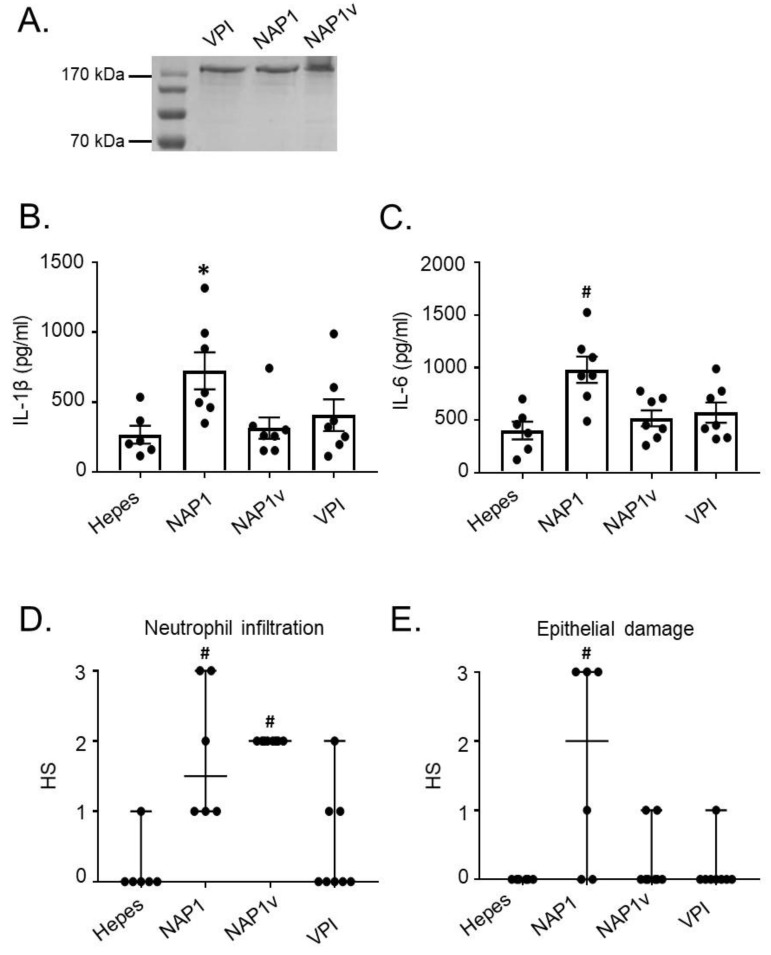
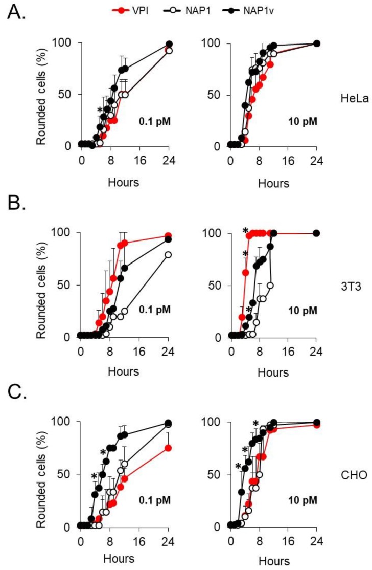
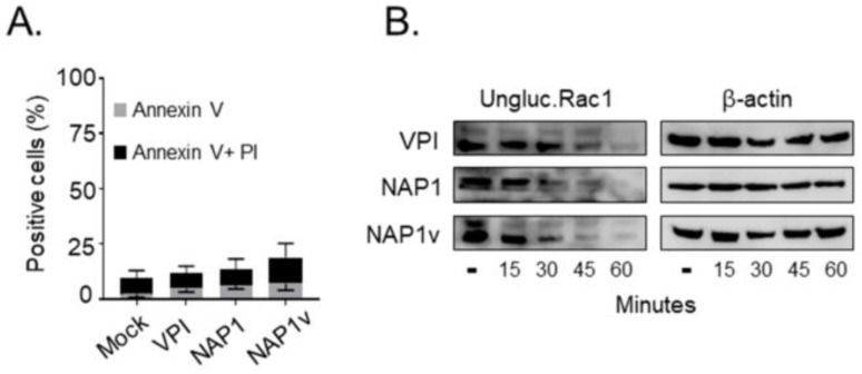
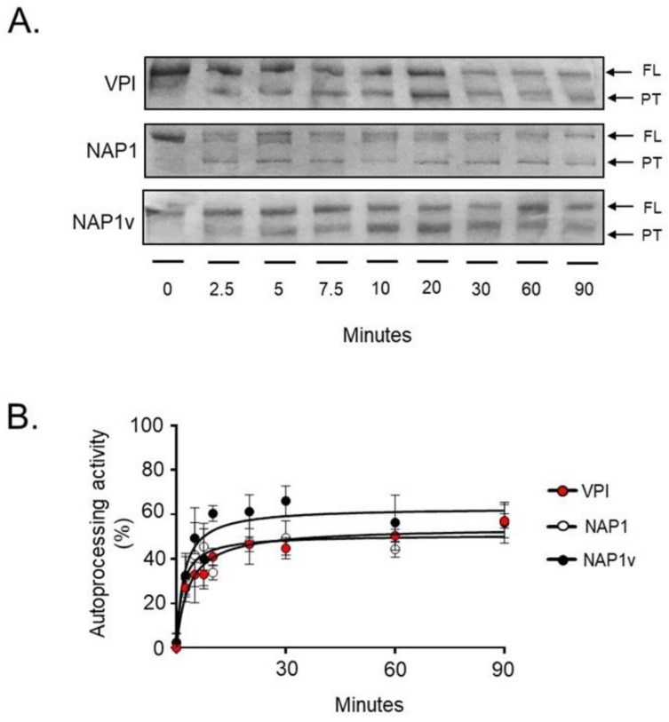
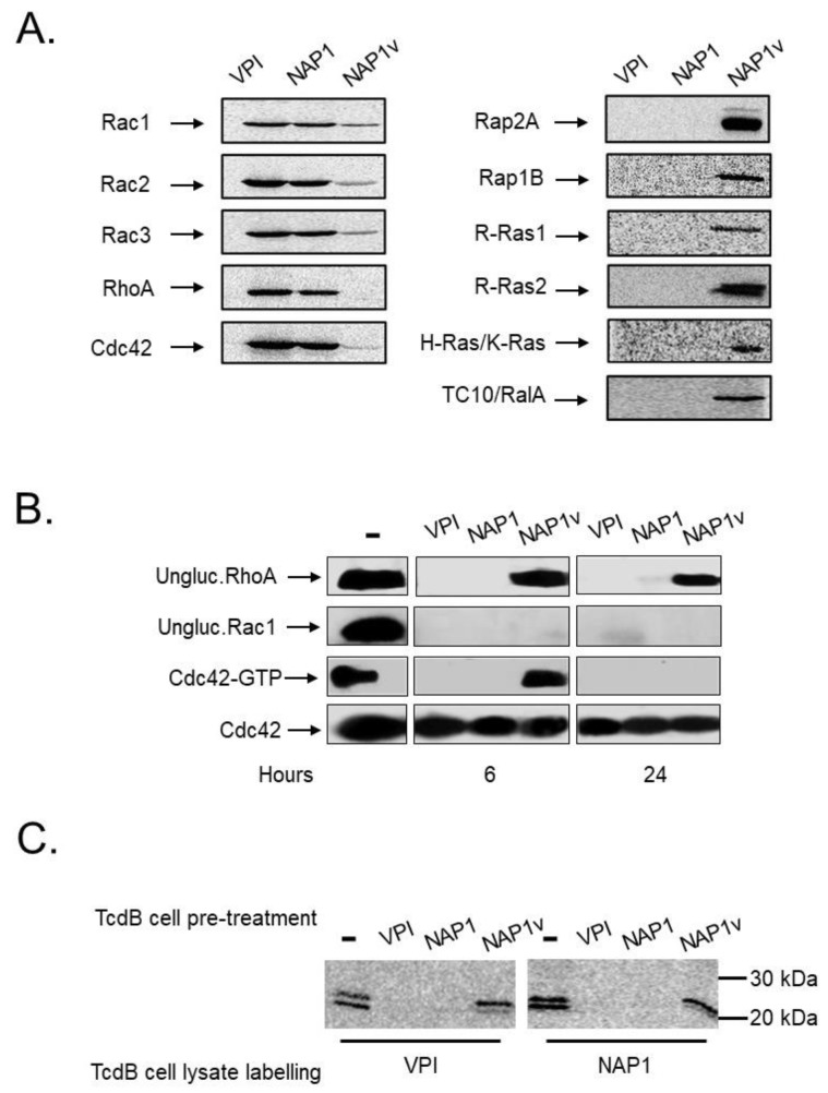
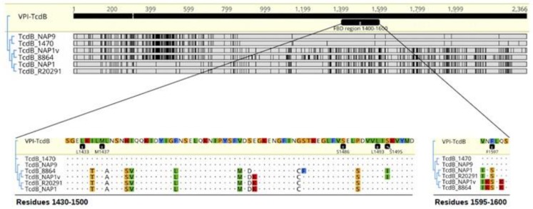
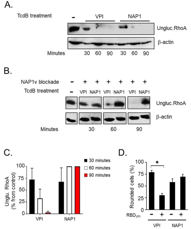
Clostridium difficile induces antibiotic-associated diarrhea due to the release of toxin A (TcdA) and toxin B (TcdB), the latter being its main virulence factor. The epidemic strain NAP1/027 has an increased virulence attributed to different factors. We compared cellular intoxication by TcdBNAP1 with that by the reference strain VPI 10463 (TcdBVPI). In a mouse ligated intestinal loop model, TcdBNAP1 induced higher neutrophil recruitment, cytokine release, and epithelial damage than TcdBVPI. Both toxins modified the same panel of small GTPases and exhibited similar in vitro autoprocessing kinetics. On the basis of sequence variations in the frizzled-binding domain (FBD), we reasoned that TcdBVPI and TcdBNAP1 might have different receptor specificities. To test this possibility, we used a TcdB from a NAP1 variant strain (TcdBNAP1v) unable to glucosylate RhoA but with the same receptor-binding domains as TcdBNAP1. Cells were preincubated with TcdBNAP1v to block cellular receptors, prior to intoxication with either TcdBVPI or TcdBNAP1. Preincubation with TcdBNAP1v blocked RhoA glucosylation by TcdBNAP1 but not by TcdBVPI, indicating that the toxins use different host factors for cell entry. This crucial difference might explain the increased biological activity of TcdBNAP1 in the intestine, representing a contributing factor for the increased virulence of the NAP1/027 strain.

Keywords: Clostridium difficile; NAP1/027 toxin B; frizzled receptors; receptor binding.


Conflict of interest statement

The authors declare no conflict of interest.

Figures

Figure 1
Figure 1
Toxin B (TcdB) from NAP1 induces a higher inflammatory influx and ileal disruption. The in vivo effect of various TcdBs was assayed in a murine ligated ileal loop model. (A) TcdBs were obtained from the supernatants of strains grown in a dialysis system culture and purified by ion-exchange chromatography and gel filtration. The purity of the toxins was assessed after SDS-PAGE by Coomassie staining of a 7.5% gel loaded with 1.5 µg of TcdBVPI (VPI), TcdBNAP1 (NAP1), and TcdBNAP1v (NAP1v). Then, ileal loops were treated for 4 h with 10 μg of purified TcdBVPI (VPI), TcdBNAP1 (NAP1), and TcdBNAP1v (NAP1v); (B,C) The effect of the toxins on inflammatory cytokines was measured; (D,E) Neutrophil infiltration and epithelial damage induced by the toxins was also determined using a histopathological score (HS) scale from 1 (mild) to 3 (severe). Hepes was used as a negative control. Error bars represent means ± SEM (A,B) and median ± range (C,D), n ≥ 5; * p < 0.05 compared to Hepes, # p < 0.05 compared to other groups (One-way ANOVA with Bonferroni’s correction, Kruskal–Wallis test, and Dunn’s multiple-comparison test).
Figure 2
Figure 2
Cell rounding of distinct cell lines induced by TcdBs. (A) HeLa, (B) 3T3 fibroblasts, and (C) CHO cells were treated with the indicated concentrations of TcdBVPI (VPI), TcdBNAP1 (NAP1), and TcdBNAP1v (NAP1v). The percentage of round cells in each well was evaluated every hour for a period of 12 h and then after 24 h from the start of the experiment. Error bars represent means ± SD of 100 cells in three independent experiments; * p < 0.05 (One-way ANOVA with Bonferroni´s correction).
Figure 3
Figure 3
Cytotoxicity of TcdBs in HeLa cells. (A) HeLa cells were treated with 100 pM of TcdBVPI (VPI), TcdBNAP1 (NAP1), and TcdBNAP1v (NAP1v) for 24 h. Cytotoxicity was analyzed by flow cytometry using propidium iodide (PI)/anexin V double staining. Control cells were left untreated (mock). Error bars represent means ± SD of three independent experiments; (B) Following the addition of 100 pM of TcdB, the cells were lysed at the indicated time points, and the dynamics of Rac1 glucosylation was monitored by immunoblot with a specific anti-Rac1 antibody that only recognizes the unmodified form of this protein (ungluc.Rac1). Untreated cells (-) were included as a positive control for unglucosylated Rac1, and immunodetection of beta-actin served as a loading control (β-actin). Shown are representative western blot images from three independent experiments.
Figure 4
Figure 4
In vitro autoprocessing of TcdB. The in vitro processing of TcdBVPI (VPI), TcdBNAP1 (NAP1), and TcdBNAP1v (NAP1v) was evaluated in the presence of 10 mM of the co-factor inositol hexakisphosphate (InsP6). The reactions were incubated at 37°C for the indicated times, and toxins or toxin fragments were then detected by Coomassie Blue staining. The enzymatic activity was determined by the presence of full-length (FL) toxin (1–2366), which decreased over time, and processed toxin (544–2366, PT), which increased over time. Shown in (A) is one representative SDS-PAGE gel from three independent experiments; (B) The bands corresponding to the FL toxin and the PT were quantified by densitometry. The autoprocessing activity of each toxin was determined by comparing the relative amounts of both forms of the toxin, and the percentage obtained was plotted using nonlinear regression. Error bars represent means ± SD of three independent experiments.
Figure 5
Figure 5
Glucosyltransferase activity of TcdBs. (A) The in vitro glucosylation of distinct recombinant GTPases by TcdBVPI (VPI), TcdBNAP1 (NAP1), and TcdBNAP1v (NAP1v) was determined using UDP-[14C] glucose as a co-substrate. The reactions were incubated for 1 h and then subjected to SDS-PAGE. The radiolabeled bands were detected by phosphor imaging; (B) Following the addition of 100 pM of TcdB, HeLa cells were lysed at the indicated times, and the glucosylation of Rac1 and RhoA was monitored by immunoblot with specific anti-Rac1 and anti RhoA antibodies that only recognize the unmodified form of the proteins (ungluc.Rac1 and ungluc.RhoA, respectively). The effect of TcdBs on the activation state of Cdc42 was also evaluated. After intoxication, one part of the lysate was used as a control for the total amount of GTPase, and the rest was incubated with Rho Binding Domain–GST-sepharose beads. Cdc42 was detected by immunoblot with anti-Cdc42 antibodies; (C) HeLa cells were treated with 10 pM of TcdBVPI (VPI), TcdBNAP1 (NAP1), and TcdBNAP1v (NAP1v) for 12 h (TcdB cell pre-treatment). The cells were lysed, and the lysate proteins were glucosylated in vitro by TcdBVPI (VPI) or TcdBNAP1 (NAP1), according to the conditions stated in (A) (TcdB cell lysate labelling). Untreated cells (-) in (B) and (C) were included as a positive control for unmodified or activated proteins.
Figure 6
Figure 6
Comparison of TcdB residues involved in frizzled receptor binding. Tree alignment of TcdB sequences of strains VPI 10493, 1470 (reference TcdB variant strain), NAP9 (TcdB variant strain), 8864 (TcdB variant strain), NAP1v, R20291 (NAP1 reference strain), and NAP1. TcdBVPI was used as a reference. Discrepancies are highlighted with black bars or colored-residue letters.
Figure 7
Figure 7
Uptake of TcdB from TcdBNAP1 is blocked by TcdBNAP1v. In order to determine whether the different toxins use the same host cell entry factors, an uptake competition assay was performed in HeLa cells. (A) The cells were treated with 10 pM of TcdBVPI (VPI) or TcdBNAP1 (NAP1) for the indicated times at 37 °C. Following cell lysis, the uptake of each toxin was evaluated by western blot using an antibody that only binds to the unglucosylated form of RhoA GTPase (ungluc.RhoA); (B) The cells were treated with 1 nM of TcdBNAP1v (NAP1v) for 30 min at 4 °C to induce binding of TcdBNAP1v to the cell but not its uptake (NAP1v blockade +). The cells were then treated with 10 pM of TcdBVPI or TcdBNAP1 and incubated for 30, 60, and 90 min at 37 °C (TcdB treatment). Toxin uptake was determined by immunodetection of unmodified RhoA, as stated in (A). Untreated cells (-) were included as a positive control for unglucosylated Rac1, and immunoblot of β-actin served as a loading control (β-actin). Shown are representative western blot images from three independent experiments; (C) The bar graph shows the amount of unglucosylated RhoA in intoxicated cells relative to the unglucosylated RhoA amount in untreated cells, which was set to 100%. Error bars represent means ± SD of three independent experiments; (D) 10 nM of recombinant Receptor Binding Domain (RBDVPI) corresponding to TcdB1349-1811 of VPI 10463 was added to the cells, and these were incubated for 30 min at 4 °C to block toxin receptors. Control cells were mocked-treated with the corresponding volume of buffer and also incubated for 30 min at 4 °C (RBDVPI -). The cells were then treated with 100 pM of TcdBVPI or TcdBNAP1, and the percentage of round cells was evaluated after 60 min as a parameter of full-length toxin uptake. Error bars represent means ± SD of 1000 cells in triplicate samples; * p < 0.05 (Student t test). This experiment is representative of three independent experiments with similar results.

Similar articles

Cited by

References

    1. Voth D.E., Ballard J.D. Clostridium difficile toxins: Mechanism of action and role in disease. Clin. Microbiol. Rev. 2005;18:247–263. doi: 10.1128/CMR.18.2.247-263.2005. - DOI - PMC - PubMed
    1. Carter G.P., Chakravorty A., Pham Nguyen T.A., Mileto S., Schreiber F., Li L., Howarth P., Clare S., Cunningham B., Sambol S.P., et al. Defining the roles of TcdA and TcdB in localized gastrointestinal disease, systemic organ damage, and the host response during Clostridium difficile Infections. mBio. 2015;6:e00551-15. doi: 10.1128/mBio.00551-15. - DOI - PMC - PubMed
    1. Lyras D., O’Connor J.R., Howarth P.M., Sambol S.P., Carter G.P., Phumoonna T., Poon R., Adams V., Vedantam G., Johnson S., et al. Toxin B is essential for virulence of Clostridium difficile. Nature. 2009;458:1176–1179. doi: 10.1038/nature07822. - DOI - PMC - PubMed
    1. Jank T., Aktories K. Structure and mode of action of clostridial glucosylating toxins: The ABCD model. Trends Microbiol. 2008;16:222–229. doi: 10.1016/j.tim.2008.01.011. - DOI - PubMed
    1. LaFrance M.E., Farrow M.A., Chandrasekaran R., Sheng J., Rubin D.H., Lacy D.B. Identification of an epithelial cell receptor responsible for Clostridium difficile TcdB-induced cytotoxicity. Proc. Natl. Acad. Sci. USA. 2015;112:7073–7078. doi: 10.1073/pnas.1500791112. - DOI - PMC - PubMed

Publication types

MeSH terms

LinkOut - more resources