Case Reports
. 2019 Aug 5;216(8):1777-1790.
doi: 10.1084/jem.20190669. Epub 2019 Jun 18.

Inherited IL-18BP deficiency in human fulminant viral hepatitis

Affiliations
Case Reports

Inherited IL-18BP deficiency in human fulminant viral hepatitis

Serkan Belkaya et al. J Exp Med. .

Abstract

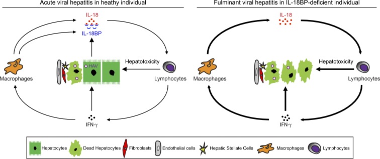
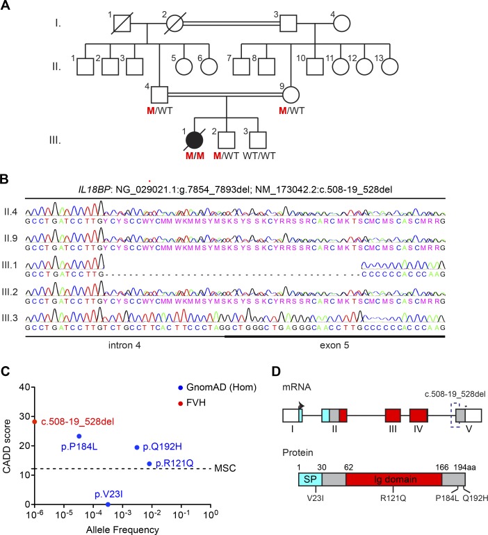
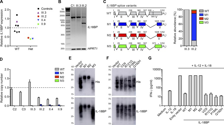
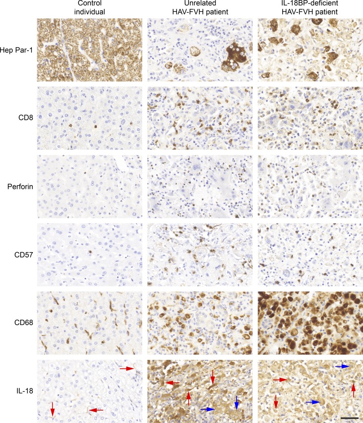
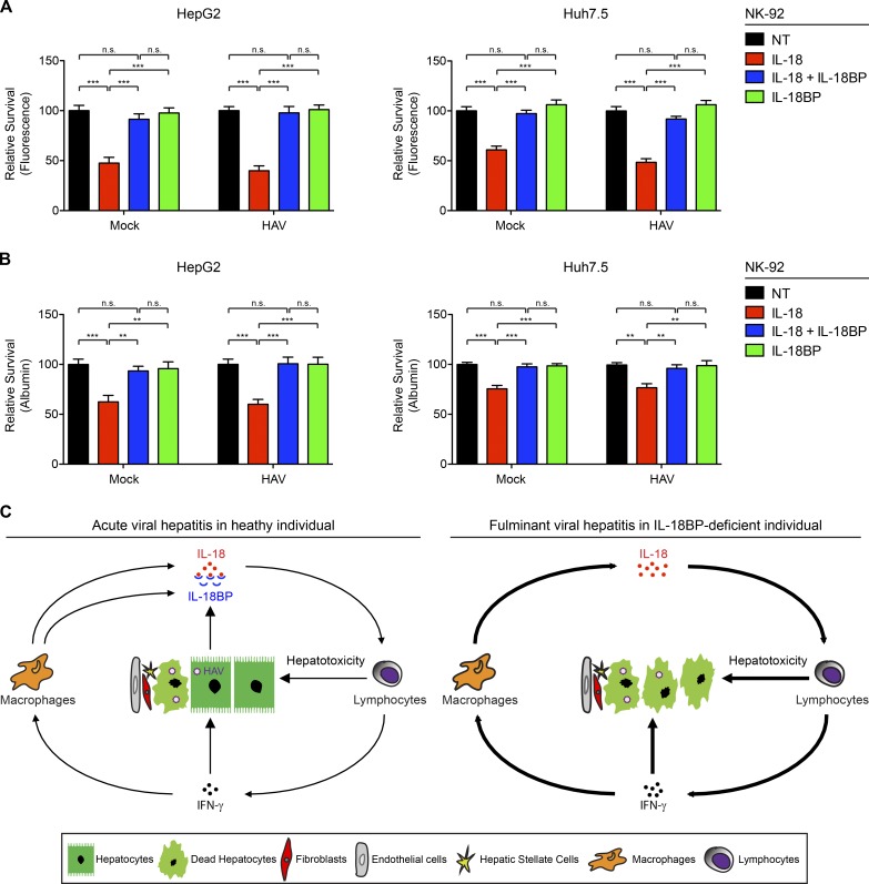
Fulminant viral hepatitis (FVH) is a devastating and unexplained condition that strikes otherwise healthy individuals during primary infection with common liver-tropic viruses. We report a child who died of FVH upon infection with hepatitis A virus (HAV) at age 11 yr and who was homozygous for a private 40-nucleotide deletion in IL18BP, which encodes the IL-18 binding protein (IL-18BP). This mutation is loss-of-function, unlike the variants found in a homozygous state in public databases. We show that human IL-18 and IL-18BP are both secreted mostly by hepatocytes and macrophages in the liver. Moreover, in the absence of IL-18BP, excessive NK cell activation by IL-18 results in uncontrolled killing of human hepatocytes in vitro. Inherited human IL-18BP deficiency thus underlies fulminant HAV hepatitis by unleashing IL-18. These findings provide proof-of-principle that FVH can be caused by single-gene inborn errors that selectively disrupt liver-specific immunity. They also show that human IL-18 is toxic to the liver and that IL-18BP is its antidote.


Figures

None
Graphical abstract
Figure 1.
Figure 1.
Homozygous 40-nt deletion in IL18BP. (A) Pedigree of the family affected by FVH due to HAV. The patient is shown in black, whereas healthy individuals are shown in white. Where available, IL18BP mutation (NM_173042.2:c.508-19_528del) status is indicated in red. M, mutant. (B) Familial segregation of the mutation and its homozygous state in the patient were confirmed by Sanger sequencing. (C) Graph showing the predicted CADD scores and global AFs of the mutation found in the patient with FVH (red circle) and missense variants of IL18BP (blue circles) for which homozygotes were reported in GnomAD. The CADD-MSC score (90% confidence interval) for IL18BP is indicated by a dashed line. (D) The upper panel shows the exons (1–5) of the canonical IL18BP transcript; the bottom panel shows a diagram for IL-18BP. The signal peptide is highlighted in blue; the Ig domain is shown in red. Start and stop codons are indicated by an arrow and an asterisk, respectively. The c.508-19_528del is shown as a dashed box on the mRNA. The locations of IL18BP alleles from GnomAD are also shown on the protein diagram.
Figure 2.
Figure 2.
Impact of the IL18BP:c.508-19_528del on gene expression and function. (A) RT-qPCR showing IL18BP levels normalized against endogenous GAPDH expression in EBV-B cell lines from six healthy controls (black), the WT sibling (III.3, purple), and heterozygous family members: brother (III.2, red), father (II.4, blue), and mother (II.9, green). Relative IL18BP expression was determined by normalization against the mean value for WT cells, set to 1 (indicated by a dashed line). The values shown are the means of two independent experiments performed in duplicate. (B) Agarose gel electrophoresis showing aberrant splicing of the IL18BP mRNA in 3′ RACE on EBV-B cells from the heterozygous sibling (III.2), relative to a control cell line (C1) and the WT sibling (III.3). HPRT1 was used as the housekeeping gene control. (C) The nested PCR products from B were cloned, and colonies were sequenced. Diagram (left) and percentages (right) of WT (gray) and mutant (M1 in blue, M2 in red, and M3 in green) splice variants of the IL18BP transcript are shown. The start codon is located at position 1, and the stop codon is at 585, shown by an asterisk, on the WT transcript. The polyadenylation site is at position 1,252 and indicated by An. (D) Expression levels of each splice variant (WT in gray, M1 in blue, M2 in red, and M3 in green) were determined and normalized against endogenous GAPDH expression levels by RT-qPCR on EBV-B cells from two healthy controls (C2 and C3) and family members. Graph shows the copy numbers of the mutant splice variants relative to the mean copy number for the WT allele in EBV-B cells from C2, C3, and III.3, which was set to 1 (indicated by a dashed line). The values are the means ± SEM of two independent experiments performed in duplicate. (E and F) Representative immunoblot images showing levels of the WT and mutant IL-18BP alleles, M1–M3 (E), and four missense alleles from GnomAD (F) in concentrated supernatants from transiently transfected COS7 cells. Immunoblotting was performed with the His tag antibody (top), and the membrane was then stripped and probed with the IL18BP antibody (bottom). (G) IL-18BP bioassay: IFN-γ production was measured in NK-92 cells stimulated with recombinant human IL-12 (100 pg/ml), IL-18 (10 ng/ml), and/or concentrated supernatants (100 µg/ml of total protein) of COS7 cells transiently transfected with either empty vector or the constructs expressing indicated IL-18BP variants. Graph is presented on a logarithmic scale with base of 10. The data are the means ± SEM of two independent experiments performed in duplicate using the supernatants shown in E and F and Fig. S1, F and G.
Figure 3.
Figure 3.
Liver immunohistochemical profile of the patient. Liver tissue sections from a control individual, an unrelated patient with FVH due to HAV, and the deceased IL-18BP–deficient FVH patient reported in this study were subjected to immunohistochemical staining with the following markers: Hep Par-1, CD8, perforin, CD57, CD68, and IL-18. Representative zoom-in views of the original images at 400× magnification (Fig. S4) are shown. Hep Par-1 staining of IL-18BP–deficient patient’s liver tissue section displayed a background staining of macrophages, with lower intensity than hepatocytes. Some IL-18–positive hepatocytes and macrophages are indicated with blue and red arrows, respectively. Scale bar represents 50 µm.
Figure 4.
Figure 4.
IL-18/IL-18BP–mediated hepatotoxicity. (A and B) Coculture of mock- or HAV-infected hepatocytes (HepG2 and Huh7.5 cells) with NK-92 cells pretreated with IL-18, IL-18 + IL-18BP, or IL-18BP. HAV infection efficiencies in HepG2 and Huh7.5 cells were ∼40% and ~100%, respectively (Fig. S5 E; Materials and methods). The relative survival of calcein-AM–stained HepG2 or Huh7.5 cells was calculated based on the measurement of fluorescence retention within cells (A) and the amount of secreted albumin (B). Relative fluorescence and albumin levels were determined by normalization against the mean value for hepatocytes cocultured with NK92 cells without pretreatment (not treated [NT]), set to 100. A decrease in the fluorescence or in albumin levels indicates an increase in NK cell–induced hepatotoxicity. The data shown are the means ± SEM of three independent experiments performed in quadruplicate (n.s., not significant; **, P < 0.01; ***, P < 0.001; one-way ANOVA with Bonferroni correction for multiple comparisons). (C) A proposed model for IL-18BP deficiency underlying FVH. During the course of acute HAV infection in an otherwise healthy individual (left), IL-18 is secreted by macrophages in the liver. This cytokine activates lymphocytes, such as NK cells, inducing IFN-γ production and cytotoxicity to eliminate HAV-infected cells. IFN-γ also induces IL-18BP secretion by hepatocytes, macrophages, and other nonparenchymal cells (endothelial cells, fibroblasts, and hepatic stellate cells), to buffer IL-18 activity. However, in the absence of IL-18BP (right), excessive IL-18 activity leads to uncontrolled, massive immune-mediated hepatotoxicity and severe liver injury, as in the IL-18BP–deficient individual with FVH.

Comment in

  • No IL-18BP? Avoid HAV.
    Diamond MS. Diamond MS. J Exp Med. 2019 Aug 5;216(8):1728-1729. doi: 10.1084/jem.20190841. Epub 2019 Jun 26. J Exp Med. 2019. PMID: 31243052 Free PMC article.

Similar articles

Cited by

References

    1. Aizawa Y., Akita K., Taniai M., Torigoe K., Mori T., Nishida Y., Ushio S., Nukada Y., Tanimoto T., Ikegami H., et al. . 1999. Cloning and expression of interleukin-18 binding protein. FEBS Lett. 445:338–342. 10.1016/S0014-5793(99)00148-9 - DOI - PubMed
    1. Ajmera V., Xia G., Vaughan G., Forbi J.C., Ganova-Raeva L.M., Khudyakov Y., Opio C.K., Taylor R., Restrepo R., Munoz S., et al. . Acute Liver Failure Study Group . 2011. What factors determine the severity of hepatitis A-related acute liver failure? J. Viral Hepat. 18:e167–e174. 10.1111/j.1365-2893.2010.01410.x - DOI - PMC - PubMed
    1. Bachmann M., Pfeilschifter J., and Mühl H.. 2018. A Prominent Role of Interleukin-18 in Acetaminophen-Induced Liver Injury Advocates Its Blockage for Therapy of Hepatic Necroinflammation. Front. Immunol. 9:161 10.3389/fimmu.2018.00161 - DOI - PMC - PubMed
    1. Belkaya S., Kontorovich A.R., Byun M., Mulero-Navarro S., Bajolle F., Cobat A., Josowitz R., Itan Y., Quint R., Lorenzo L., et al. . 2017. Autosomal Recessive Cardiomyopathy Presenting as Acute Myocarditis. J. Am. Coll. Cardiol. 69:1653–1665. 10.1016/j.jacc.2017.01.043 - DOI - PMC - PubMed
    1. Blechingberg J., Lykke-Andersen S., Jensen T.H., Jørgensen A.L., and Nielsen A.L.. 2007. Regulatory mechanisms for 3′-end alternative splicing and polyadenylation of the Glial Fibrillary Acidic Protein, GFAP, transcript. Nucleic Acids Res. 35:7636–7650. 10.1093/nar/gkm931 - DOI - PMC - PubMed

Publication types

MeSH terms

Substances