. 2019 Aug 2;294(31):11654-11664.
doi: 10.1074/jbc.RA119.009045. Epub 2019 Jun 18.

Human FAM173A is a mitochondrial lysine-specific methyltransferase that targets adenine nucleotide translocase and affects mitochondrial respiration

Affiliations

Human FAM173A is a mitochondrial lysine-specific methyltransferase that targets adenine nucleotide translocase and affects mitochondrial respiration

Jędrzej M Małecki et al. J Biol Chem. .

Abstract

Lysine methylation is a common posttranslational modification of nuclear and cytoplasmic proteins but is also present in mitochondria. The human protein denoted "family with sequence similarity 173 member B" (FAM173B) was recently uncovered as a mitochondrial lysine (K)-specific methyltransferase (KMT) targeting the c-subunit of mitochondrial ATP synthase (ATPSc), and was therefore renamed ATPSc-KMT. We here set out to investigate the biochemical function of its yet uncharacterized paralogue FAM173A. We demonstrate that FAM173A localizes to mitochondria, mediated by a noncanonical targeting sequence that is partially retained in the mature protein. Immunoblotting analysis using methyllysine-specific antibodies revealed that FAM173A knock-out (KO) abrogates lysine methylation of a single mitochondrial protein in human cells. Mass spectrometry analysis identified this protein as adenine nucleotide translocase (ANT), represented by two highly similar isoforms ANT2 and ANT3. We found that methylation occurs at Lys-52 of ANT, which was previously reported to be trimethylated. Complementation of KO cells with WT or enzyme-dead FAM173A indicated that the enzymatic activity of FAM173A is required for ANT methylation at Lys-52 to occur. Both in human cells and in rat organs, Lys-52 was exclusively trimethylated, indicating that this modification is constitutive, rather than regulatory and dynamic. Moreover, FAM173A-deficient cells displayed increased mitochondrial respiration compared with FAM173A-proficient cells. In summary, we demonstrate that FAM173A is the long-sought KMT responsible for ANT methylation at Lys-52, and point out the functional significance of Lys-52 methylation in ANT. Based on the established naming nomenclature for KMTs, we propose to rename FAM173A to ANT-KMT (gene name ANTKMT).

Keywords: ADP/ATP carrier; ADP/ATP translocase; ANT-KMT; FAM173A; KMT; adenine nucleotide translocase; bioenergetics; cardiolipin; lysine methylation; membrane enzyme; methyltransferase; mitochondria; mitochondrial transport; nucleoside/nucleotide transport; posttranslational modification (PTM); protein methylation; proteomics; solute carrier protein; transmembrane domain; transmembrane protein.


Conflict of interest statement

The authors declare that they have no conflicts of interest with the contents of this article

Figures
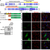
Figure 1.
Figure 1.
A noncanonical MTS targets human FAM173A to mitochondria. A, alignment of N-terminal parts of FAM173A and ATPSc-KMT (alias FAM173B) showing sequence elements present in their N-terminal region. Colors indicate the position of N-terminal sequence (NTS, gray), predicted TMD (yellow), preMT (orange), and the N-terminal portion of the MTase domain (green). Hallmark motifs of 7BS MTase domain are indicated by black boxes. The conserved acidic residue in motif Post I (Glu-105 in FAM173A), crucial for AdoMet binding, is marked with an asterisk. B and C, subcellular localization of FAM173A-derived GFP-fusion proteins. B, a schematic representation of the FAM173A-derived sequences used in (C), with the various domains denoted as in (A). C, confocal fluorescence microscopy images of live HeLa cells, stably transfected with plasmid encoding: FAM173A-GFP, (Δ42)-FAM173A-GFP, (Δ76)-FAM173A-GFP, or (amino acids 43–77)-FAM173A-GFP. Cells were counterstained with MitoTracker Orange to visualize mitochondria, and with Hoechst 33258 to visualize nuclei. Data were acquired through green (GFP), red (MitoTracker), and blue (Hoechst) channels and merged. D, Western blot analysis of proteins present in mitoplast extracts from HAP1 FAM173A KO cells complemented with FAM173A-FLAG, either WT or E105A-mutated, detected with anti-FLAG antibody. Expression of cytochrome c oxidase subunit IV (COX IV) is shown as a loading control.
Figure 2.
Figure 2.
Human FAM173A mediates methylation of Lys-52 in ANT inside cells. A and B, Western blot analysis of methyllysine-containing proteins present in mitoplast extracts from HAP1 cells, either unmodified (WT) or FAM173A KO. Extracts enriched in mitochondrial membrane proteins were prepared from HAP1 WT or KO cells and resolved by SDS-PAGE. Proteins were transferred by Western blotting to a membrane, which was probed with antibodies against pan-trimethyllysine (A) or pan-methyllysine (B). In parallel, detection with anti-COX IV antibodies was performed (loading control). C, FAM173A KO abrogates ANT methylation in HAP1 cells. Extracts enriched in mitochondrial membrane proteins were prepared from HAP1 WT, FAM173A KO, and ATPSc-KMT KO cells, resolved by SDS-PAGE and the portion of the gel corresponding to the ∼32 kDa region was chymotrypsin-digested and analyzed by MS. Shown are representative, normalized extracted ion chromatograms, gated for different methylation states of ANT-derived, chymotrypsin-generated peptides, encompassing residues 52–68 of human ANT2 and ANT3, present in indicated cells, with Lys-52 marked in magenta. Differences between ANT2 and ANT3 sequences are indicated in cyan. Percentages indicate the area under each peak, relative to the total area of all peaks. A.U., arbitrary units. D, MS/MS fragmentation spectra demonstrating absence of Lys-52 methylation in ANT2 from FAM173A KO cells (top) and presence of Lys-52 trimethylation in HAP1 WT cells (bottom). E, Same as in (D), but for ANT3. CMe, carbamidomethyl.
Figure 3.
Figure 3.
Complementation of FAM173A KO cells with FAM173A restores the methylation of Lys-52 in ANT. Extracts enriched in mitochondrial membrane proteins were prepared from HAP1 WT, FAM173A KO, or KO cells expressing FLAG-tagged FAM173A, either nonmutated or E105A-mutated (expression of FLAG-tagged proteins was verified in Fig. 1D), and analyzed by MS as in Fig. 2. Shown are the mean relative intensities of MS signals, gated for different methylation states of the indicated, ANT-derived peptides, encompassing residues 52–68 of human ANT2 and ANT3, with Lys-52 marked in magenta. Differences between ANT2 and ANT3 sequences are indicated in cyan. Error bars indicate the range of values from three independent analyses of each cell line.
Figure 4.
Figure 4.
ANT1 and ANT2 from rat are fully trimethylated at Lys-52. A, Extracts enriched in mitochondrial membrane proteins were prepared from indicated rat organs, resolved by SDS-PAGE and a portion of gel corresponding to ANT was chymotrypsin-digested and analyzed by MS. Shown are representative, normalized extracted ion chromatograms, gated for different methylation states of ANT-derived, chymotrypsin-generated peptides, encompassing residues 52–68 of ANT1 and ANT2 present in indicated organs. MS signals and amino acid sequences corresponding to ANT1 and ANT2-derived peptides are indicated, with Lys-52 marked in magenta. Differences between ANT1 and ANT2 sequences are indicated in cyan. B, MS/MS fragmentation spectra demonstrating trimethylation of Lys-52 in ANT1 from rat heart. C, MS/MS fragmentation spectra demonstrating trimethylation of Lys-52 in ANT2 from rat kidney. CMe, carbamidomethyl.
Figure 5.
Figure 5.
Lack of active FAM173A increases mitochondrial respiration in HAP1-derived cells. A, mitochondrial respiration is increased in FAM173A-deficient cells. Mitochondria were isolated from unmodified HAP1 (WT), FAM173A KO, or KO cells complemented with FLAG-tagged FAM173A, either nonmutated or E105A-mutated. OCRs of isolated mitochondria (15 μg of mitochondrial protein) were measured by Seahorse analyzer, in the presence of succinate and rotenone, under basal conditions, and after sequential addition of ADP, oligomycin, FCCP, and AntA. Left, typical OCR traces from a representative experiment, with arrows indicating the time of addition of the indicated compounds. Error bars represent the S.D. (n = 5). Right, mitochondrial respiration dissected into individual states, i.e. State II (basal respiration), State III (respiration after addition of ADP, because of ATP synthesis), State IVo (respiration in presence of oligomycin, because of proton leak), and State IIIu (respiration in presence of mitochondrial uncoupler FCCP). Shown are the average values from two independent experiments. Error bars represent the S.D. (n = 10). *, p value < 0.1; **, p value < 0.01; ***, p value < 0.001. B, Western blot analysis of mitoplast extracts from HAP1-derived cells. Extracts enriched in mitochondrial membrane proteins were prepared from cells indicated as in (A). 40 μg of protein from extracts were resolved by SDS-PAGE and transferred by Western blotting (WB) to a membrane, which was probed with anti-ANT2 antibody. The membrane was then sequentially reprobed with anti-ATPSc, anti-COX IV, anti-FLAG, and anti-ATP5A antibodies. Arrow indicates the position of ANT2 band visible on the membrane probed with anti-FLAG antibody, which results from the previous probing of this membrane with anti-ANT2 antibody. Shown are images from a representative experiment.
Figure 6.
Figure 6.
The paralogous KMTs, FAM173A/ANT-KMT, and FAM173B/ATPSc-KMT, target similarly positioned lysines in ANT and ATPSc. Left, structural representation of bovine ANT, and its topology in the inner mitochondrial membrane, generated from a previously published structure (34) (PDB ID: 1OKC). Rotation and magnification (2.5×) of ANT was performed to better visualize the pore-forming structure of ANT, and the positions of Lys-52 (cyan) and cardiolipin (CL; yellow). A model for the submitochondrial localization of FAM173A/B is presented in the center, with annotations and coloring as in Fig. 1. Right, structural representation of F1-c8 subcomplex of bovine ATP synthase, and its topology in the inner mitochondrial membrane, generated from a previously published structure (45) (PDB ID: 2XND) and further adapted from Małecki et al. (21). The F1 subcomplex is shown in blue, and ATPSc are shown in green, except for one of the eight protomers of the c8-ring, which is shown in red, and Lys-43 is here indicated in cyan.

References

    1. Schubert H. L., Blumenthal R. M., and Cheng X. (2003) Many paths to methyltransfer: A chronicle of convergence. Trends Biochem. Sci. 28, 329–335 10.1016/S0968-0004(03)00090-2 - DOI - PMC - PubMed
    1. Petrossian T. C., and Clarke S. G. (2011) Uncovering the human methyltransferasome. Mol. Cell Proteomics 10, M110.000976 10.1074/mcp.M110.000976 - DOI - PMC - PubMed
    1. Schubert H. L., Blumenthal R. M., and Cheng X. (2006) 1 Protein methyltransferases: Their distribution among the five structural classes of AdoMet-dependent methyltransferases. Enzymes 24, 3–28 10.1016/S1874-6047(06)80003-X - DOI - PubMed
    1. Bedford M. T. (2007) Arginine methylation at a glance. J. Cell Sci. 120, 4243–4246 10.1242/jcs.019885 - DOI - PubMed
    1. Figaro S., Scrima N., Buckingham R. H., and Heurgué-Hamard V. (2008) HemK2 protein, encoded on human chromosome 21, methylates translation termination factor eRF1. FEBS Lett. 582, 2352–2356 10.1016/j.febslet.2008.05.045 - DOI - PubMed

Publication types

MeSH terms