. 2019 Jul 2;116(27):13330-13339.
doi: 10.1073/pnas.1902658116. Epub 2019 Jun 18.

SHOC2 complex-driven RAF dimerization selectively contributes to ERK pathway dynamics

Affiliations

SHOC2 complex-driven RAF dimerization selectively contributes to ERK pathway dynamics

Isabel Boned Del Río et al. Proc Natl Acad Sci U S A. .

Abstract

Despite the crucial role of RAF kinases in cell signaling and disease, we still lack a complete understanding of their regulation. Heterodimerization of RAF kinases as well as dephosphorylation of a conserved "S259" inhibitory site are important steps for RAF activation but the precise mechanisms and dynamics remain unclear. A ternary complex comprised of SHOC2, MRAS, and PP1 (SHOC2 complex) functions as a RAF S259 holophosphatase and gain-of-function mutations in SHOC2, MRAS, and PP1 that promote complex formation are found in Noonan syndrome. Here we show that SHOC2 complex-mediated S259 RAF dephosphorylation is critically required for growth factor-induced RAF heterodimerization as well as for MEK dissociation from BRAF. We also uncover SHOC2-independent mechanisms of RAF and ERK pathway activation that rely on N-region phosphorylation of CRAF. In DLD-1 cells stimulated with EGF, SHOC2 function is essential for a rapid transient phase of ERK activation, but is not required for a slow, sustained phase that is instead driven by palmitoylated H/N-RAS proteins and CRAF. Whereas redundant SHOC2-dependent and -independent mechanisms of RAF and ERK activation make SHOC2 dispensable for proliferation in 2D, KRAS mutant cells preferentially rely on SHOC2 for ERK signaling under anchorage-independent conditions. Our study highlights a context-dependent contribution of SHOC2 to ERK pathway dynamics that is preferentially engaged by KRAS oncogenic signaling and provides a biochemical framework for selective ERK pathway inhibition by targeting the SHOC2 holophosphatase.

Keywords: ERK; MRAS; RAF; RAS; SHOC2.


Conflict of interest statement

The authors declare no conflict of interest.

Figures

Fig. 1.
Fig. 1.
MRAS and SHOC2 expression promotes S365 BRAF/S259 CRAF dephosphorylation, BRAF-MEK dissociation, and BRAF-CRAF dimerization. (A) Calyculin A inhibits BRAF S365 dephosphorylation and ERK activation by MRAS-SHOC2 expression. Expression of MRAS L71 and SHOC2 was induced in T-17 cells stably expressing T6-BRAF by 1 μg/mL Dox treatment for 24 h. Cells were incubated with a Calyculin A dose–response for 20 min and lysates immunoprobed, as indicated. (B) Intact RBD is required for efficient S365 BRAF dephosphorylation by MRAS-SHOC2 expression: T6-BRAF WT and mutants were transiently transfected into T-17 cells and MRAS-SHOC2 expression induced for 24 h. (C) Impaired MRAS-SHOC2 induced S259 dephosphorylation by R89L RBD CRAF mutation is rescued by constitutive membrane localization. As in B but with T6-CRAF mutants. (D) MRAS-SHOC2 expression stimulates BRAF S365 dephosphorylation, MEK dissociation, and CRAF binding to BRAF. T-17 T6-BRAF cells as in A were treated with different Dox concentrations for 24 h. StrepTactin pull-downs of T6-BRAF and lysates were immunoprobed and visualized using a Li-COR Odyssey scanner. (E) Li-Cor quantification of D. (F) MRAS-SHOC2 expression stimulates MEK1 dissociation from BRAF and CRAF but not KSR1. GST-fusion genes were cotransfected into HEK293T cells, together with Myc-MEK1 and either empty vector or MRAS-L71 and SHOC2. GST-S6K was used as a control. GST pull-downs and lysates were probed as indicated.
Fig. 2.
Fig. 2.
SHOC2 is required for EGF-induced S365/S259 dephosphorylation, RAF dimerization, BRAF-MEK dissociation, and efficient ERK pathway activation. (A) SHOC2 is required for EGF-induced BRAF-S365 dephosphorylation and efficient ERK pathway activation in T-REx-293 cells. Serum-starved cells stably expressing control or SHOC2 shRNA were treated with 25 ng/mL EGF for the indicated times and lysates immunoprobed as indicated. (B) SHOC2 is required for EGF-induced BRAF-CRAF heterodimerization and dissociation of BRAF–MEK complexes but not RAS–RAF interaction. Endogenous BRAF, CRAF, or RAS (238) IPs from lysates used in A were probed for as indicated. (C) SHOC2 is required for EGF-induced KSR/BRAF dimerization. Endogenous KSR1 IPs and lysates from T-REx-293 cells were immunoprobed as in B. (D) Impaired S365 dephosphorylation and ERK pathway activation by EGF in SHOC2 KO DLD-1 cells is rescued by reexpression of WT but not SHOC2 mutants defective for complex formation with MRAS and PP1. Lysates from DLD-1 parental (P) and SHOC2 KO cells transduced with lentivirures expressing flag-SHOC2 WT, mutants, or empty vector were stimulated with 25 ng/mL EGF for 5 min after serum starvation. (E) SHOC2 is required for EGF-stimulated MEK and 14-3-3 dissociation from BRAF and BRAF dimerization with ARAF and CRAF. Serum-starved DLD-1 cells were stimulated with EGF for the indicated times and endogenous RAF IPs immunoblotted using the Li-COR Odyssey system. (F) Li-COR quantification of CRAF, MEK, 14-3-3 and P-S365 BRAF from BRAF IPs in E, relative to EGF-untreated parental cells. (G) BRAF S365A does not bind MEK and rescues ERK activation in SHOC2 KO cells. DLD-1 cells, nontransduced or stably expressing T6-BRAF WT or S365A, were treated with EGF for 10 min and Streptactin pull-downs of T6-BRAF and lysates probed as indicated.
Fig. 3.
Fig. 3.
SHOC2 is selectively required for early, but not delayed ERK pathway activation by EGF in DLD-1 cells. (A) Serum-starved DLD-1 parental or SHOC2 KO cells were stimulated with 25 ng/mL EGF for the indicated times. Lysates were probed and visualized by Li-COR. (B) Quantification of P-S365 BRAF, P-S259 CRAF, P-MEK, P-ERK, P-S380 RSK, P-S473 AKT in A (mean ± SD) (n = 3), relative to EGF-untreated parental condition. (C) Model of biphasic ERK activation by EGF with an early and transient phase that requires SHOC2 and a delayed, sustained phase that is SHOC2-independent. Based on ref. .
Fig. 4.
Fig. 4.
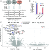
Phosphoproteomic analysis of SHOC2’s contribution to EGF-regulated dynamics. (A) Experimental strategy used for quantitative phosphoproteomics. DLD-1 parental or SHOC2 KO cells were serum-starved and left untreated or stimulated with 25 ng/mL EGF for 5 or 20 min. Parental cells were also pretreated with the MEK inhibitor Trametinib (100 nM) for 20 min. (B) Significantly regulated phosphorylated sites (cutoffs: fold-change ± 2, adjusted P < 0.05) at 5 and 20 min of EGF stimulation. Representative of n = 3 experiments. (C) Volcano plots of quantified phosphosites regulated by EGF at 5 and 20 min in parental but not SHOC2 KO cells. Blue up-regulated, red down-regulated upon EGF stimulation; gray no regulation. See SI Appendix, Fig. S4C for representative phosphosite plots.
Fig. 5.
Fig. 5.
SHOC2-independent ERK activation requires CRAF. (A) KO of individual RAF isoforms does not affect ERK pathway activation by EGF. Serum-starved DLD-1 parental (P) or ARAF, BRAF, and CRAF KO cells generated by CRISPR were stimulated with 25 ng/mL EGF. (B) ERK pathway activation by EGF in CRAF-only or BRAF-only double KO DLD-1 cells (generated by second round CRISPR of BRAF or CRAF respectively in ARAF KO cells) is normal. However, KD of CRAF in ARAF/BRAF KO (CRAF-only) cells inhibits ERK activation. DLD-1 cells transfected with SCR or CRAF siRNAs were stimulated with EGF as before. (C) CRAF KD (but not ARAF or BRAF) inhibits delayed ERK activation in SHOC2 KO cells. DLD-1 parental and SHOC2 KO cells transfected with ARAF, BRAF, or CRAF siRNAs, stimulated with 25 ng/mL EGF and lysates probed by Li-COR. (D) Li-COR quantification of P-MEK and P-ERK in C (mean ± SD) (n = 2). (E) CRAF KD inhibits growth in SHOC2 KO cells. Parental and two SHOC2 KO clones transfected with SCR or A/B/CRAF siRNAs were used for colony formation assays.
Fig. 6.
Fig. 6.
SHOC2-independent ERK activation requires palmitoylated HRAS/NRAS and CRAF N-region phosphorylation. (A) NRAS and HRAS KD (but not KRAS) inhibit ERK activation in SHOC2 KO cells. SHOC2 KO DLD-1 cells transfected with siRNAs were stimulated with 25 ng/mL EGF for the indicated times. (B) The palmitoylation inhibitor 2-BP reduces sustained ERK activation in SHOC2 KO cells. DLD-1 cells were pretreated with 2-BP (100 µM) for the indicated times before EGF stimulation. (C) PAK, FAK, and SRC family inhibitors (besides RAF and MEK inhibitors) impair sustained ERK pathway activation by EGF in SHOC2 KO DLD-1 cells. Cells were stimulated with 25 ng/mL EGF for 20 min after 30-min pretreatment with indicated kinase inhibitors (and 2-BP). Lysates were probed and P-ERK quantified by Li-COR (mean ± SD) (n = 3–7). Significance is determined using a two tailed t-test *P < 0.05, **P < 0.01, or ***P < 0.001. See SI Appendix, Fig. S6B for representative experiment. (D) Cells were pretreated with 10 μM PAK (FRAX597), SRC (SU6656), and FAK (PF-562271) inhibitors alone or in combination, 30 min before stimulation with EGF for 20 min. (E) Model of selective contribution of the SHOC2 complex to ERK pathway spatiotemporal dynamics. EGF Receptor activation leads to N/H/K-RAS and MRAS/SHOC2 complex activation at the plasma membrane and an early phase of ERK activation involving A/B/C-RAF isoforms. As a result of intracellular trafficking of palmitoylated proteins (by the constitutive de/reacylation cycle and/or receptor-mediated endocytosis and/or other nonmutually exclusive mechanisms not shown), H/N-RAS travel to endomembrane compartments from where they signal through CRAF to drive sustained ERK pathway activation. Because poly-basic motif-containing KRAS-4B and MRAS (and associated proteins) remain at the plasma membrane, this CRAF is now uncoupled from regulation by the SHOC2 complex, but is instead dependent on N-region phosphorylation by kinases, such as PAK, SFK, and FAK. See Discussion for further details. Membrane anchors represent farnesyl (red) and palmitate (black) groups. S338 and S341 residues in CRAF belong to the N-region. ERK may phosphorylate diverse substrates in different compartments, as shown by different color arrows.
Fig. 7.
Fig. 7.
SHOC2 is selectively required for ERK pathway activation under anchorage-independent conditions in KRAS mutant cells. (A) SHOC2 is dispensable for anchorage-dependent/2D growth. Incucyte growth curves of DLD-1 parental and three independent SHOC2 KO clones stably expressing WT and D175N SHOC2 were generated using the IncuCyte Live Cell imaging system. Representative of n = 2 experiments. (B) SHOC2 KO impairs growth in 3D. Cells in A were seeded in low attachment plates and growth at day 5 measured by Alamar blue staining (mean ± SD) (n = 2–4). Significance is determined using a two tailed t test *P < 0.05, **P < 0.01, or ***P < 0.001. (C) SHOC2 is preferentially required for ERK pathway activation in 3D in DLD-1 cells. DLD-1 cells were grown for 24 h on regular or poly-HEMA–coated plates and lysates immunoprobed as indicated. (D) As in C but with HCT116 KRASG13D cells. (E) As in C and D with SW480 KRASG12V cells. (F) SHOC2 is dispensable for ERK phosphorylation in 2D and 3D in the BRAF mutant (V600E) RKO and HT-29 cell lines. (G) PAK (FRAX597), SRC (SU6656), and FAK (PF-562271) family inhibitors inhibit basal ERK signaling more potently in the absence of SHOC2. DLD-1 cells growing in log phase in the presence of 10% FBS were incubated with 10-µM inhibitors for 1 h and lysates immunoblotted as indicated.

References

    1. Kholodenko B. N., Hancock J. F., Kolch W., Signalling ballet in space and time. Nat. Rev. Mol. Cell Biol. 11, 414–426 (2010). - PMC - PubMed
    1. Shaul Y. D., Seger R., The MEK/ERK cascade: From signaling specificity to diverse functions. Biochim. Biophys. Acta 1773, 1213–1226 (2007). - PubMed
    1. Rauen K. A., The RASopathies. Annu. Rev. Genomics Hum. Genet. 14, 355–369 (2013). - PMC - PubMed
    1. Simanshu D. K., Nissley D. V., McCormick F., RAS proteins and their regulators in human disease. Cell 170, 17–33 (2017). - PMC - PubMed
    1. Samatar A. A., Poulikakos P. I., Targeting RAS-ERK signalling in cancer: Promises and challenges. Nat. Rev. Drug Discov. 13, 928–942 (2014). - PubMed

Publication types