. 2019 Jul 2;116(27):13394-13403.
doi: 10.1073/pnas.1821401116. Epub 2019 Jun 18.

PFKFB3-mediated endothelial glycolysis promotes pulmonary hypertension

Affiliations

PFKFB3-mediated endothelial glycolysis promotes pulmonary hypertension

Yapeng Cao et al. Proc Natl Acad Sci U S A. .

Abstract

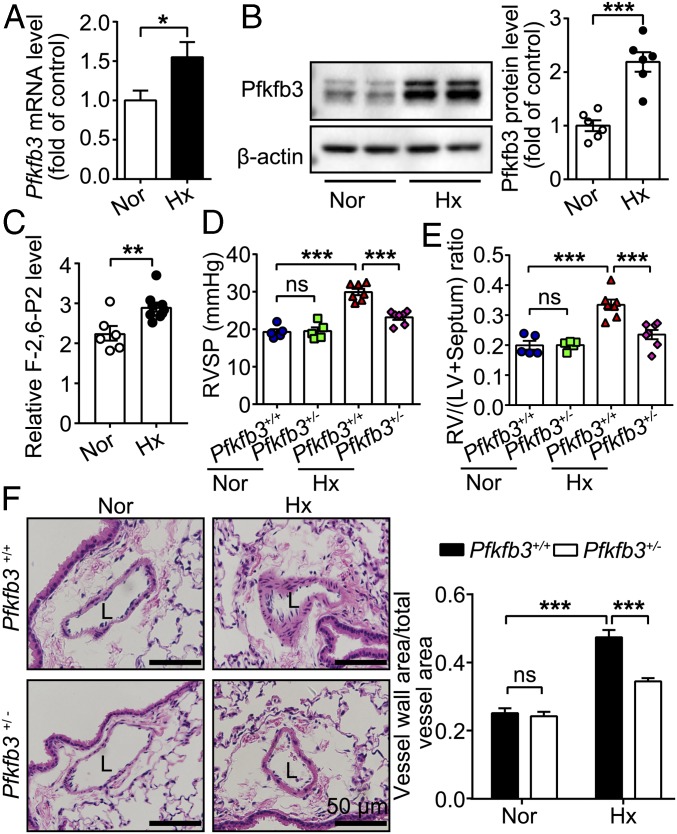
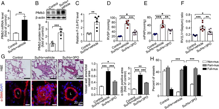
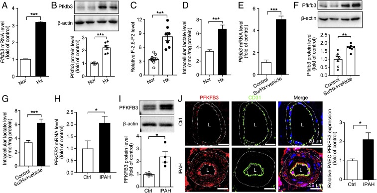
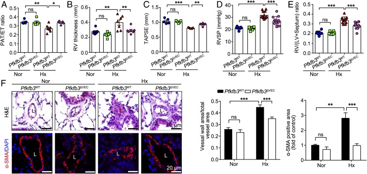
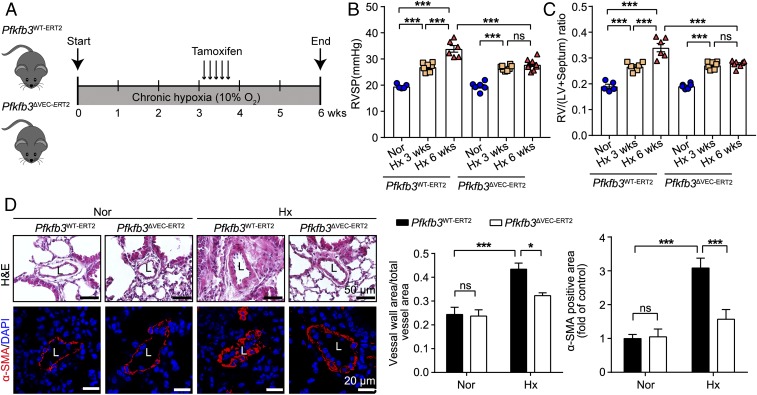
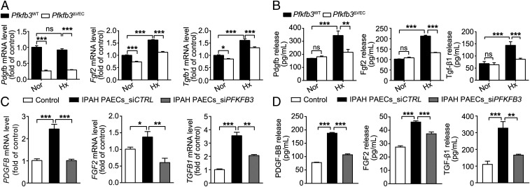
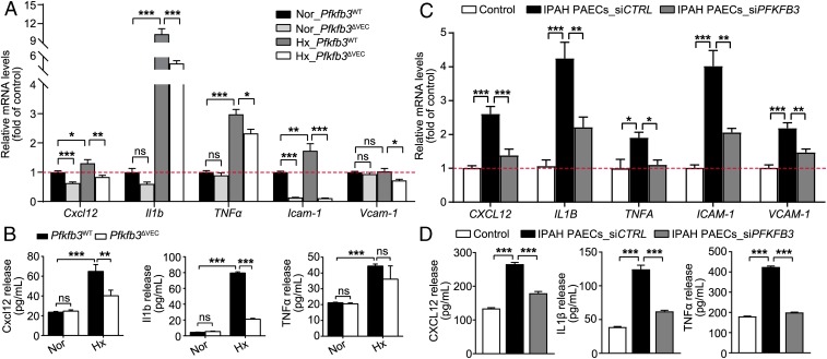
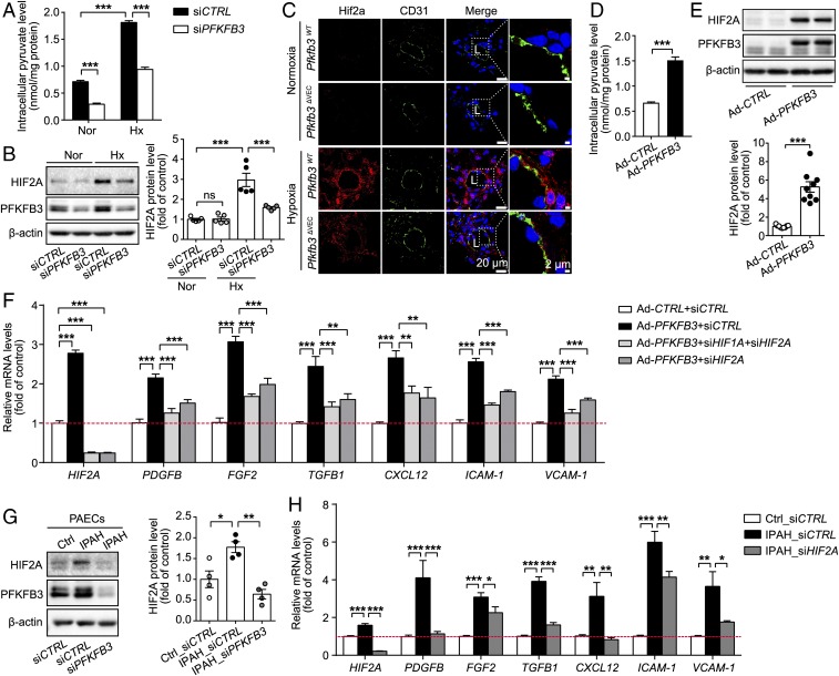
Increased glycolysis in the lung vasculature has been connected to the development of pulmonary hypertension (PH). We therefore investigated whether glycolytic regulator 6-phosphofructo-2-kinase/fructose-2, 6-bisphosphatase (PFKFB3)-mediated endothelial glycolysis plays a critical role in the development of PH. Heterozygous global deficiency of Pfkfb3 protected mice from developing hypoxia-induced PH, and administration of the PFKFB3 inhibitor 3PO almost completely prevented PH in rats treated with Sugen 5416/hypoxia, indicating a causative role of PFKFB3 in the development of PH. Immunostaining of lung sections and Western blot with isolated lung endothelial cells showed a dramatic increase in PFKFB3 expression and activity in pulmonary endothelial cells of rodents and humans with PH. We generated mice that were constitutively or inducibly deficient in endothelial Pfkfb3 and found that these mice were incapable of developing PH or showed slowed PH progression. Compared with control mice, endothelial Pfkfb3-knockout mice exhibited less severity of vascular smooth muscle cell proliferation, endothelial inflammation, and leukocyte recruitment in the lungs. In the absence of PFKFB3, lung endothelial cells from rodents and humans with PH produced lower levels of growth factors (such as PDGFB and FGF2) and proinflammatory factors (such as CXCL12 and IL1β). This is mechanistically linked to decreased levels of HIF2A in lung ECs following PFKFB3 knockdown. Taken together, these results suggest that targeting PFKFB3 is a promising strategy for the treatment of PH.

Keywords: endothelial cells; glycolysis; pulmonary hypertension.


Conflict of interest statement

The authors declare no conflict of interest.

Figures

Fig. 1.
Fig. 1.
Heterozygous Pfkfb3 deficiency in mice inhibits the development of hypoxia (Hx)-induced PH. (A) Real-time PCR analysis of Pfkfb3 mRNA levels in lung homogenates of mice exposed to hypoxia (10% O2) or ambient oxygen levels (21% O2) for 4 wk (n = 6). (B) Western blot analysis and densitometric quantification of Pfkfb3 protein levels in lung homogenates of mice exposed to hypoxia (10% O2) or ambient oxygen levels (21% O2) for 4 wk (n = 6 mice per group). (C) Relative F-2,6-P2 levels in lung homogenates of mice exposed to normoxia (Nor) or hypoxia (10% O2) for 4 wk (n = 6 mice for normoxia group, n = 9 mice for hypoxia group). (D) RVSP and (E) RV hypertrophy assessed by the ratio of RV/LV+septum. (F) (Left) Representative images of H&E staining of distal pulmonary arteries from control mice and Pfkfb3+/− mice exposed to normoxia or hypoxia (10% O2) for 4 wk. L, lumen. (Scale bars, 50 µm.) (Right) Quantification of pulmonary artery thickness as measured by the ratio of vessel wall area to total vessel area (n = 3 for control mice under normoxic or hypoxic condition, n = 4 for normoxic Pfkfb3+/− mice, and n = 7 for hypoxic Pfkfb3+/− mice). All data are expressed as mean ± SEM. Statistical significance was determined by unpaired Student’s t test (AC) and one-way ANOVA followed by Bonferroni test (D–F). *P < 0.05 was considered significant, **P < 0.01, ***P < 0.001. ns, no significance.
Fig. 2.
Fig. 2.
PFKFB3 inhibitor 3PO reduces Sugen 5416/hypoxia (Su/Hx)-induced PH in rats. (A) Real-time PCR analysis of Pfkfb3 mRNA levels in lung homogenates of control rats and Su/Hx-treated rats (n = 6). (B) Western blot analysis and densitometric quantification of Pfkfb3 protein levels in lung homogenates of control rats and Su/Hx-treated rats (n = 5). (C) Relative F-2,6-P2 levels in lung homogenates of control rats and Su/Hx-treated rats (n = 5 for control group, n = 6 for Su/Hx-treated group). (D) Quantification of RVSP, (E) mean pulmonary arterial pressure (mPAP), and (F) RV hypertrophy assessed by the ratio of RV/LV+septum. (G) (Left) Representative images of H&E staining and α-SMA immunostaining of the distal pulmonary arteries of control and treated (Su/Hx + vehicle and Su/Hx + 3PO) rats. (Right) Quantification of the ratio of vessel wall area to total vessel area and α-SMA immunostaining-positive area. (H) Quantitative assessment of nonmuscularized, partially muscularized, and fully muscularized arteries as percentages of total assessed arteries (n = 6–8). All data are expressed as mean ± SEM. Statistical significance was determined by unpaired Student’s t test (A–C) and one-way ANOVA followed by Bonferroni test (DH). *P < 0.05 was considered significant, **P < 0.01, ***P < 0.001. ns, no significance.
Fig. 3.
Fig. 3.
Pfkfb3/PFKFB3 expression level is significantly increased in PAECs isolated from mice, rats, and humans with PH. (A) Real-time PCR analysis of Pfkfb3 mRNA levels in PAECs of control and PH mice (n = 6). (B) Western blot analysis and densitometric quantification of Pfkfb3 protein levels in PAECs of control and PH mice (n = 6). (C) Relative F-2,6-P2 levels in PAECs of control and PH mice (n = 9 for normoxia [Nor] group, n = 7 for hypoxia [Hx] group). (D) Intracellular lactate levels of PAECs isolated from control and PH mice (n = 10). (E) Real-time PCR analysis of Pfkfb3 mRNA levels in PAECs of control and Su/Hx-treated rats (n = 5). (F) Western blot analysis and densitometric quantification of Pfkfb3 protein levels in PAECs of control and Su/Hx-treated rats (n = 7). (G) Intracellular lactate levels of PAECs isolated from control and Su/Hx-treated rats (n = 8). (H) Real-time PCR analysis of PFKFB3 mRNA levels in PAECs of control subjects or patients with IPAH (n = 6). Experiments were repeated four times independently with cells from four patients or control subjects. (I) Western blot analysis and densitometric quantification of PFKFB3 protein levels in PAECs of control subjects or patients with IPAH (n = 4). (J) (Left) Representative micrographs of PFKFB3 expression in the pulmonary endothelium of control (Ctrl) or IPAH patients. Sections were costained for PFKFB3 (red) and CD31 (green). (Right) Quantification of the relative PFKFB3 fluorescence intensity in the pulmonary endothelium of Ctrl or IPAH patients. L, lumen. (Scale bar, 20 μm.) All data are expressed as mean ± SEM. Statistical significance was determined by unpaired Student’s t test (A, H, and J) and Mann–Whitney U test (I). *P < 0.05 was considered significant, **P < 0.01, ***P < 0.001.
Fig. 4.
Fig. 4.
Endothelial-specific Pfkfb3 deficiency in mice ameliorates the development of hypoxia-induced PH. (AC) Quantification of PAT/ET ratio (A), RV thickness (in millimeters) (B), and TAPSE (in millimeters) (C). (D) Quantification of RVSP and (E) RV hypertrophy as assessed by RV/LV+septum. (F) (Left) Representative images of H&E staining and α-SMA immunostaining of the distal pulmonary arteries of Pfkfb3WT and Pfkfb3ΔVEC mice under normoxic (Nor) or hypoxic (Hx) conditions for 4 wk and (Right) quantification of pulmonary artery thickness as measured by the ratio of vessel wall area to total vessel area and α-SMA immunostaining-positive area. L, lumen (n = 6). All data are expressed as mean ± SEM. Statistical significance was determined by one-way ANOVA followed by Bonferroni test. *P < 0.05 was considered significant, **P < 0.01, ***P < 0.001. ns, no significance.
Fig. 5.
Fig. 5.
Inducible deficiency of endothelial-specific Pfkfb3 in mice suppresses the progression of hypoxia-induced PH. (A) Schematic diagram of the experimental design. Inducible endothelial-specific Pfkfb3-deficient mice (Pfkfb3ΔVEC-ERT2) and control mice (Pfkfb3WT-ERT2) were exposed to chronic hypoxia (Hx; 10% O2) for 3 wk followed by 75 mg/kg of tamoxifen injection for 5 d under hypoxia condition. Mice continued to be exposed to hypoxia for 2 wk before assessment. (B) Quantification of RVSP and (C) RV hypertrophy as assessed by RV/LV+septum. (D) (Left) Representative images of H&E staining and α-SMA immunostaining of the distal pulmonary arteries of Pfkfb3WT-ERT2 and Pfkfb3ΔVEC-ERT2 mice under normoxic (Nor) or hypoxic conditions for 6 wk and quantification (Right) of pulmonary artery thickness as measured by the ratio of vessel wall area to total vessel area and the α-SMA immunostaining-positive area. L, lumen (n = 6–7). All data are expressed as mean ± SEM. Statistical significance was determined by one-way ANOVA followed by Bonferroni test. *P < 0.05 was considered significant, ***P < 0.001. ns, no significance.
Fig. 6.
Fig. 6.
Endothelial Pfkfb3/PFKFB3 deficiency or knockdown decreases the hypoxia (Hx)-induced gene expression and protein release of growth factors. (A) Real-time PCR analysis of mRNA levels of Pdgfb, Fgf2, and Tgfb1 in PAECs of Pfkfb3ΔVEC and Pfkfb3WT mice exposed to normoxia (Nor) or hypoxia (10% O2) for 4 wk (n = 6). (B) Levels of released Pdgfb, Fgf2, and Tgf-β1 in the culture supernatants of mouse PAECs exposed to hypoxia (1% O2) for 24 h (n = 4–6). (C) Real-time PCR analysis of mRNA levels of PDGFB, FGF2, and TGFB1 in normal PAECs or siCTRL- and siPFKFB3-transfected PAECs of patients with IPAH (n = 6). Experiments were repeated four times independently with cells from four patients with IPAH or control subjects. (D) Levels of released PDGF-BB, FGF2, and TGF-β1 in the culture supernatants of normal PAECs or siCTRL- and siPFKFB3-transfected PAECs of patients with IPAH (n = 4–6). All data are expressed as mean ± SEM. Statistical significance was determined by one-way ANOVA followed by Bonferroni test. *P < 0.05 was considered significant, **P < 0.01, ***P < 0.001. ns, no significance.
Fig. 7.
Fig. 7.
Endothelial Pfkfb3/PFKFB3 deficiency or knockdown decreases inflammatory responses. (A) Real-time PCR analysis of mRNA levels of Cxcl12, Il1b, TNFα, Icam-1, and Vcam-1 in PAECs of Pfkfb3ΔVEC and Pfkfb3WT mice exposed to normoxia (Nor) or hypoxia (Hx; 10% O2) for 4 wk (n = 6). (B) Levels of released Cxcl12, Il1b, and TNFα in the culture supernatants of mouse PAECs exposed to hypoxia (1% O2) for 24 h (n = 4–6). (C) Real-time PCR analysis of mRNA levels of CXCL12, IL1B, TNFA, ICAM-1, and VCAM-1 in normal PAECs or siCTRL- and siPFKFB3-transfected PAECs of patients with IPAH (n = 6). Experiments were repeated four times independently with cells from four patients with IPAH or control subjects. (D) Levels of released CXCL12, IL1β, and TNFα in the culture supernatants of normal PAECs or siCTRL- and siPFKFB3-transfected PAECs of patients with IPAH (n = 8–9). All data are expressed as mean ± SEM. Statistical significance was determined by one-way ANOVA followed by Bonferroni test. *P < 0.05 was considered significant, **P < 0.01, ***P < 0.001. ns, no significance.
Fig. 8.
Fig. 8.
Expression of growth factors and inflammatory cytokines induced by PFKFB3/Pfkfb3 in endothelial cells is mediated by HIF2A/Hif2a. (A) Intracellular pyruvate levels in human PAECs transfected with siCTRL and siPFKFB3 under normoxic (Nor) and hypoxic (Hx; 1% O2) conditions for 24 h (n = 5). (B) Western blot analysis and densitometric quantification of HIF2A protein levels in human PAECs transfected with siCTRL and siPFKFB3 under normoxic and hypoxic (1% O2) conditions for 6 h (n = 5). (C) Representative micrographs of Hif2a expression in the distal pulmonary arteries of Pfkfb3WT and Pfkfb3ΔVEC mice under normoxic or hypoxic conditions for 4 wk. Sections were costained for Hif2a (red) and CD31 (green). L, lumen. (D) Intracellular pyruvate levels in human PAECs transfected with Ad-CTRL and Ad-PFKFB3 (n = 9). (E) Western blot analysis and densitometric quantification of HIF2A protein levels in human PAECs transfected with Ad-CTRL and Ad-PFKFB3 (n = 9). (F) Real-time PCR analysis of mRNA levels of HIF2A, PDGFB, FGF2 and TGFB1, CXCL12, ICAM-1, and VCAM-1 in human PAECs transfected with Ad-CTRL-siCTRL, Ad-PFKFB3-siCTRL, Ad-PFKFB3-siHIF1A+siHIF2A, and Ad-PFKFB3-siHIF2A (n = 6). (G) (Left) Representative Western blot images and (Right) densitometric quantification of HIF2A expression in normal or IPAH PAECs with or without PFKFB3 KD (n = 4). (H) Real-time PCR analysis of mRNA levels of HIF2A, PDGFB, FGF2 and TGFB1, CXCL12, ICAM-1, and VCAM-1 in normal or IPAH PAECs transfected with siCTRL or siHIF2A (n = 6). Experiments were repeated four times independently with cells from four patients with IPAH or control subjects. All data are expressed as mean ± SEM. Statistical significance was determined by one-way ANOVA followed by Bonferroni test (A, B, F, and H) and unpaired Student’s t test (D and E). *P < 0.05 was considered significant, **P < 0.01, ***P < 0.001. ns, no significance.

References

    1. Rabinovitch M., Molecular pathogenesis of pulmonary arterial hypertension. J. Clin. Invest. 118, 2372–2379 (2008). - PMC - PubMed
    1. Tuder R. M., Pathology of pulmonary arterial hypertension. Semin. Respir. Crit. Care Med. 30, 376–385 (2009). - PubMed
    1. Rabinovitch M., Guignabert C., Humbert M., Nicolls M. R., Inflammation and immunity in the pathogenesis of pulmonary arterial hypertension. Circ. Res. 115, 165–175 (2014). - PMC - PubMed
    1. Elinoff J. M., et al. , Challenges in pulmonary hypertension: Controversies in treating the tip of the iceberg. A joint national institutes of health clinical center and pulmonary hypertension association symposium report. Am. J. Respir. Crit. Care Med. 198, 166–174 (2018). - PMC - PubMed
    1. Xu W., et al. , Alterations of cellular bioenergetics in pulmonary artery endothelial cells. Proc. Natl. Acad. Sci. U.S.A. 104, 1342–1347 (2007). - PMC - PubMed

Publication types

Substances