. 2019 Jun 18;10(3):e01094-19.
doi: 10.1128/mBio.01094-19.

Integration of Fungus-Specific CandA-C1 into a Trimeric CandA Complex Allowed Splitting of the Gene for the Conserved Receptor Exchange Factor of CullinA E3 Ubiquitin Ligases in Aspergilli

Affiliations

Integration of Fungus-Specific CandA-C1 into a Trimeric CandA Complex Allowed Splitting of the Gene for the Conserved Receptor Exchange Factor of CullinA E3 Ubiquitin Ligases in Aspergilli

Anna M Köhler et al. mBio. .

Abstract

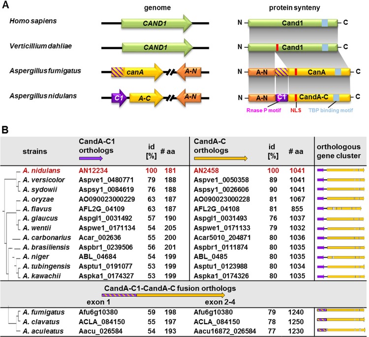
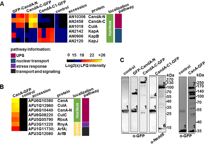
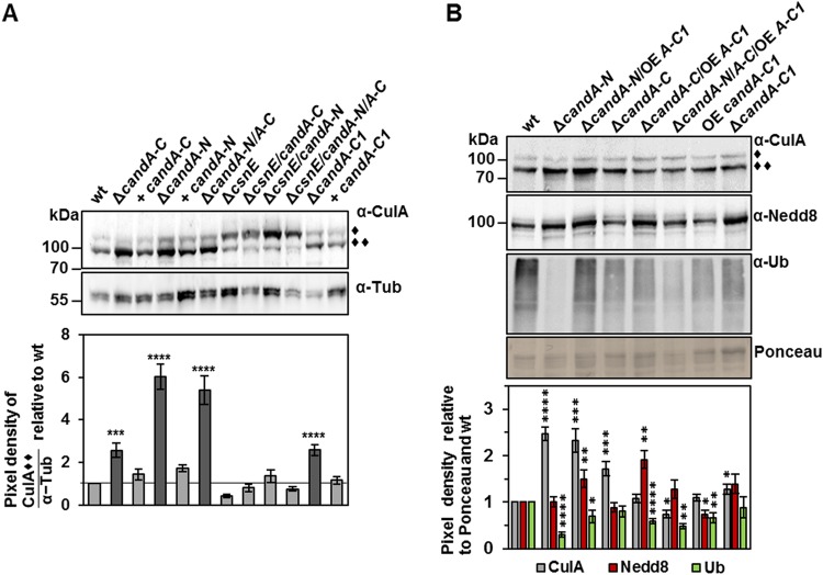
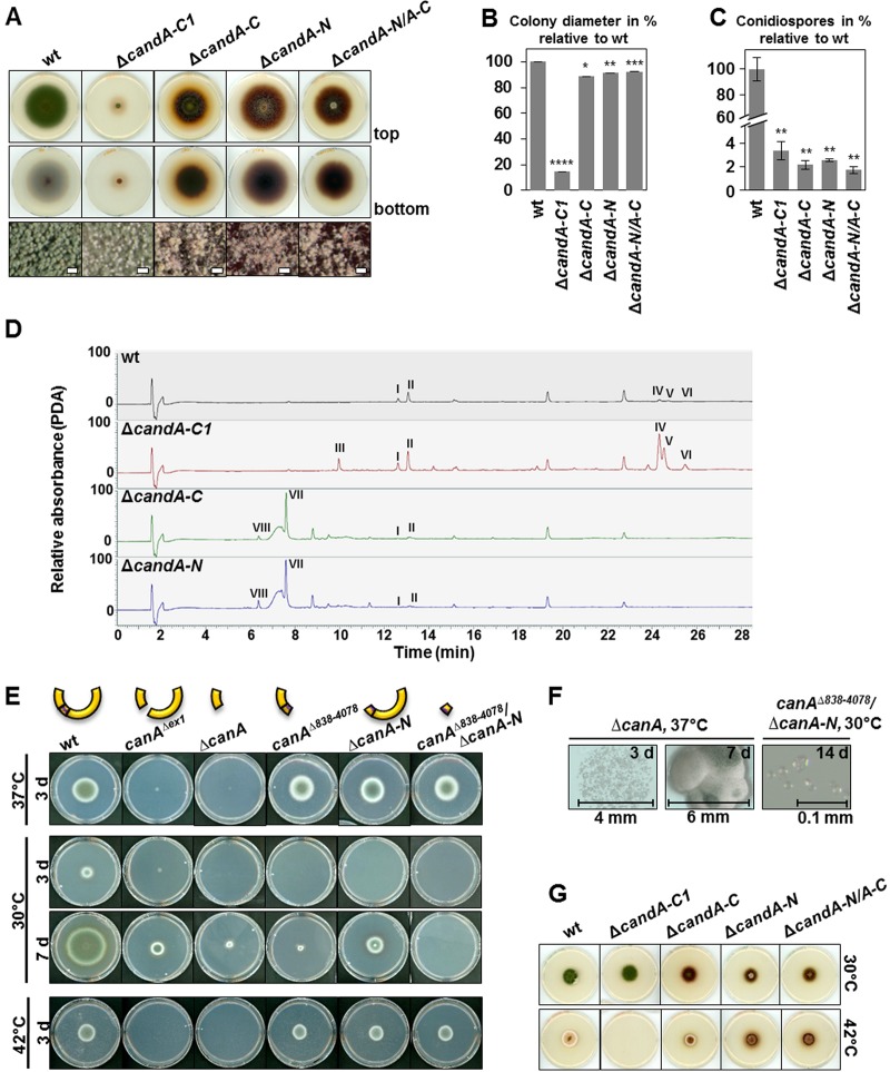
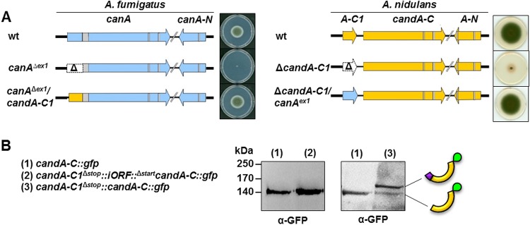
E3 cullin-RING ubiquitin ligase (CRL) complexes recognize specific substrates and are activated by covalent modification with ubiquitin-like Nedd8. Deneddylation inactivates CRLs and allows Cand1/A to bind and exchange substrate recognition subunits. Human as well as most fungi possess a single gene for the receptor exchange factor Cand1, which is split and rearranged in aspergilli into two genes for separate proteins. Aspergillus nidulans CandA-N blocks the neddylation site, and CandA-C inhibits the interaction to the adaptor/substrate receptor subunits similar to the respective N-terminal and C-terminal parts of single Cand1. The pathogen Aspergillus fumigatus and related species express a CandA-C with a 190-amino-acid N-terminal extension domain encoded by an additional exon. This extension corresponds in most aspergilli, including A. nidulans, to a gene directly upstream of candA-C encoding a 20-kDa protein without human counterpart. This protein was named CandA-C1, because it is also required for the cellular deneddylation/neddylation cycle and can form a trimeric nuclear complex with CandA-C and CandA-N. CandA-C and CandA-N are required for asexual and sexual development and control a distinct secondary metabolism. CandA-C1 and the corresponding domain of A. fumigatus control spore germination, vegetative growth, and the repression of additional secondary metabolites. This suggests that the dissection of the conserved Cand1-encoding gene within the genome of aspergilli was possible because it allowed the integration of a fungus-specific protein required for growth into the CandA complex in two different gene set versions, which might provide an advantage in evolution.IMPORTANCEAspergillus species are important for biotechnological applications, like the production of citric acid or antibacterial agents. Aspergilli can cause food contamination or invasive aspergillosis to immunocompromised humans or animals. Specific treatment is difficult due to limited drug targets and emerging resistances. The CandA complex regulates, as a receptor exchange factor, the activity and substrate variability of the ubiquitin labeling machinery for 26S proteasome-mediated protein degradation. Only Aspergillus species encode at least two proteins that form a CandA complex. This study shows that Aspergillus species had to integrate a third component into the CandA receptor exchange factor complex that is unique to aspergilli and required for vegetative growth, sexual reproduction, and activation of the ubiquitin labeling machinery. These features have interesting implications for the evolution of protein complexes and could make CandA-C1 an interesting candidate for target-specific drug design to control fungal growth without affecting the human ubiquitin-proteasome system.

Keywords: Aspergillus fumigatus; Aspergillus nidulans; COP9 signalosome; Cand1; Cullin-RING ubiquitin ligase; Nedd8; asexual development; protein complex; protein degradation; secondary metabolism; sexual development; spore germination.


Figures

FIG 1
FIG 1
Comparison of single and split Cand proteins. (A) Comparison of human, Verticillium dahliae, Aspergillus fumigatus, and A. nidulans candA genes and encoding proteins. Human (Homo sapiens) genes and numerous fungal genes, such as those of V. dahliae, express a single Cand1 protein. The counterpart of human or fungal Cand1 is split in aspergilli in an N-terminal CandA-N/CanA-N (A-N) and C-terminal CandA-C/CanA part encoded by separate genes. Aspergilli require an additional Cand polypeptide CandA-C1 (A-C1) which is not present in humans. The CandA-C1 polypeptide represents the N-terminal part of the CanA protein of A. fumigatus but is encoded by a separate candA-C1 gene (546 bp, 181 amino acids [aa], 19 kDa) in A. nidulans, which is located next and upstream to candA-C (3,254 bp, 1,041 aa, 113.5 kDa), which is separated from candA-N (1,055 bp, 313 aa, 33.6 kDa) by five open reading frames. CandA-C1 has an RNase P Rpr2/Rpp21 motif (pink), and V. dahliae Cand1 and Aspergillus CanA/CandA-C have an NLS (red) and a TATA binding protein (TBP) interaction motif (blue), which is conserved in all eukaryotic CandA C-terminal ends. H. sapiens Cand1, UniProt ID Q86VP6-1; V. dahliae Cand1, MycoCosm ID VDAG_05065T0; A. fumigatus CanA, UniProt ID Q4WMC6, and CanA-N, UniProt ID Q4WMC0; A. nidulans CandA-N, UniProt ID C8VP82; and CandA-C1, AspGD/FungiDB ID AN12234, CandA-C, UniProt ID Q5BAH2). (B) Comparative analysis of CandA-C1 and CandA-C orthologs in different aspergilli. The protein sequence of A. nidulans CandA-C1 (AN12234) was compared to those of different Aspergillus spp. by a BLASTp search in the Joint Genome Institute (JGI) MycoCosm genome portal. Genomic clusters of orthologs of separated candA-C1 (purple) followed by intergenic ORF (iORF; black line) and downstream-located candA-C (yellow) genes are depicted. Three corresponding fused genes with the CandA-C1-like domain marked in purple-yellow stripes followed by an intron (gray) instead of the iORF are depicted in the bottom. Phylogenetic relationships are based on protein identities of the CandA-C1 orthologs in the genus Aspergillus, similar to relations described by de Vries et al. (2). id, protein identity.
FIG 2
FIG 2
The intergenic ORF (iORF) contains the terminator for candA-C1 and promoter for candA-C expression in A. nidulans. (A) Quantitative real-time PCR (qRT-PCR) measurements show the gene expression of candA-C1, candA-C, and candA-N in wild type (wt) compared to the overexpression (OE) candA-C1::gfp mutant strain. candA-C1::gfp is significantly overexpressed compared to wild-type expression but does not influence the transcription levels of candA-C or candA-N (****, P ≤ 0.0001; n = 3). rel., relative. (B) cDNA amplification assay showing different PCR setups used to amplify candA-C1 (primer 1 + 2 [oAMK120/121]), candA-C (primer 3 + 4 [oAMK03b/04b]), and candA-C1::candA-C (primer 1 + 4 [oAMK120/04b]). The table shows expected sizes in kilobase pairs from gDNA or cDNA. The amplification of candA-N, which contains two introns, was used as control to exclude gDNA contamination in cDNA samples (primer oAMK01/02). (C) Agarose gel pictures of PCR products are depicted, and sizes are indicated. (D) qRT-PCR experiments show the expression levels after 20 h of vegetative growth of candA-C1 and candA-C in the wild type compared to the candA-C1, iORF, candA-C1/iORF, and candA-C deletion mutants. The expression of candA-C1 is significantly upregulated in a ΔcandA-C mutant sample, and candA-C is significantly downregulated when the iORF is deleted (*, P ≤ 0.05; ***, P ≤ 0.001; ****, P ≤ 0.0001; n = 3, except for ΔcandA-C1/iORF mutant, n = 2). (E) Western hybridization of 20-h-old vegetative A. nidulans and A. fumigatus CandA/CanA subunits probed with anti-HA (left panel), anti-GFP (middle panels), and anti-GFP and anti-RFP (right panels) antibodies shows that A. fumigatus CanA-GFP (163 kDa) and A. nidulans CandA-C-GFP (142 kDa) exhibit different molecular weights. GFP-CandA-N and RFP-CanA-N have the same weight (59 kDa) and show a double band. CandA-C1-GFP runs at 47 kDa, and a fusion of CandA-N∼A-C1∼A-C-HA runs at 170 kDa.
FIG 3
FIG 3
A. nidulans CandA-C1 interacts with CandA-C and CandA-N. (A) Localization of native CandA-C-GFP, GFP-CandA-N, and overexpression (OE) CandA-C1-GFP proteins. All three CandA proteins colocalize with monomeric RFP (mRFP)-tagged histone H2A (RFP-H2A). CandA-C1-GFP is also localized to nucleoli and colocalizes with MitoTracker Red (MT) that stains mitochondria. (B) Bimolecular fluorescence complementation (BiFC) microscopy of CandA-C1 with CandA-C and CandA-N in a wild-type background as well as in the csnE deletion strain. BiFC signals are visible in green and localized to the nuclei, which are stained with H2A-RFP or 4′,6-diamidino-2-phenylindole (DAPI). DIC, differential interference contrast. (C) Nuclear localization of CandA-C and CandA-N is dependent on CandA-C NLS, as both CandA proteins are absent from RFP-H2A-stained nuclei when the NLS is deleted. OE CandA-C1-GFP is localized to the nuclei and nucleoli (white arrows indicate colocalization to the nuclei; scale bars = 10 μm).
FIG 4
FIG 4
GFP-pulldown coupled to LC-MS analysis of A. nidulans and A. fumigatus CandA proteins. (A) Comparison of putative interaction partners of CandA-C and CandA-N with CandA-C1 from pulldown experiments. An excerpt of the heatmap, generated with Perseus (version 1.6.0.7), depicts label free-quantification (LFQ) intensities of three biological replicates of GFP control, CandA-N, CandA-C, and four replicates of CandA-C1. Log2(x) LFQ intensities range from 0 to 15, not considered to be identified (black); 15 to 18, low intensity (blue); and 18 to 26/32, low to high LFQ intensities (gradient from red to orange to yellow). Colored bars indicate cellular localization, based on KEGG and UniProt databases (green, nucleus; light yellow, cytosol). The molecular pathway of putative interaction partners is labeled as follows: berry, ubiquitin-proteasome system (UPS); dark blue, nuclear transport; purple, stress response; and dark gray, transport and signaling. (B) Heatmap of identified proteins from A. fumigatus CanA-GFP pulldown compared to overexpression GFP control strain. (C) Western hybridization of pulldown elution samples probed with anti-GFP antibody, which shows free GFP (1), GFP-CandA-N (2), CandA-C-GFP (3), CandA-C1-GFP (4), and CanA-GFP (7). The elution sample of CandA-C1-GFP was reprobed with anti-Nedd8 antibody, which highlights neddylated cullins (5) and free Nedd8 (6).
FIG 5
FIG 5
CandA is required for CulA neddylation in A. nidulans. Western hybridization was performed with crude extracts from 20-h-grown vegetative mycelium. (A) Western hybridization probed with anti-CulA antibody to observe CulA neddylation ratios. In the wild type, most CulA is deneddylated (∼96 kDa, ◆◆), which is different from a ΔcsnE strain, which is defective in cullin deneddylation and has most CulA bound to Nedd8 (∼106 kDa, ◆). In the ΔcandA and ΔcandA-C1 deletion strains, deneddylated CulA accumulates. Double and triple deletions of csnE, candA-N, and/or candA-C show an accumulation of neddylated CulA as observed for the ΔcsnE mutant. Signals were quantified with pixel density measurements using BIO1D software (Peqlab) for total 12 replicates (three biological replicates each with four technical replicates). Tubulin (Tub) served as a loading control. (B) Western hybridization probed with anti-CulA (gray), anti-Nedd8 (red), and anti-ubiquitin (α-Ub) (green) antibodies. Ponceau served as loading control. The ΔcandA mutant strains were compared to candA-N and candA-C deletion strains overexpressing candA-C1. The pixel density ratio was determined with the BIO1D software (Peqlab), quantified against Ponceau, and normalized to wild-type signals. CulA and Ub used three biological with three technical replicates each, and Nedd8 used four biological with three technical replicates each; error bars represent the standard error of the mean. *, P ≤ 0.05; **, P ≤ 0.01; ***, P ≤ 0.001; ****, P ≤ 0.0001.
FIG 6
FIG 6
A. nidulans CandA-C1 and A. fumigatus CanA are required for growth and asexual development. (A) A. nidulans conidia (4 × 103) were point inoculated on solid minimal medium supplemented with para-aminobenzoic acid and incubated at 37°C in light for 5 days. Pictures were taken from the top and bottom views of the plate. Binocular pictures show asexual spores (scale bars = 100 μm). (B and C) Quantification of colony diameter (B) and amount of spores after 5 days of asexual growth (C) in percentage (%) relative to the wild type. Error bars represent the standard error of the mean (SEM) (n = 3). (D) LC-MS combined with photodiode array detection (PDA) analysis of secondary metabolites extracted from 7-day-old asexually developed mycelium revealed differences in all tested strains. The wild type and ΔcandA-C1 mutant produce similar amounts of austinol (I) and dehydroaustinol (II). The ΔcandA-C1 mutant produces asperthecin (III) and greater amounts of emericellin (IV) and shamixanthone/epishamixanthone (V and VI) than does the wild type. Metabolites III, IV, V, and VI were absent in the ΔcandA-C and ΔcandA-N mutants, but both produced cichorine (VII) and a metabolite (VIII) with high-resolution–electrospray ionization–MS (HR-ESI-MS) at m/z 210.0761 [M+H]+ (calculated for C10H12NO4, m/z 210.0766). (E) A. fumigatus conidia (4 × 103) were point inoculated on solid modified minimal medium and incubated at 37°C, 30°C, and 42°C for 3 to 7 days. The ΔcanA mutant strains are growth defective at all tested temperatures. (F) Micrographs of ΔcanA mutant colonies after 3 and 7 days of growth at 37°C, and micrograph of ΔcanA838-4078/ΔcanA-N mutant after 14 days at 30°C shows colorless conidia, which did not germinate. (G) A. nidulans conidia (4 × 103) were point inoculated on solid minimal medium supplemented with para-aminobenzoic acid and incubated at 30°C and 42°C in light for 5 days. The candA mutant strains grew like the wild type at 30°C and 42°C, except for the ΔcandA-C1 mutant, which grew better at 30°C than at 37°C and was unable to germinate at 42°C.
FIG 7
FIG 7
A. fumigatus and A. nidulans gene orthologs for candA-C1 are interchangeable with each other. (A) Genome map of A. fumigatus (blue) wild-type canA and canA-N, a mutant strain carrying a canAexon1 deletion (Δ), and a mutant strain with the replacement of canAexon1 with the A. nidulans candA-C1 sequence (yellow). Introns are colored in gray. Genome map of A. nidulans (yellow) wild-type candA-C1, candA-C, and candA-N, a mutant strain with candA-C1 deletion (Δ), and a mutant strain where candA-C1 is replaced with A. fumigatus canAexon1 (blue). Solid modified minimal medium was point inoculated with 4 × 103 conidia of A. fumigatus strains for 3 days at 37°C in darkness. Solid minimal medium was point inoculated with 4 × 103 conidia of A. nidulans strains and incubated at 37°C with illumination for 5 days. (B) Western hybridization with anti-GFP antibody of A. nidulans protein crude extracts from a strain expressing CandA-C-GFP [140 kDa (1)] and two strains carrying candA-C1::candA-C::gfp fusion constructs with (2) and without (3) the iORF sequence. A CandA-C1–CandA-C–GFP fusion protein (161 kDa) was expressed from the construct without iORF also showing a signal for CandA-C-GFP.
FIG 8
FIG 8
Ascospore formation is dependent on candA-C1 in A. nidulans. Solid minimal medium was point inoculated with 4 × 103 conidia and incubated in the dark and with limited oxygen supply for seven and 14 days. (A) Pictures of sexual phenotypes were taken after 7 days. (B) Micrograph pictures show cleistothecia (c) covered by Hülle cells (h) for wild-type (wt) and the candA-C1 complementation strain. The candA-C1 deletion strain has nests (n) after 7 days which develop to empty cleistothecia (c*) after 14 days. candA-C and candA-N deletion strains only produce early nests (en) but cannot undergo a complete sexual life cycle. (C) Micrograph pictures of Hülle cell-free cleistothecia. candA-C1 has soft cleistothecia with dents, indicated with red arrows. (D and E) Microscopic pictures of squeezed cleistothecia never showed any mature ascospores for the candA-C1 deletion strain (D) but did show ascogenous hyphae (ah) (E). (F) Closeup view of the ΔcandA-C1 mutant colony. Black scale bars = 100 μm; white scale bar = 1,000 μm. d, days. (G) LC-MS combined with photodiode array detection (PDA) analysis of secondary metabolites extracted from 7-day-old sexually developed mycelium revealed that the ΔcandA-C1 mutant produces asperthecin (III). The wild type and ΔcandA-C1 mutant produce similar amounts of emericellin (IV) and shamixanthone/epishamixanthone (V and VI). Small amounts of cichorine (VII) were detected in the ΔcandA-C mutant, which were increased in the ΔcandA-N mutant. The ΔcandA-C and ΔcandA-N mutants produced compound VIII with HR-ESI-MS at m/z 210.0761 [M+H]+ (calculated for C10H12NO4, m/z 210.0766).
FIG 9
FIG 9
A trimeric CandA is required for growth, development, a coordinated secondary metabolism (Sec. Met.), and the CRL cycle in Aspergillus spp. (A) Scheme of a putative Aspergillus ancestor and DNA rearrangement of the candA loci. The ancestor of all Aspergillus spp. presumably had one gene containing sequence information of candA-N and candA-C. A DNA double-strand break, followed by ligation, has changed the position of candA-C five open reading frames upstream of candA-N and directly downstream of candA-C1 in A. nidulans. In A. fumigatus, the candA-C1-like sequence fused to the rearranged canA. (B) A. fumigatus CanA N-terminal extension is essential for properly timed spore germination and vegetative growth. CanA and CanA-N promote germination and vegetative growth during low-temperature stress and promote conidiophore development. (C) A. nidulans CandA-C1 promotes germination and vegetative growth. CandA-N and CandA-C are essential for multicellular sexual fruiting bodies from the stage of early nest formation. CandA-C1 is required for properly timed cleistothecia formation and is essential for the development of ascospores. All three CandA proteins support the CRL cycle and conidiophore development. Furthermore, CandA contributes to secondary metabolism control.

Similar articles

Cited by

References

    1. Riquelme M, Aguirre J, Bartnicki-García S, Braus GH, Feldbrügge M, Fleig U, Hansberg W, Herrera-Estrella A, Kämper J, Kück U, Mouriño-Pérez RR, Takeshita N, Fischer R. 2018. Fungal morphogenesis, from the polarized growth of hyphae to complex reproduction and infection structures. Microbiol Mol Biol Rev 82:e00068-17. doi:10.1128/MMBR.00068-17. - DOI - PMC - PubMed
    1. de Vries RP, Riley R, Wiebenga A, Aguilar-Osorio G, Amillis S, Uchima CA, Anderluh G, Asadollahi M, Askin M, Barry K, Battaglia E, Bayram Ö, Benocci T, Braus-Stromeyer SA, Caldana C, Cánovas D, Cerqueira GC, Chen F, Chen W, Choi C, Clum A, Dos Santos RA, Damásio AR, Diallinas G, Emri T, Fekete E, Flipphi M, Freyberg S, Gallo A, Gournas C, Habgood R, Hainaut M, Harispe ML, Henrissat B, Hildén KS, Hope R, Hossain A, Karabika E, Karaffa L, Karányi Z, Kraševec N, Kuo A, Kusch H, LaButti K, Lagendijk EL, Lapidus A, Levasseur A, Lindquist E, Lipzen A, Logrieco AF, et al. . 2017. Comparative genomics reveals high biological diversity and specific adaptations in the industrially and medically important fungal genus Aspergillus. Genome Biol 18:28. doi:10.1186/s13059-017-1151-0. - DOI - PMC - PubMed
    1. Alberts AW, Chen J, Kuron G, Hunt V, Huff J, Hoffman C, Rothrock J, Lopez M, Joshua H, Harris E, Patchett A, Monaghan R, Currie S, Stapley E, Albers-Schonberg G, Hensens O, Hirshfield J, Hoogsteen K, Liesch J, Springer J. 1980. Mevinolin: a highly potent competitive inhibitor of hydroxymethylglutaryl-coenzyme A reductase and a cholesterol-lowering agent. Proc Natl Acad Sci U S A 77:3957–3961. doi:10.1073/pnas.77.7.3957. - DOI - PMC - PubMed
    1. Pontecorvo G, Roper JA, Chemmons LM, Macdonald KD, Bufton AWJ. 1953. The genetics of Aspergillus nidulans, p 141–238. In Demerec M. (ed), Advances in genetics. Academic Press, Cambridge, MA. - PubMed
    1. Challa S. 2018. Pathogenesis and pathology of invasive aspergillosis. Curr Fungal Infect Rep 12:23–32. doi:10.1007/s12281-018-0310-4. - DOI

Publication types

MeSH terms

LinkOut - more resources