. 2019 Jun 18;9(1):8719.
doi: 10.1038/s41598-019-44855-3.

Meniscus-Derived Matrix Scaffolds Promote the Integrative Repair of Meniscal Defects

Affiliations

Meniscus-Derived Matrix Scaffolds Promote the Integrative Repair of Meniscal Defects

Jacob C Ruprecht et al. Sci Rep. .

Abstract

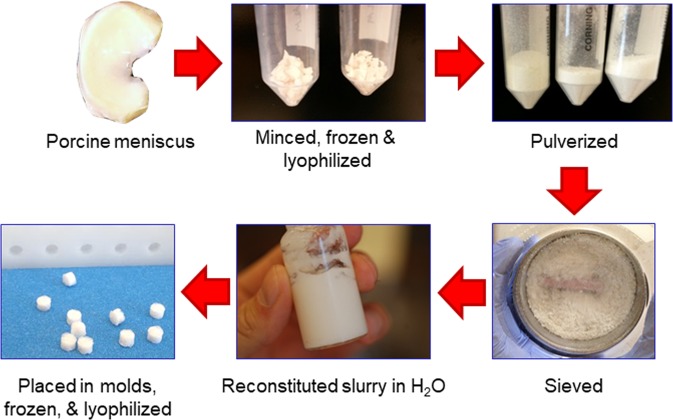
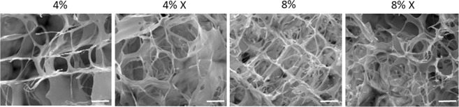
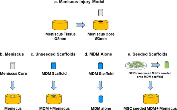
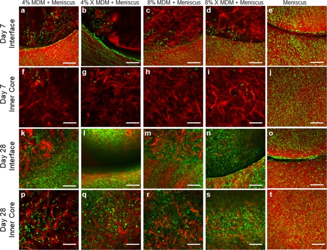
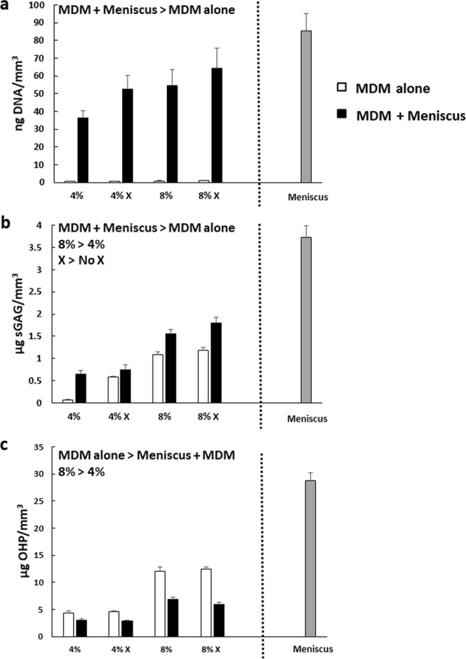
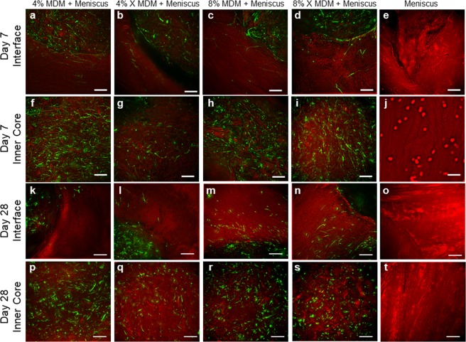
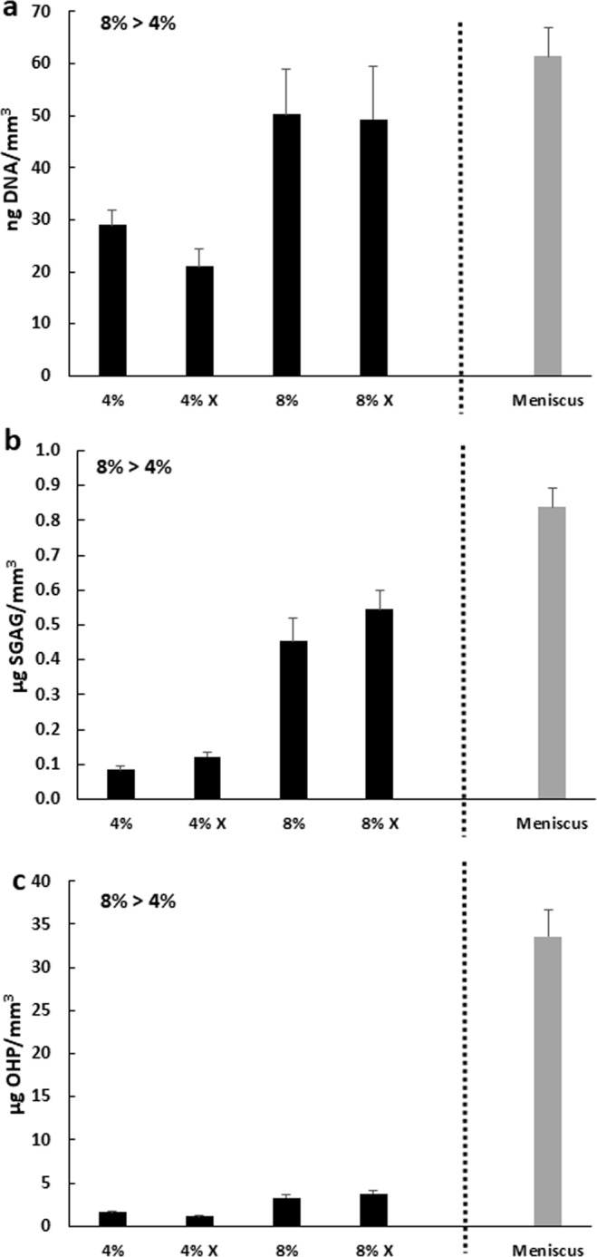
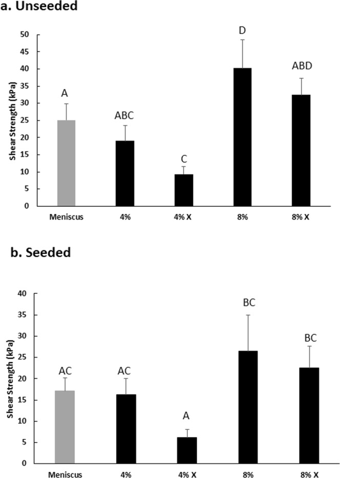
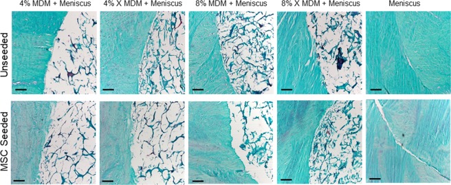
Meniscal tears have a poor healing capacity, and damage to the meniscus is associated with significant pain, disability, and progressive degenerative changes in the knee joint that lead to osteoarthritis. Therefore, strategies to promote meniscus repair and improve meniscus function are needed. The objective of this study was to generate porcine meniscus-derived matrix (MDM) scaffolds and test their effectiveness in promoting meniscus repair via migration of endogenous meniscus cells from the surrounding meniscus or exogenously seeded human bone marrow-derived mesenchymal stem cells (MSCs). Both endogenous meniscal cells and MSCs infiltrated the MDM scaffolds. In the absence of exogenous cells, the 8% MDM scaffolds promoted the integrative repair of an in vitro meniscal defect. Dehydrothermal crosslinking and concentration of the MDM influenced the biochemical content and shear strength of repair, demonstrating that the MDM can be tailored to promote tissue repair. These findings indicate that native meniscus cells can enhance meniscus healing if a scaffold is provided that promotes cellular infiltration and tissue growth. The high affinity of cells for the MDM and the ability to remodel the scaffold reveals the potential of MDM to integrate with native meniscal tissue to promote long-term repair without necessarily requiring exogenous cells.


Conflict of interest statement

F.G. is a founder and shareholder of Cytex Therapeutics, Inc. The other authors declare no competing interests.

Figures

Figure 1
Figure 1
Meniscus-derived matrix (MDM) was prepared from porcine medial menisci that were minced, frozen, and lyophilized. Next, the tissue was pulverized and sieved, and then resuspended at 4% or 8% in water. The MDM slurry was pipetted into 3 mm diameter molds, which were frozen and lyophilized to generate scaffolds.
Figure 2
Figure 2
Scanning electron microscope images of the MDM scaffolds showing the porosity and structure of the 4%, 4% crosslinked (X), 8%, and 8% X scaffolds. Scale bar is 50 μm.
Figure 3
Figure 3
Schematic showing the experimental model and groups used in these experiments. (a) For the meniscus injury model, 8 mm diameter explants were isolated from porcine meniscus tissue. A 3 mm biopsy was removed from the center of the explant. (b) For meniscus tissue controls, the 3 mm inner core of meniscus tissue was reinserted into the meniscus explant (Meniscus). (c) For experiments with unseeded scaffolds, the inner core was filled with an acellular MDM scaffold (MDM + Meniscus). (d) MDM scaffolds were also cultured alone to assess the biochemical composition of the scaffold alone (MDM alone). (e) In order to generate seeded scaffolds, MSCs were transduced with enhanced green fluorescent protein (eGFP) and seeded on the MDM scaffolds. These MSC seeded MDM scaffolds were used in the inner core of the meniscus tissue (MSC seeded MDM + Meniscus) to assess the effects of MSCs on the MDM scaffold and meniscus repair.
Figure 4
Figure 4
Fluorescent images of the unseeded MDM scaffolds in meniscus tissue (MDM + Meniscus) and meniscus tissue controls (Meniscus) at day 7 (aj) and day 28 (kt). Initially, the meniscus tissue contains abundant cells throughout (e,j) and by day 28 cells are bridging the interface between the inner core and outer ring of meniscus tissue (o). For the samples containing the MDM scaffold inner cores (Meniscus + MDM), the meniscus outer ring contains abundant cells (ad), and these cells are starting to populate the MDM scaffold at day 7 (fi). By day 28, the meniscus cells fully bridge the interface (kn) and have filled the MDM scaffold inner cores (ps). All meniscus cells are stained green (calcein AM) and the matrix is stained red (Alexa fluor 633 NHS ester). Scale bar is 200 μm.
Figure 5
Figure 5
Meniscus tissue surrounding the MDM scaffolds (MDM + Meniscus) leads to increased DNA content (a) and sulfated glycosaminoglycan (sGAG) content (b) but decreased collagen (OHP) content (c) in the scaffolds. Data is expressed as the mean + SEM for the MDM alone (white bars) and the MDM + Meniscus (black bars). For reference the mean + SEM values for control meniscus tissue is indicated by the gray bar.
Figure 6
Figure 6
Fluorescent images of the MSC seeded MDM scaffolds in meniscus tissue (MSC seeded MDM + Meniscus) and meniscus tissue controls (Meniscus) at day 7 (aj) and day 28 (kt). Initially, the enhanced green fluorescent protein-expressing MSCs (eGFP-MSC) are localized to the MDM scaffold inner core (fi). By day 28 the eGFP-MSCs have bridged the interface between the scaffold and outer ring of meniscus tissue and infiltrated into the meniscus tissue outer ring (kn), particularly in the 8% groups (m,n). There are no eGFP-expressing cells in the meniscus tissue control (e,j,o,t). EGFP-MSCs are green and the matrix is stained red (Alexa fluor 633 NHS ester). Scale bar is 100 μm.
Figure 7
Figure 7
The 8% MDM MSC seeded scaffolds have higher DNA content (a), sulfated glycosaminoglycan (sGAG) content (b), and collagen (OHP) content (c) as compared to the 4% MDM scaffolds. Data is expressed as the mean + SEM for MSC seeded MDM + Meniscus (black bars). For reference the mean + SEM values for control meniscus tissue is indicated by the gray bar.
Figure 8
Figure 8
(a) The unseeded 8% MDM scaffolds have a higher integrative shear strength of repair with the meniscus tissue as compared to the control meniscus tissue. (b) The 8% and 8% X MSC seeded MDM scaffolds have improved repair strength over the 4% X MSC seeded MDM scaffolds. Data is expressed as the mean + SEM. Groups not sharing the same letter have p-values ≤ 0.05.
Figure 9
Figure 9
Histological staining of the MDM scaffolds cultured with meniscus tissue reveals the tissue structure and composition at the interface between the meniscus tissue and MDM scaffolds. Sections were stained with Safranin-O (red: proteoglycans), fast green (blue: collagen), and hematoxylin (black: nuclei). Scale bar is 100 μm.

Similar articles

Cited by

References

    1. Ahmed AM, Burke DL. In-vitro measurement of static pressure distribution in synovial joints–Part I: Tibial surface of the knee. J Biomech Eng. 1983;105:216–225. doi: 10.1115/1.3138409. - DOI - PubMed
    1. Markolf KL, Bargar WL, Shoemaker SC, Amstutz HC. The role of joint load in knee stability. J Bone Joint Surg Am. 1981;63:570–585. doi: 10.2106/00004623-198163040-00007. - DOI - PubMed
    1. Wojtys EM, Chan DB. Meniscus Structure and Function. AAOS Instructional Course Lectures. 2005;54:323–330. - PubMed
    1. Carter TE, et al. In vivo cartilage strain increases following medial meniscal tear and correlates with synovial fluid matrix metalloproteinase activity. J Biomech. 2015;48:1461–1468. doi: 10.1016/j.jbiomech.2015.02.030. - DOI - PMC - PubMed
    1. Badlani JT, Borrero C, Golla S, Harner CD, Irrgang JJ. The effects of meniscus injury on the development of knee osteoarthritis: data from the osteoarthritis initiative. The American journal of sports medicine. 2013;41:1238–1244. doi: 10.1177/0363546513490276. - DOI - PubMed

Publication types