. 2019 Aug;29(8):641-654.
doi: 10.1038/s41422-019-0193-0. Epub 2019 Jun 18.

Interferon-inducible cytoplasmic lncLrrc55-AS promotes antiviral innate responses by strengthening IRF3 phosphorylation

Affiliations

Interferon-inducible cytoplasmic lncLrrc55-AS promotes antiviral innate responses by strengthening IRF3 phosphorylation

Yumei Zhou et al. Cell Res. 2019 Aug.

Abstract

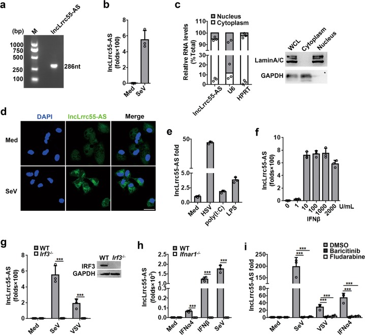
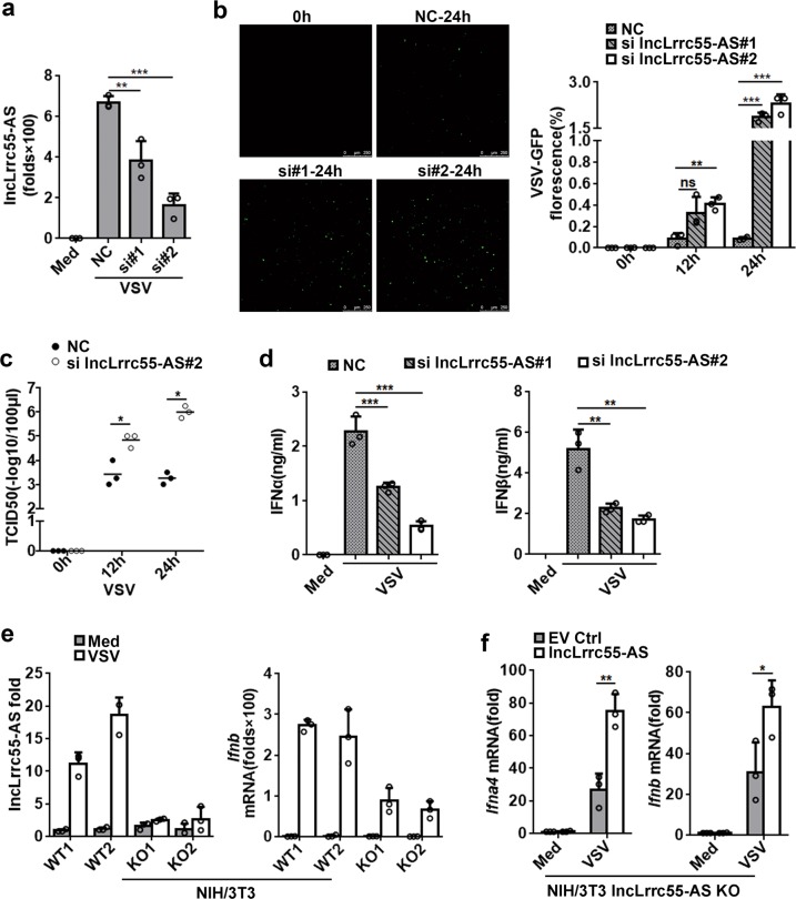
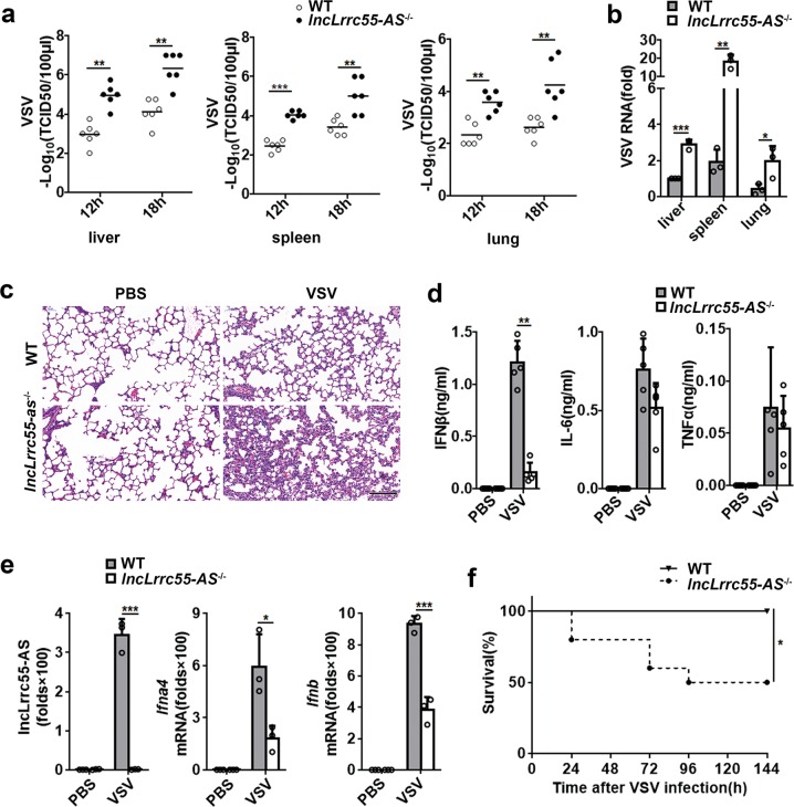
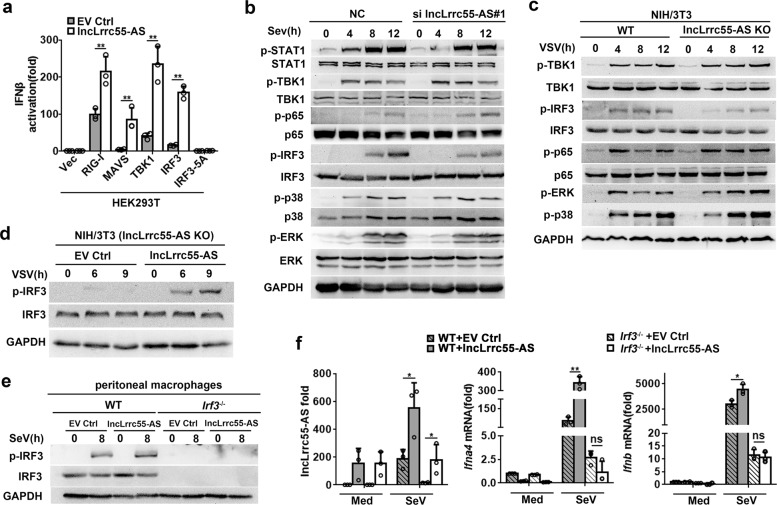
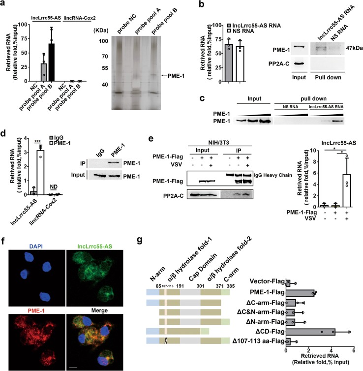
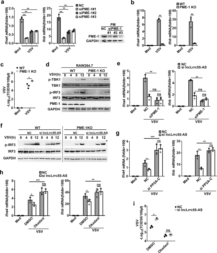
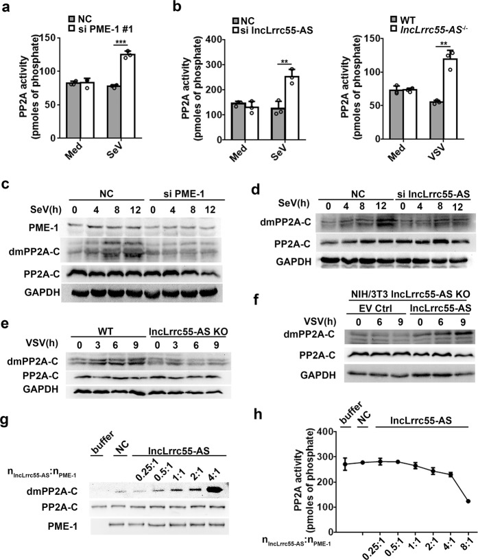
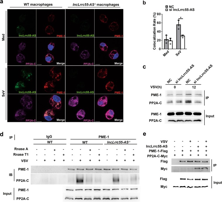
Type I interferon (IFN-I) production is efficiently induced to ensure a potent innate immune response to viral infection. How this response can be enhanced, however, remains to be explored. Here, we identify a new cytoplasmic long non-coding RNA (lncRNA), lncLrrc55-AS, that drives a positive feedback loop to promote interferon regulatory factor 3 (IRF3) signaling and IFN-I production. We show that lncLrrc55-AS is virus-induced in multiple cell types via the IFN-JAK-STAT pathway. LncLrrc55-AS-deficient mice display a weakened antiviral immune response and are more susceptible to viral challenge. Mechanistically, lncLrrc55-AS binds phosphatase methylesterase 1 (PME-1), and promotes the interaction between PME-1 and the phosphatase PP2A, an inhibitor of IRF3 signaling. LncLrrc55-AS supports PME-1-mediated demethylation and inactivation of PP2A, thereby enhancing IRF3 phosphorylation and signaling. Loss of PME-1 phenocopies lncLrrc55-AS deficiency, leading to diminished IRF3 phosphorylation and IFN-I production. We have identified an IFN-induced lncRNA as a positive regulator of IFN-I production, adding mechanistic insight into lncRNA-mediated regulation of signaling in innate immunity and inflammation.


Conflict of interest statement

The authors declare no competing interests.

Figures

Fig. 1
Fig. 1
Identification of virus-induced IFN-I-dependent lncLrrc55-AS in macrophages. a cDNA of lncLrrc55-AS in VSV-infected mouse peritoneal macrophages. b RT-qPCR analysis of lncLrrc55-AS in macrophages infected with SeV for 12 h. c RT-qPCR analysis (left) of the distribution of different RNAs following nucleus/cytoplasm fractionation of macrophages infected with SeV for 12 h. Western blot analysis (right) of nucleus/cytoplasm fractionation. d FISH analysis of lncLrrc55-AS in macrophages infected with SeV for 12 h. Scale bar, 20 µm. e, f RT-qPCR analysis of lncLrrc55-AS expression in macrophages infected with HSV for 12 h, or stimulated with poly(I:C) for 12 h, LPS for 3 h (e), or IFNβ with the indicated dose for 8 h (f). g, h RT-qPCR analysis of lncLrrc55-AS expression in WT and Irf3−/− macrophages infected with SeV or VSV for 12 h (g), in WT and Ifnar1−/− macrophages treated with IFNα4, IFNβ (500 U/mL) for 8 h or infected with SeV for 12 h (h). i RT-qPCR analysis of lncLrrc55-AS expression in macrophages pretreated for 1 h with the JAK inhibitor Baricitinib (2 μM) or the STAT1 inhibitor Fludarabine (1 μM), then infected with SeV or VSV for 12 h or treated with IFNα4 for 8 h. Data are from three independent experiments (b, c left, ei, means ± SEM) or are representative of three independent experiments with similar results (a, c right, d). ***P < 0.005 (Student’s t-test or ANOVA)
Fig. 2
Fig. 2
LncLrrc55-AS enhances IFN-I production in response to viral infection. a RT-qPCR analysis of lncLrrc55-AS silencing efficiency by two siRNAs (#1, #2). NC, a non-targeting control siRNA. b Fluorescence analysis of VSV replication in NC- or lncLrrc55-AS-silenced peritoneal macrophages infected with VSV-GFP for the indicated hours. c Determination of virus loads by TCID50 assay of the supernatant from macrophages infected with VSV for the indicated hours. d ELISA of IFNα and IFNβ in supernatant of control or lncLrrc55-AS-silenced macrophages infected with VSV for 12 h. e RT-qPCR analysis of lncLrrc55-AS or Ifnb mRNA expression in the selected WT or lncLrrc55-AS KO NIH/3T3 cell clones infected with VSV for 12 h. f RT-qPCR analysis of Ifna4 and Ifnb mRNA expression in empty vector control (EV Ctrl) or lncLrrc55-AS transfected lncLrrc55-AS KO NIH/3T3 cells infected with VSV for 12 h. Data are from three independent experiments (a, b right, cf, means ± SEM) or are representative of three independent experiments with similar results (b left). *P < 0.05, **P < 0.01 and ***P < 0.005; ns, no significance (Student’s t-test or ANOVA)
Fig. 3
Fig. 3
LncLrrc55-AS protects mice against viral infection. a Determination of VSV loads by TCID50 in organs of WT or lncLrrc55-AS−/− mice (n = 6 per group, 7 weeks old) 12 or 18 h after intraperitoneal injection of VSV (5 × 107 pfu/g). b Determination of VSV replication by RT-qPCR in organs of WT or lncLrrc55-AS−/− mice 18 h after infection with VSV (as in a). c Hematoxylin and eosin staining of lung sections from WT or lncLrrc55-AS−/− mice 18 h after infection with VSV (as in a). Scale bar, 50 µm. d ELISA of cytokines in serum from WT or lncLrrc55-AS−/− mice 12 h after infection with VSV (as in a, n = 4). e RT-qPCR analysis of lncLrrc55-AS, Ifna4 and Ifnb mRNA expression in peritoneal macrophages from WT or lncLrrc55-AS−/− mice 12 h after infection with VSV (as in a, n = 3). f Survival of 7-week-old WT or lncLrrc55-AS−/− mice (n = 10) after intraperitoneal injection of VSV (1 × 108 pfu/g). Kaplan–Meier curve was used to evaluate survival rate. Data are shown as means ± SEM (a, b, d, e), or are representative of three independent experiments with similar results (c). *P < 0.05, **P < 0.01 and ***P < 0.005 (Student’s t-test)
Fig. 4
Fig. 4
LncLrrc55-AS enhances IRF3 signaling in antiviral innate responses. a Dual luciferase analysis of the activity of the IFNβ promoter after co-transfecting plasmids expressing lncLrrc55-AS (or empty vector control, EV Ctrl), an IFNβ reporter and TK-renilla with RIG-I, MAVS, TBK1, IRF3 or IRF3-5A in 293T cells. b, c Western blot analysis of phosphorylated (p-) or total protein in lysates of NC- and lncLrrc55-AS-silenced peritoneal macrophages infected with SeV (b) or WT control and lncLrrc55-AS KO NIH/3T3 cells infected with VSV (c) for the indicated hours. d Western blot analysis of phosphorylated (p-) or total IRF3 in lysates of lncLrrc55-AS KO NIH/3T3 cells ectopically expressing EV Ctrl or lncLrrc55-AS after VSV infection for the indicated hours. e Western blot analysis of phosphorylated (p-) or total IRF3 in lysates of WT and Irf3−/− peritoneal macrophages ectopically expressing lncLrrc55-AS or EV Ctrl after SeV infection for 8 h. f RT-qPCR analysis of lncLrrc55-AS, Ifna4 and Ifnb mRNA expression of WT and Irf3−/− peritoneal macrophages ectopically expressing lncLrrc55-AS or EV Ctrl after SeV infection for 8 h. Data are from three independent experiments (a, f, means ± SEM) or are representative of three independent experiments with similar results (be). *P < 0.05, **P < 0.01; ns, no significance (Student’s t-test or ANOVA)
Fig. 5
Fig. 5
Identification of protein phosphatase methylesterase PME-1 as a binding partner of lncLrrc55-AS. a RT-qPCR analysis of the RNA retrieval ratio of lncLrrc55-AS purified by hybridizing with antisense lncLrrc55-AS probes from whole cell extracts of peritoneal macrophages infected with SeV for 12 h (left). SDS-PAGE with a silver nitrate staining analysis of proteins co-purified with lncLrrc55-AS (right). The arrow indicates protein bands that were subjected to mass spectrometry analysis. NC, no-targeting probe control. b, c RNA pull-down experiments using in vitro transcribed biotinylated lncLrrc55-AS or negative strand RNA (NS RNA) as a control to retrieve PME-1 from HEK293T cell extracts (b) or recombinant His-tagged PME-1 protein collections (c). RNA retrieval ratio of the RNA pull-down assay was assessed by RT-qPCR (b left). PME-1 retrieval by the RNA pull-down assay was assessed by western blot (b right and c). d RNA immunoprecipitation (RIP) experiments were performed with anti-PME-1 antibody in macrophages infected with SeV for 12 h. Immunoprecipitation of PME-1 was assessed by western blot. Co-purified RNAs were assessed by RT-qPCR analysis. e RIP experiments were performed with anti-PME-1 antibody in NIH/3T3 cells infected with VSV for 10 h. Immunoprecipitation of PME-1 and co-immunoprecipitation of PP2A-C were assessed by western blot. Co-purified RNA was assessed by RT-qPCR analysis. f RNA FISH-immunofluorescence detection of endogenous lncLrrc55-AS molecules and PME-1 in macrophages infected with SeV for 12 h. Scale bar, 10 µm. g RIP experiments were performed with anti-Flag antibody-coupled beads in NIH/3T3 cells transfected with full-length or truncated PME-1-Flag plasmids. LncLrrc55-AS retrieval was assessed by RT-qPCR. Schematic of PME-1 and truncated PME-1 used in the study (left). ΔC-arm, carboxyl-terminal arm (residues 371–385) truncated; ΔN-arm, amino-terminal arm (residues 1–64) truncated; ΔC&N-arm, residues 1–64 and 371–385 truncated; ΔCD, cap domain (residues 191–301) deleted; Δ107–113 aa, residues 107–113 deleted. Data are from three independent experiments (a, b left, d left, e, g means ± SEM) or are representative of three independent experiments with similar results (a, b, d right, c, e left, f). *P < 0.05, ***P < 0.005, ND, not detected (Student’s t-test or ANOVA)
Fig. 6
Fig. 6
LncLrrc55-AS inhibits PP2A-mediated negative regulation of IRF3-induced IFN-I production via PME-1. a RT-qPCR analysis of Ifna4 and Ifnb mRNA expression in non-target siRNA control (NC)- or PME-1-silenced peritoneal macrophages infected with VSV for 12 h. The silencing efficiency of three siRNAs targeting PME-1 was confirmed by western blot (right). b RT-qPCR analysis of Ifna4 and Ifnb mRNA expression in WT or PME-1 KO RAW264.7 cells infected with VSV for 12 h. c Determination of virus loads by TCID50 assay in the supernatant from VSV-infected WT or PME-1 KO RAW264.7 cells for 12 h. d Western blot analysis of phosphorylated (p-) or total proteins in lysates of WT and PME-1 KO RAW264.7 cells infected with VSV for the indicated hours. e RT-qPCR analysis of Ifna4 and Ifnb mRNA expression in NC- and PME-1-, lncLrrc55-AS- or double-silenced peritoneal macrophages infected with VSV for 12 h. f Western blot analysis of p-IRF3 or total IRF3 proteins in lysates of NC- or lncLrrc55-AS-silenced WT and PME-1 KO RAW264.7 cells infected with VSV for the indicated hours. g RT-qPCR analysis of Ifna4 and Ifnb mRNA expression in NC- and PP2A-C-, lncLrrc55-AS- or double-silenced peritoneal macrophages infected with VSV for 12 h. h RT-qPCR analysis of Ifna4 and Ifnb mRNA expression in NC- or lncLrrc55-AS-silenced macrophages pre-treated with Okadaol (50 nM) or DMSO 1 h before infection with VSV for 12 h. i Determination of virus loads by TCID50 in the supernatant from NC- or lncLrrc55-AS-silenced macrophages pre-treated with Okadaol (50 nM) or DMSO 1 h before infection with VSV for 12 h. Data are from three independent experiments (ac, e, gi, means ± SEM) or are representative of three independent experiments with similar results (d, f). *P < 0.05, **P < 0.01, ***P < 0.005; ns, no significance (Student’s t-test or ANOVA)
Fig. 7
Fig. 7
LncLrrc55-AS promotes the deactivation and demethylation of PP2A. a Phosphatase activity analysis of PP2A in lysates from non-target siRNA control (NC)- or PME-1-silenced macrophages infected with SeV for 12 h. b Phosphatase activity analysis of PP2A in lysates of NC- or lncLrrc55-AS-silenced macrophages infected with SeV for 12 h (left), and in WT or lncLrrc55-AS−/− macrophages infected with VSV for 12 h (right). c, d Western blot analysis of demethylated PP2A-C (dmPP2A-C) or total PP2A-C proteins in lysates of NC- and PME-1- or lncLrrc55-AS-silenced peritoneal macrophages with SeV infection for the indicated hours. e, f Western blot analysis of dmPP2A-C or total PP2A-C proteins in lysates from WT and lncLrrc55-AS KO NIH/3T3 cells (e), and from EV Ctrl- or lncLrrc55-AS-transfected lncLrrc55-AS KO NIH/3T3 cells (f) infected with VSV for the indicated hours. g, h Demethylation level by western blot analysis (g) and phosphatase activity assay (h) of recombinant PP2A-C proteins catalyzed by PME-1 in vitro, with increasing amount of lncLrrc55-AS added to the catalytic reaction system. The nlncLrrc55-AS: nPME-1 indicates the molar ratio of lncLrrc55-AS to PME-1. Data are from three independent experiments (a, b, h means ± SEM) or are representative of three independent experiments with similar results (cg). **P < 0.01, ***P < 0.005 (Student’s t-test)
Fig. 8
Fig. 8
LncLrrc55-AS promotes the interaction between PME-1 and PP2A. a RNA FISH-immunofluorescence detection of endogenous lncLrrc55-AS, PME-1, and PP2A-C in peritoneal macrophages from WT or lncLrrc55-AS−/− non-infected mice and mice infected with SeV for 12 h. Scale bars, 10 µm. b Quantitative analysis of colocalization rate for PME-1 and PP2A in NC- or lncLrrc55-AS-silenced peritoneal macrophages after SeV infection for 12 h. c Interaction between PME-1 and PP2A in RAW264.1 cells. NC- or lncLrrc55-AS-silenced cells were infected with VSV for the indicated hours, followed by immunoprecipitation with anti-PME-1 antibody. The immunoprecipitated PME-1 and PP2A were detected by western blot. d Interaction between PME-1 and PP2A in WT and lncLrrc55-AS−/− peritoneal macrophages infected with SeV for the indicated hours. Samples isolated from cells were equally divided into three parts. All parts were digested with Rnase A or Rnase T1 (10 U/mL), followed by immunoprecipitation with anti-PME-1 antibody (two parts) or with normal IgG antibody (one part; control). The immunoprecipitated PME-1 and PP2A were detected by western blot. e Interactions of PME-1 and PP2A with lncLrrc55-AS in an overexpression system. HEK293T cells were transfected with plasmids encoding PME-1-Flag, PP2A-C-Myc, and lncLrrc55-AS, followed by immunoprecipitation with anti-Flag antibody-coupled beads. The immunoprecipitated PME-1-Flag and PP2A-C-Myc were detected by western blot analysis. HEK293T cells transfected with empty vector plasmids were used as controls. Data are from three independent experiments (b, means ± SEM) or are representative of three independent experiments with similar results (a, ce). *P < 0.05 (Student’s t-test)

Comment in

Similar articles

Cited by

References

    1. McNab F, et al. Type I interferons in infectious disease. Nat. Rev. Immunol. 2015;15:87–103. doi: 10.1038/nri3787. - DOI - PMC - PubMed
    1. Hoffmann HH, Schneider WM, Rice CM. Interferons and viruses: an evolutionary arms race of molecular interactions. Trends Immunol. 2015;36:124–138. doi: 10.1016/j.it.2015.01.004. - DOI - PMC - PubMed
    1. Wu J, Chen ZJ. Innate immune sensing and signaling of cytosolic nucleic acids. Annu. Rev. Immunol. 2014;32:461–488. doi: 10.1146/annurev-immunol-032713-120156. - DOI - PubMed
    1. Liu S, et al. Phosphorylation of innate immune adaptor proteins MAVS, STING, and TRIF induces IRF3 activation. Science. 2015;347:aaa2630. doi: 10.1126/science.aaa2630. - DOI - PubMed
    1. Liu J, Qian C, Cao X. Post-translational modification control of innate immunity. Immunity. 2016;45:15–30. doi: 10.1016/j.immuni.2016.06.020. - DOI - PubMed

Publication types

MeSH terms