Glutamate Imaging Reveals Multiple Sites of Stochastic Release in the CA3 Giant Mossy Fiber Boutons
- PMID: 31213985
- PMCID: PMC6558140
- DOI: 10.3389/fncel.2019.00243
Glutamate Imaging Reveals Multiple Sites of Stochastic Release in the CA3 Giant Mossy Fiber Boutons
Abstract
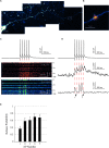
One of the most studied central synapses which have provided fundamental insights into cellular mechanisms of neural connectivity is the "giant" excitatory connection between hippocampal mossy fibers (MFs) and CA3 pyramidal cells. Its large presynaptic bouton features multiple release sites and is densely packed with thousands of synaptic vesicles, to sustain a highly facilitating "detonator" transmission. However, whether glutamate release sites at this synapse act independently, in a stochastic manner, or rather synchronously, remains poorly understood. This knowledge is critical for a better understanding of mechanisms underpinning presynaptic plasticity and postsynaptic signal integration rules. Here, we use the optical glutamate sensor SF-iGluSnFR and the intracellular Ca2+ indicator Cal-590 to monitor spike-evoked glutamate release and presynaptic calcium entry in MF boutons. Multiplexed imaging reveals that distinct sites in individual MF giant boutons release glutamate in a probabilistic fashion, also showing use-dependent short-term facilitation. The present approach provides novel insights into the basic mechanisms of neurotransmitter release at excitatory synapses.
Keywords: CA3 pyramidal cell; action potential; dentate gyrus; giant mossy fiber bouton; glutamate release; short-term plasticity.
Figures


Similar articles
-
Target-cell specificity of kainate autoreceptor and Ca2+-store-dependent short-term plasticity at hippocampal mossy fiber synapses.J Neurosci. 2008 Dec 3;28(49):13139-49. doi: 10.1523/JNEUROSCI.2932-08.2008. J Neurosci. 2008. PMID: 19052205 Free PMC article.
-
Short-Term Facilitation at a Detonator Synapse Requires the Distinct Contribution of Multiple Types of Voltage-Gated Calcium Channels.J Neurosci. 2017 May 10;37(19):4913-4927. doi: 10.1523/JNEUROSCI.0159-17.2017. Epub 2017 Apr 14. J Neurosci. 2017. PMID: 28411270 Free PMC article.
-
Differential mechanisms of transmission at three types of mossy fiber synapse.J Neurosci. 2000 Nov 15;20(22):8279-89. doi: 10.1523/JNEUROSCI.20-22-08279.2000. J Neurosci. 2000. PMID: 11069934 Free PMC article.
-
Timing and efficacy of transmitter release at mossy fiber synapses in the hippocampal network.Pflugers Arch. 2006 Dec;453(3):361-72. doi: 10.1007/s00424-006-0093-2. Epub 2006 Jun 27. Pflugers Arch. 2006. PMID: 16802161 Review.
-
Quantal analysis of excitatory postsynaptic currents at the hippocampal mossy fiber-CA3 pyramidal cell synapse.Adv Second Messenger Phosphoprotein Res. 1994;29:235-60. doi: 10.1016/s1040-7952(06)80019-4. Adv Second Messenger Phosphoprotein Res. 1994. PMID: 7848714 Review.
Cited by
-
Early changes in synaptic and intrinsic properties of dentate gyrus granule cells in a mouse model of Alzheimer's disease neuropathology and atypical effects of the cholinergic antagonist atropine.Neurobiol Dis. 2021 May;152:105274. doi: 10.1016/j.nbd.2021.105274. Epub 2021 Jan 20. Neurobiol Dis. 2021. PMID: 33484828 Free PMC article.
-
Recruitment of release sites underlies chemical presynaptic potentiation at hippocampal mossy fiber boutons.PLoS Biol. 2021 Jun 21;19(6):e3001149. doi: 10.1371/journal.pbio.3001149. eCollection 2021 Jun. PLoS Biol. 2021. PMID: 34153028 Free PMC article.
-
Mossy fiber expression of αSMA in human hippocampus and its relevance to brain evolution and neuronal development.Sci Rep. 2025 May 6;15(1):15834. doi: 10.1038/s41598-025-00094-3. Sci Rep. 2025. PMID: 40328887 Free PMC article.
-
Population imaging of synaptically released glutamate in mouse hippocampal slices.STAR Protoc. 2021 Nov 9;2(4):100877. doi: 10.1016/j.xpro.2021.100877. eCollection 2021 Dec 17. STAR Protoc. 2021. PMID: 34816125 Free PMC article.
References
Grants and funding
LinkOut - more resources
Full Text Sources
Miscellaneous

