. 2019 May 31:10:1138.
doi: 10.3389/fmicb.2019.01138. eCollection 2019.

Simultaneous Colorimetric Detection of a Variety of Salmonella spp. in Food and Environmental Samples by Optical Biosensing Using Oligonucleotide-Gold Nanoparticles

Affiliations

Simultaneous Colorimetric Detection of a Variety of Salmonella spp. in Food and Environmental Samples by Optical Biosensing Using Oligonucleotide-Gold Nanoparticles

Irwin A Quintela et al. Front Microbiol. .

Abstract

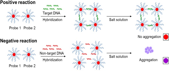
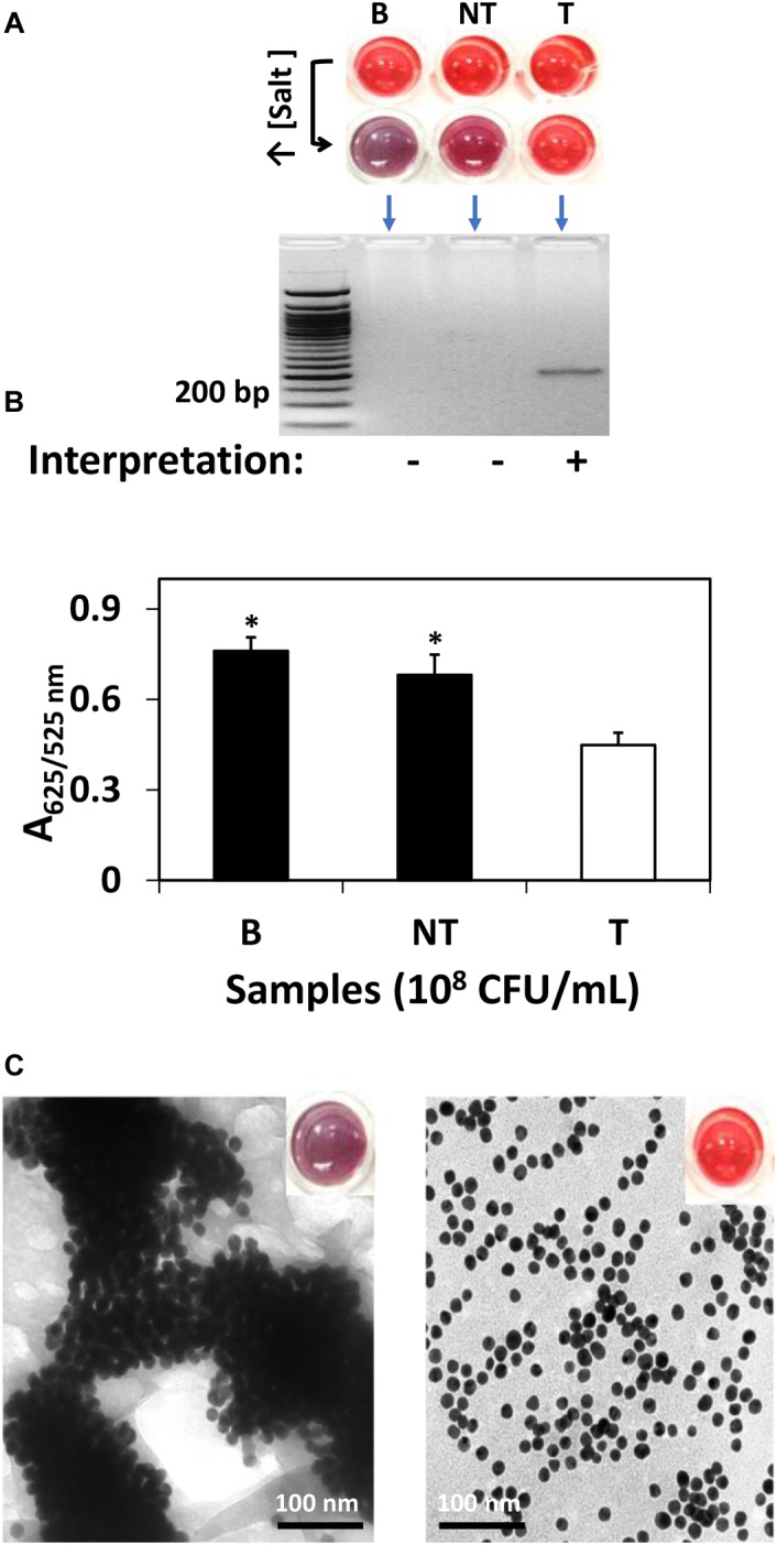
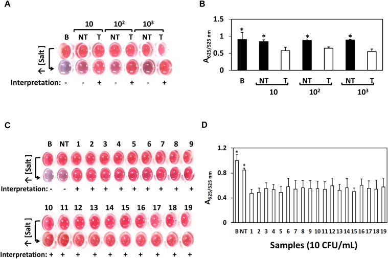
Optical biosensors for rapid detection of significant foodborne pathogens are steadily gaining popularity due to its simplicity and sensitivity. While nanomaterials such as gold nanoparticles (AuNPs) are commonly used as signal amplifiers for optical biosensors, AuNPs can also be utilized as a robust biosensing platform. Many reported optical biosensors were designed for individual pathogen detection in a single assay and have high detection limit (DL). Salmonella spp. is one of the major causative agents of foodborne sickness, hospitalization and deaths. Unfortunately, there are around 2,000 serotypes of Salmonella worldwide, and rapid and simultaneous detection of multiple strains in a single assay is lacking. In this study, a comprehensive and highly sensitive simultaneous colorimetric detection of nineteen (19) environmental and outbreak Salmonella spp. strains was achieved by a novel optical biosensing platform using oligonucleotide-functionalized AuNPs. A pair of newly designed single stranded oligonucleotides (30-mer) was displayed onto the surface of AuNPs (13 nm) as detection probes to hybridize with a conserved genomic region (192-bases) of ttrRSBCA found on a broad range of Salmonella spp. strains. The sandwich hybridization (30 min, 55°C) resulted in a structural formation of highly stable oligonucleotide/AuNPs-DNA complexes which remained undisturbed even after subjecting to an increased salt concentration (2 M, final), thus allowing a direct discrimination via color change of target (red color) from non-target (purplish-blue color) reaction mixtures by direct observation using the naked eye. In food matrices (blueberries and chicken meat), nineteen different Salmonella spp. strains were concentrated using immunomagnetic separation and then simultaneously detected in a 96-well microplate by oligonucleotide-functionalized AuNPs after DNA preparation. Successful oligonucleotide/AuNPs-DNA hybridization was confirmed by gel electrophoresis while AuNPs aggregation in non-target and control reaction mixtures was verified by both spectrophotometric analysis and TEM images. Results showed that the optical AuNP biosensing platform can simultaneously screen nineteen (19) viable Salmonella spp. strains tested with 100% specificity and a superior detection limit of <10 CFU/mL or g for both pure culture and complex matrices setups. The highly sensitive colorimetric detection system can significantly improve the screening and detection of viable Salmonella spp. strains present in complex food and environmental matrices, therefore reducing the risks of contamination and incidence of foodborne diseases.

Keywords: Salmonella; colorimetric; gold nanoparticles; oligonucleotides; optical biosensor; ttrRSBCA.


Figures

SCHEME 1
SCHEME 1
The novel principles behind AuNPs optical biosensing and the schematic representation of DNA sandwich hybridization targeting ttrRSBCA locus of Salmonella spp. The high complementarity between the AuNPs-oligonucleotide probes (Probes 1 and 2) and ttrRSBCA regions allows the occurrence of DNA sandwich hybridization. Probe-target complexes form with high specificity and retain in the solution even after increasing its salt concentration. Positive reaction (ttrRSBCA locus) shows no AuNPs-oligonucleotide probes aggregation thus no color change (original red) is observed. Formation of probe-target complexes did not occur in blank or non-target DNA samples; therefore, the salt-induced AuNPs-oligonucleotide probes aggregation occurs that eventually changes the color of the reaction mixtures (from the original red to purplish blue). Color differentiation facilitates direct detection of positive and negative samples by observation using the naked eye. Size not to scale (reproduced with some modifications and with permissions from Quintela et al. (2015); copyright 2015, Royal Society of Chemistry).
SCHEME 2
SCHEME 2
A workflow showing rapid Salmonella spp. strains detection in complex matrices using the novel AuNPs optical biosensor coupled with a robust sample pooling plan and an IMS system. (A) Twenty-five different individual enriched food or environmental samples are randomly pooled into five groups: Pools 1–5. (B) Each pool is circulated in the IMS system for capture and concentration of Salmonella spp. strains. Genomic DNA are extracted from the concentrated IMS samples and amplified (C) DNA sandwich hybridization reaction mixtures are transfered into a 96-well microplate for color challenge test by increasing its salt concentration. If pools change from red to purplish blue after adding salt, reactions are interpreted as negative for ttRSBCA; all of the individual subsamples are therefore ttrRSBCA-negative. If pools remain red after adding salt, reaction mixtures are interpreted as positive for ttrRSBCA and all of the individual subsamples are further tested. (D) AuNPs optical biosensing is conducted for all individual subsamples belonging to the ttrRSBCA-positive pools. S = Subsamples.
FIGURE 1
FIGURE 1
AuNPs optical biosensing targeting ttrRSBCA region of S. Agona in pure culture setup. (A) AuNPs optical biosensing showing three reaction mixtures before and after adding salt solution (50 μL, 2 M NaCl–Na2HPO4 in final reaction). Both B and NT samples changed from red to bluish-purple color while no color change was observed in T reaction mixture. DNA sandwich hybridization between ttrRSBCA and AuNPs-probes was confirmed by a gel band (192-bp). (B) A625/525nm of the reaction mixtures after increasing its salt concentration showed significant differences at p < 0.05 between B, NT and T (C) TEM images confirmed salt induced AuNPs-probe aggregation in B or NT (left image with a bluish-purple solution as inserted) sample while no significant AuNPs aggregation was observed in S. Agona (right image with a red color solution as inserted). Both NT and T had 108 CFU/mL bacterial concentration. B = Blank, nuclease-free water; NT = non-target, STEC O157:H7 ATCC 35150; T = target, S. Agona. Significant difference at p < 0.05.
FIGURE 2
FIGURE 2
AuNPs optical biosensing – pure culture setup using various Salmonella spp. strains. (A) AuNPs optical biosensing a 10, 102 and 103 CFU/mL bacterial count levels, showing positive results for T and negative for both B and NT reaction mixtures after adding salt (B) A625/525nm of the reaction mixtures after increasing its salt concentration showing significant differences at p < 0.05 between B, NT and T (C) AuNPs optical biosensing of various Salmonella spp. strains at 10 CFU/mL bacterial counts. Reaction mixtures containing B and NT changed from red to purplish blue after adding salt solution while the original color of all target numbered samples remained unchanged. (D) A625/525nm of the reaction mixtures presented in panel (C) after increasing its salt concentration showing significant differences at p < 0.05 between B, NT and T. B = Blank, nuclease-free water; NT = non-target, STEC O157:H7 ATCC 35150, T = target, Salmonella spp. strains as numbered 1–19 in Table 1. Significant difference at p < 0.05.
FIGURE 3
FIGURE 3
Detection of various Salmonella spp. strains in complex matrices. (A) After IMS system, five pools (P1-P5) of chicken meat with skin were tested for the presence of ttrRSBCA. Among these five pools, P2 tested negative so as its five subsamples. The other 4 pools were positive: P1, P3, P4 and P5. (B) From the four positive pools, a total of 8 individual subsamples tested positive for ttrRSBCA. (C) Three out of 5 blueberry pools: P2, P3 and P4 were positive for ttrRSBCA. (D) Individual subsamples from the three positive blueberry pools were tested and 12 out of 15 these individual subsamples showed positive for ttrRSBCA. (E) All five soil sample pools: P1-P5 were negative for ttrRSBCA. B = Blank, nuclease-free water; NT = non-target, STEC O157:H7 ATCC 35150.

References

    1. Abdelhaseib M. U., Singh A. K., Bailey M., Singh M., El-Khateib T., Bhunia A. K. (2016). Fiber optic and light scattering sensors: complimentary approaches to rapid detection of Salmonella enterica in food samples. Food Control 61 135–145.
    1. Afonso A. S., Pérez-López B., Faria R. C., Mattoso L. H. C., Hernández-Herrero M., Roig-Sagués A. X., et al. (2013). Electrochemical detection of Salmonella using gold nanoparticles. Biosens. Bioelectron. 40 121–126. 10.1016/j.bios.2012.06.054 - DOI - PubMed
    1. Ahn S., Walt D. R. (2005). Detection of Salmonella spp. using microsphere-based, fiber-optic DNA microarrays. Anal. Chem. 77 5041–5047. - PubMed
    1. Arnold M., Carrique-Mas J., Mclaren I., Davies R. (2011). A comparison of pooled and individual bird sampling for detection of Salmonella in commercial egg laying flocks. Prev. Vet. Med. 99 176–184. 10.1016/j.prevetmed.2010.12.007 - DOI - PubMed
    1. Arnold M., Cook A. (2009). Estimation of sample sizes for pooled faecal sampling for detection of Salmonella in pigs. Epidemiol. Infect. 137 1734–1741. 10.1017/S0950268809002702 - DOI - PubMed

LinkOut - more resources