. 2019 Jun 4:10:1268.
doi: 10.3389/fimmu.2019.01268. eCollection 2019.

Thrombospondin-1/CD47 Interaction Regulates Th17 and Treg Differentiation in Psoriasis

Affiliations

Thrombospondin-1/CD47 Interaction Regulates Th17 and Treg Differentiation in Psoriasis

Pedro Rodríguez-Jiménez et al. Front Immunol. .

Abstract

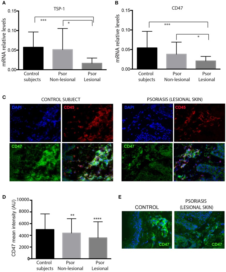
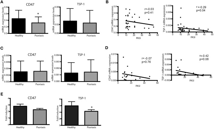
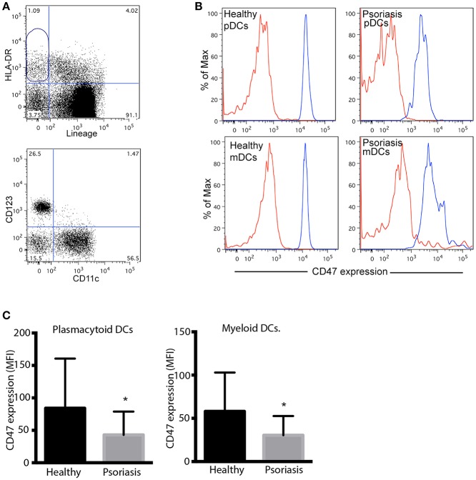
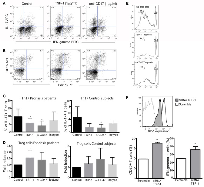
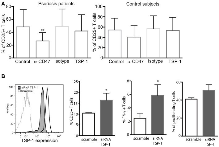
Accumulating evidence on the role of Thrombospondin-1 (TSP-1) in the immune response has emerged during the last years. In spite of the importance of TSP-1 not only as anti-angiogenic factor but also as an immunomodulatory molecule, studies on the role of TSP-1 in psoriasis have been neglected. TSP-1 and CD47 expression were analyzed in skin samples from psoriasis patients and control subjects using RT-PCR and immunofluorescence. Expression of these molecules was also evaluated in peripheral blood CD4+ T cells, moDCs, and circulating primary DCs. The functional role of TSP-1/CD47 signaling axis in psoriasis was assessed in Th17 and Treg differentiation assays. Additionally, small interfering RNA assays specific to TSP-1 were performed in CD4+ T cells and monocyte derived DC to specifically evaluate the function of this protein. Lesional skin of psoriasis patients expressed lower TSP-1 and CD47 mRNA levels compared to non-lesional skin or skin from controls. Immunofluorescence staining revealed decreased expression of CD47 in CD45+ dermal cells from psoriasis samples compared to control subjects. Peripheral CD4+ T cells and circulating primary DCs from psoriasis also expressed lower levels of CD47 compared to controls. Although no significant differences were detected in TSP-1 expression in CD4+ T cells and moDCs between patients and controls, TSP-1 expression in psoriasis patients inversely correlated with disease activity evaluated by the Psoriasis Area and Index Activity. Furthermore, exogenous TSP-1 inhibited Th17 differentiation and stimulated the differentiation of CD4+ T cells toward Treg cells. Furthermore, RNA interference specific for TSP-1 confirmed the role of this molecule as a negative regulator of T cell activation. Because of the impact of TSP-1/CD47 signaling axis in Th17 and Treg differentiation, a dysregulated expression of these molecules in the immune cells from psoriasis patients may favor the exacerbated inflammatory response in this disease.

Keywords: CD47; TSP-1; Th17; Treg cells; psoriasis.


Figures

Figure 1
Figure 1
Skin samples from psoriasis patients express lower levels of TSP-1, and CD47 compared with healthy controls. mRNA levels of TSP-1 (A) and its receptor CD47 (B) were analyzed by RT-PCR in skin samples from 26 psoriasis patients and 20 healthy controls. GAPDH expression was used to normalize gene expression. Data were analyzed by one-way ANOVA followed by Tukey's multiple comparisons test, ***p < 0.001, *p < 0.05. (C) Double immunofluorescence staining of CD47 (green) and CD45 (red) in a representative skin sample from control subjects (left panels) and lesional skin from psoriasis patients (right panels) is shown. Nuclei were counterstained with DAPI (blue). (D) For quantification of immunofluorescence staining, fluorescence intensity of CD47 in CD45+ dermal cells was calculated using Image J software. (E) Representative expression of CD47 (green) in skin samples from control subjects and psoriasis patients. Graphs represent mean ± SD. Differences between groups were determined by one-way ANOVA followed by Tukey's multiple comparisons test, ****p < 0.0001, **p ≤ 0.001.
Figure 2
Figure 2
Dysregulated expression of CD47 and TSP-1 in peripheral blood CD4+ T lymphocytes and moDCs. (A,B) CD4+ T cells from psoriasis patients (n = 30) and healthy controls (n = 18) were isolated from peripheral blood using magnetic microbeads. (A) CD47 and TSP-1 expression was analyzed using real-time PCR. GAPDH expression was used to normalize data. Differences between groups were analyzed using Mann-Whitney U-test. (B) Correlation between the expression of CD47 and TSP-1 in CD4+ T cells and PASI determined by Spearman test. (C) Basal expression of CD47 and TSP-1 in non-stimulated moDCS from patients and controls. Monocytes were isolated from peripheral blood using immune-magnetic bead and differentiated to DCs during 5 days in the presence of IL-4 and GM-CSF. (D) Correlation between the expression of CD47 and TSP-1 in moDCs cells and PASI determined by Spearman test. (E) Fold induction of CD47 and TSP-1 expression levels in moDCs in response to LPS. moDCs were stimulated for 24 h in the presence of LPS (10 ng/ml), then RNA was isolated and the expression of TSP-1 and CD47 analyzed by RT-PCR, calculating their fold induction as LPS/basal expression (n = 18). Differences were tested by Mann-Whitney U-test, *p < 0.05.
Figure 3
Figure 3
Peripheral blood plasmacytoid and myeloid DCs from psoriasis patients express low levels of CD47. (A) Gating strategy for the detection of pDCs and mDCs. Dendritic cells were identified as HLA-DR positive, Lineage (CD3, CD14, CD20, CD56) negative, CD47 expression was then determined in CD123+ and CD11c+ by flow cytometry. (B) Representative histograms of CD47 expression in pDCs and mDCs from one patient and one control subject. Red and blues lines indicates isotype control and CD47 expression, respectively. (C) CD47 expression was calculated as mean fluorescence intensity (MFI) of CD47/MFI isotype control. Bars represent mean ± SD from 18 psoriasis patients and 10 control subjects. Differences were analyzed by the Mann-Whitney U-test, *p < 0.05.
Figure 4
Figure 4
Functional effects of TSP-1 in CD4+ T lymphocytes from psoriasis patients. (A,B) Representative dot plots from Th17 (A) and Treg differentiation (B) assays in the presence of TSP-1 and anti-CD47 mAb. CD4+ T cells were isolated from peripheral venous blood from psoriasis patients using magnetic beads. Cells (1 × 10⁶/ml) were differentiated to Th17 in the presence or absence of human anti-CD47 mAb 1 μg/ml (left) or human TSP-1 5 μg/ml (center). (C) Percent of IL- 17+ T cells from psoriasis patients (n = 13) and control subjects (n = 5) evaluated by flow cytometry after 10 days of culture in the presence of TSP-1, anti-CD47 mAb, or IgG1 isotype control (1 μg/ml). Data were analyzed using Friedman test and Dunn's multiple comparison test, *p < 0.05. (D) Differentiation of Treg cells in the presence of anti-CD47 mAb (1 μg/ml), TSP-1 (5 μg/ml) or IgG1 isotype control (1 μg/ml) from psoriasis patients (n = 9) and control subjects (n = 6). Graphs show the fold induction of FoxP3+ CD25+ cells in the presence of the indicated stimuli compared to the cells cultured only with the differentiation cocktail. (E) Treg suppression assay, Treg cells differentiated in the presence of TSP-1 or anti-CD47 mAb were isolated by immunomagnetic beads and added to mixed leukocytes reaction (MLR) cultures. Upper histogram corresponds to control culture (moDCs plus responder T cells), central and lower histograms correspond to MLR cultures plus Treg cells that were differentiated in the presence of TSP-1 or anti-CD47 mAb (histograms are representative of two independent experiments). (F) Knockdown of TSP-1 favors T cell activation via TCR. Histogram showing the knockdown efficiency of TSP-1 siRNA. Briefly, unstimulated CD4+ T cells were transfected by nucleofection with TSP-1 specific or control siRNA (50 pmol), 24 h later T cells were stimulated with anti-CD3/CD28 + IL-2 during 48 h. CD25 expression and IFN-γ production were evaluated by flow cytometry data. Data correspond to one out of two independent experiments performed by triplicate. Differences were tested by Mann-Whitney test, *p < 0.05.
Figure 5
Figure 5
CD47/TSP-1 signaling prevents CD4+ T cell activation in an antigen presentation assay. (A) CD25 expression from CD4+ T cells after antigen presentation, monocyte-derived DCs from psoriasis patients (n = 8) and healthy controls (n = 3) were cultured for 3 days with autologous peripheral blood CD4+ T cells in the presence of staphylococcal enterotoxin E. Where indicated, anti-CD47 (1 μg/ml), IgG1 isotype control (1 μg/ml), or TSP-1 (5 μg/ml) were added. Then, expression of CD25 was evaluated using flow cytometry. Data correspond to mean ± SD and differences were tested using Friedman test, **p < 0.01. (B) Knockdown of TSP-1 of moDCs augments the activation of T cells following Ag presentation. Histogram showing the knockdown efficiency of TSP-1 siRNA. Briefly, immature moDCs were transfected by nucleofection with TSP-1 specific or control siRNA (50 pmol), after 48 h moDCs were stimulated with LPS (10 ng/ml) during 12 h and then co-cultured with heterologous CD4+ T cells. Data correspond to one out of three experiments performed by triplicate. Differences were analyzed using Mann-Whitney test, *p < 0.05.

References

    1. Perera GK, Di Meglio P, Nestle FO. Psoriasis. Annu Rev Pathol. (2012) 7:385–422. 10.1146/annurev-pathol-011811-132448 - DOI - PubMed
    1. Hawkes JE, Chan TC, Krueger JG. Psoriasis pathogenesis and the development of novel targeted immune therapies. J Allergy Clin Immunol. (2017) 140:645–53. 10.1016/j.jaci.2017.07.004 - DOI - PMC - PubMed
    1. Lowes MA, Kikuchi T, Fuentes-Duculan J, Cardinale I, Zaba LC, Haider AS, et al. . Psoriasis vulgaris lesions contain discrete populations of Th1 and Th17 T cells. J Invest Dermatol. (2008) 128:1207–11. 10.1038/sj.jid.5701213 - DOI - PubMed
    1. Kryczek I, Bruce AT, Gudjonsson JE, Johnston A, Aphale A, Vatan L, et al. . Induction of IL-17+ T cell trafficking and development by IFN-gamma: mechanism and pathological relevance in psoriasis. J Immunol. (2008) 181:4733–41. 10.4049/jimmunol.181.7.4733 - DOI - PMC - PubMed
    1. Res PC, Piskin G, de Boer OJ, van der Loos CM, Teeling P, Bos JD, et al. . Overrepresentation of IL-17A and IL-22 producing CD8 T cells in lesional skin suggests their involvement in the pathogenesis of psoriasis. PLoS ONE. (2010) 5:e14108. 10.1371/journal.pone.0014108 - DOI - PMC - PubMed

Publication types

MeSH terms