

Rapid Growth of Pelvic Cyst during Pregnancy: A Case Report
- PMID: 31214368
- PMCID: PMC6535882
- DOI: 10.1155/2019/3120921
Rapid Growth of Pelvic Cyst during Pregnancy: A Case Report
Abstract
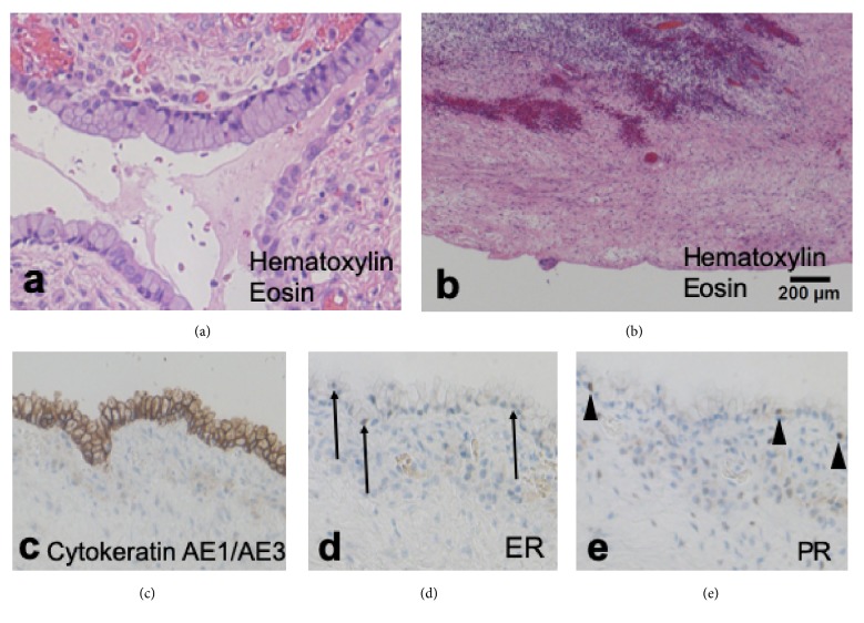
We describe a patient with bilateral cystic tumors of the pelvis. The left one rapidly grew during pregnancy and combined with the right one, whose clinical course made diagnosis difficult. A pregnant woman with a history of laparotomy was referred to us due to suspected bilateral pelvic cysts. The left-sided cyst had rapidly grown to 27 cm in diameter and merged with the right cyst, forming a large cyst occupying the entire pelvic cavity in the third trimester. Considering this rapid growth, cesarean section and resection of the cyst were performed at 37th week. The resected cyst consisted of two components: a large unilocular cyst containing serous fluid and a multilocular cyst suggestive of ovarian mucinous cystadenoma in the right ovary. The wall of the former largely lacked lining epithelium, but it was partly continuous with the latter mucinous epithelium. Immunohistochemically, estrogen and progesterone receptors were focally positive in the cyst wall, suggesting that pregnancy-associated sex-hormones may have contributed to the rapid growth of the cyst. We diagnosed this condition as a peritoneal inclusion cyst margining with a right ovarian mucinous cystadenoma. Peritoneal inclusion cyst should be considered in the differential diagnosis of a rapidly growing pelvic mass during pregnancy.
Figures


Similar articles
-
Giant Ovarian Serous Cystadenoma in an Infant: Report of a Rare Case.J Lab Physicians. 2021 Jul 14;13(4):388-390. doi: 10.1055/s-0041-1732487. eCollection 2021 Dec. J Lab Physicians. 2021. PMID: 34975262 Free PMC article.
-
[A case of large Mucinous Cystadenoma of the Ovary at the Regional Teaching Hospital of Ouahigouya (Burkina Faso)].Med Trop Sante Int. 2022 Jun 20;2(2):mtsi.v2i2.2022.187. doi: 10.48327/mtsi.v2i2.2022.187. eCollection 2022 Jun 30. Med Trop Sante Int. 2022. PMID: 35919249 Free PMC article. French.
-
Extracutaneous seborrheic inclusion cyst: an unusual presentation.Pathologica. 2010 Oct;102(5):420-2. Pathologica. 2010. PMID: 21361125
-
Mucinous cystadenoma of the ovary with functioning stroma and virilization in pregnancy: a case report and review of the literature.Clin Exp Obstet Gynecol. 2003;30(4):248-52. Clin Exp Obstet Gynecol. 2003. PMID: 14664425 Review.
-
Full-term pregnancy with retroperitoneal giant mucinous cyst: A case report and literature review.Medicine (Baltimore). 2024 Mar 8;103(10):e36979. doi: 10.1097/MD.0000000000036979. Medicine (Baltimore). 2024. PMID: 38457602 Free PMC article. Review.
Cited by
-
A case of huge ovarian cyst in second trimester: A rare case report.Ann Med Surg (Lond). 2022 Sep 27;82:104765. doi: 10.1016/j.amsu.2022.104765. eCollection 2022 Oct. Ann Med Surg (Lond). 2022. PMID: 36268389 Free PMC article.
References
-
- Usui R., Minakami H., Kosuge S., Iwasaki R., Ohwada M., Sato I. A retrospective survey of clinical, pathologic, and prognostic features of adnexal masses operated on during pregnancy. Journal of Obstetrics and Gynaecology Research. 2000;26(2):89–93. doi: 10.1111/j.1447-0756.2000.tb01289.x. - DOI - PubMed
Publication types
LinkOut - more resources
Full Text Sources
