

The Ectopic Expression of Meiosis Regulatory Genes in Cutaneous T-Cell Lymphomas (CTCL)
- PMID: 31214493
- PMCID: PMC6554469
- DOI: 10.3389/fonc.2019.00429
The Ectopic Expression of Meiosis Regulatory Genes in Cutaneous T-Cell Lymphomas (CTCL)
Abstract
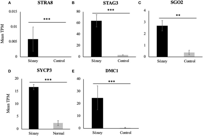
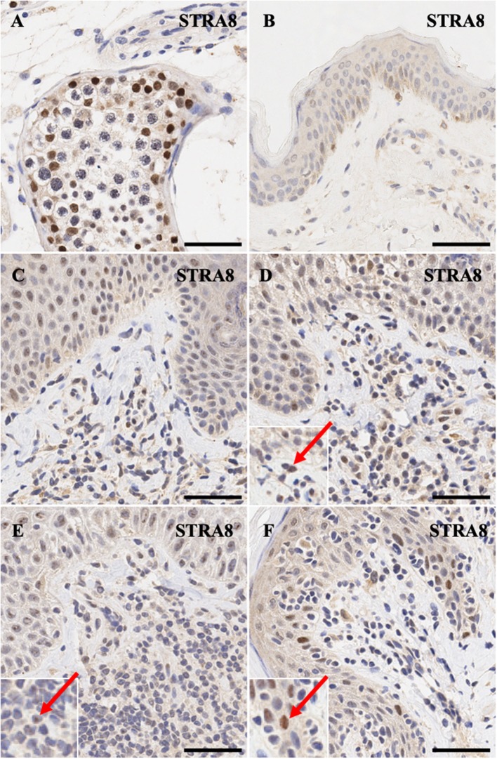
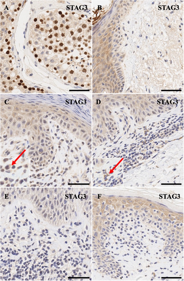
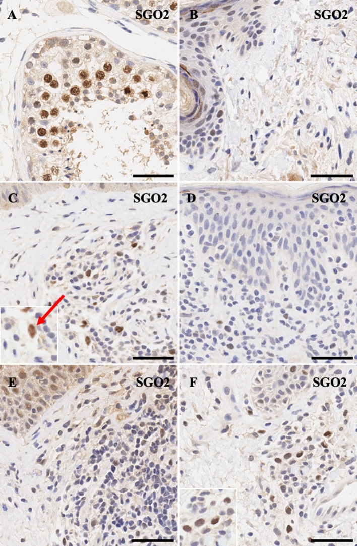
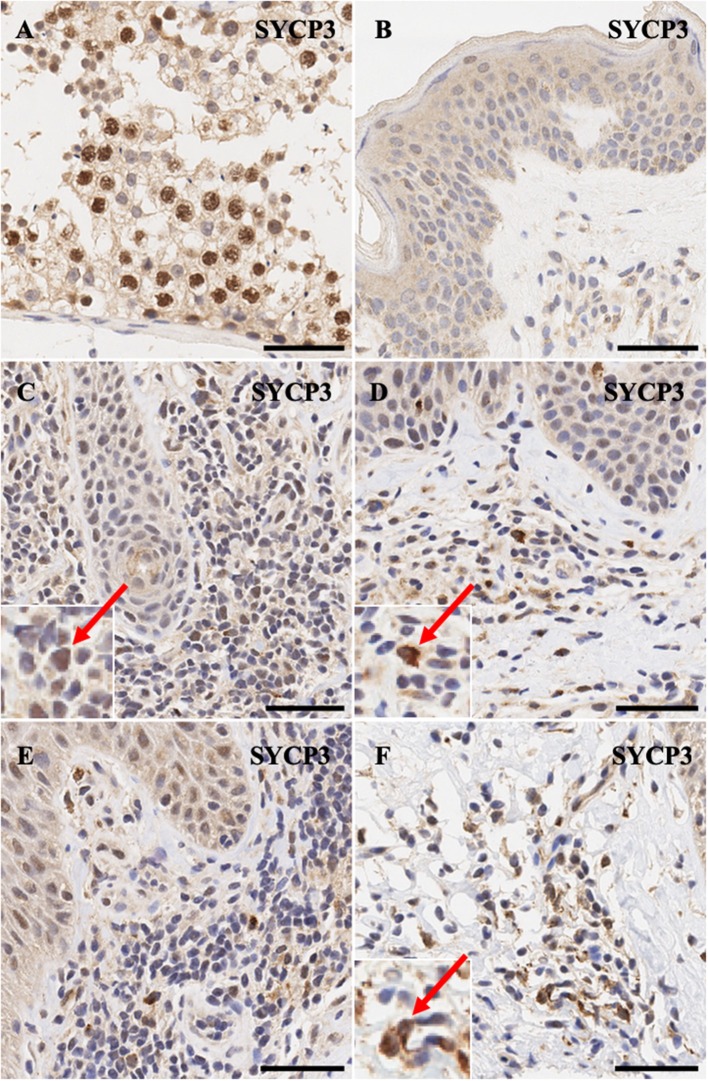
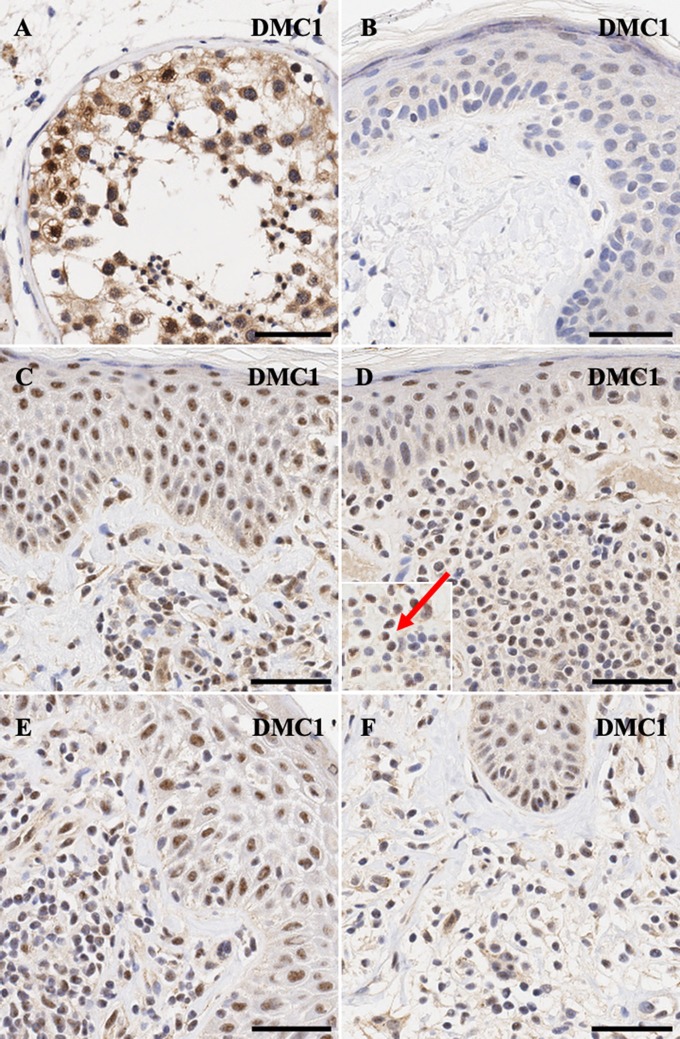
Cancer testis (CT) antigens, under normal circumstances are uniquely expressed in testicular germ cells. Recent research has shown that meiosis-specific CT (meiCT) antigens are ectopically expressed in cutaneous T-cell lymphoma (CTCL) and may contribute to increased genomic instability. The aberrant activation of meiosis genes in a mitotic cell is now recognized as a distinctive process, "meiomitosis." We have previously demonstrated the ectopic expression of several meiCT antigens in nine patient-derived CTCL cell lines and in expanded peripheral T lymphocytes isolated from Sézary Syndrome patients. In this study we analyzed the transcriptional expression of meiCT genes in Sézary Syndrome patients and healthy controls using publicly-available RNA sequencing (RNA-Seq) data. We corroborated our in silico analysis by examining the expression of 5 meiCT proteins in formalin-fixed, paraffin-embedded (FFPE) lesional samples from CTCL patients. Our results show significant differential gene expression of STAG3, SGO2, SYCP3, and DMC1 in a cohort of Sézary Syndrome patients when compared to healthy controls. Additionally, our study demonstrates a heterogenous expression of meiCT genes involved in initiation (STRA8), sister chromatin cohesion (STAG3, SGO2), homologous chromosome synapsis (SYCP3) and homologous recombination (DMC1) in atypical lymphocytes in FFPE samples. Our results further confirm the ectopic expression of meiCT genes in CTCL which indicates that CTCL malignant cells likely undergo the process of cancer meiomitosis, as opposed to a typical mitotic division. The ectopic expression of meiCT genes together with investigations into the functional mechanisms of cancer meiomitosis will help provide a foundation to develop novel diagnostic tests to distinguish CTCL from benign inflammatory dermatoses and may enable us to develop additional targeted therapies for patients with this malignancy.
Keywords: Sézary Syndrome (SS); cancer/testis antigens; cutaneous T-cell lymphomas (CTCL); germ cell proteins; meiCT; meiomitosis; mycosis fungoides (MF).
Figures
Similar articles
-
The ectopic expression of meiCT genes promotes meiomitosis and may facilitate carcinogenesis.Cell Cycle. 2020 Apr;19(8):837-854. doi: 10.1080/15384101.2020.1743902. Epub 2020 Mar 30. Cell Cycle. 2020. PMID: 32223693 Free PMC article. Review.
-
Ectopic expression of embryonic stem cell and other developmental genes in cutaneous T-cell lymphoma.Oncoimmunology. 2014 Dec 21;3(11):e970025. doi: 10.4161/21624011.2014.970025. eCollection 2014 Nov. Oncoimmunology. 2014. PMID: 25941598 Free PMC article.
-
A study of meiomitosis and novel pathways of genomic instability in cutaneous T-cell lymphomas (CTCL).Oncotarget. 2018 Dec 28;9(102):37647-37661. doi: 10.18632/oncotarget.26479. eCollection 2018 Dec 28. Oncotarget. 2018. PMID: 30701021 Free PMC article.
-
Ectopic expression of cancer-testis antigens in cutaneous T-cell lymphoma patients.Clin Cancer Res. 2014 Jul 15;20(14):3799-808. doi: 10.1158/1078-0432.CCR-14-0307. Epub 2014 May 21. Clin Cancer Res. 2014. PMID: 24850846 Free PMC article.
-
Primary cutaneous T-cell lymphoma (mycosis fungoides and Sézary syndrome): part I. Diagnosis: clinical and histopathologic features and new molecular and biologic markers.J Am Acad Dermatol. 2014 Feb;70(2):205.e1-16; quiz 221-2. doi: 10.1016/j.jaad.2013.07.049. J Am Acad Dermatol. 2014. PMID: 24438969 Review.
Cited by
-
Targeting PRAME directly or via EZH2 inhibition overcomes retinoid resistance and represents a novel therapy for keratinocyte carcinoma.Mol Oncol. 2025 May;19(5):1471-1492. doi: 10.1002/1878-0261.13820. Epub 2025 Mar 18. Mol Oncol. 2025. PMID: 40101298 Free PMC article.
-
Insights Into the Molecular and Cellular Underpinnings of Cutaneous T Cell Lymphoma.Yale J Biol Med. 2020 Mar 27;93(1):111-121. eCollection 2020 Mar. Yale J Biol Med. 2020. PMID: 32226341 Free PMC article. Review.
-
Patterns of Gene Expression in Cutaneous T-Cell Lymphoma: Systematic Review of Transcriptomic Studies in Mycosis Fungoides.Cells. 2021 Jun 6;10(6):1409. doi: 10.3390/cells10061409. Cells. 2021. PMID: 34204115 Free PMC article.
-
Comprehensive analysis of transcriptome data stemness indices identifies key genes for controlling cancer stem cell characteristics in gastric cancer.Transl Cancer Res. 2020 Oct;9(10):6050-6061. doi: 10.21037/tcr-20-704. Transl Cancer Res. 2020. PMID: 35117216 Free PMC article.
-
SGOL2 is a novel prognostic marker and fosters disease progression via a MAD2-mediated pathway in hepatocellular carcinoma.Biomark Res. 2022 Nov 15;10(1):82. doi: 10.1186/s40364-022-00422-z. Biomark Res. 2022. PMID: 36380369 Free PMC article.
References
-
- Olsen E, Vonderheid E, Pimpinelli N, Willemze R, Kim Y, Knobler R, et al. . Revisions to the staging and classification of mycosis fungoides and Sezary syndrome: a proposal of the International Society for Cutaneous Lymphomas (ISCL) and the cutaneous lymphoma task force of the European Organization of Research and Treatment of Cancer (EORTC). Blood. (2007) 110:1713–22. 10.1182/blood-2007-03-055749 - DOI - PubMed
LinkOut - more resources
Full Text Sources