Crucial Breakthrough of Functional Persistent Luminescence Materials for Biomedical and Information Technological Applications
- PMID: 31214570
- PMCID: PMC6554534
- DOI: 10.3389/fchem.2019.00387
Crucial Breakthrough of Functional Persistent Luminescence Materials for Biomedical and Information Technological Applications
Abstract
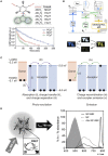
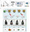
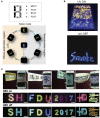
Persistent luminescence is a phenomenon in which luminescence is maintained for minutes to hours without an excitation source. Owing to their unique optical properties, various kinds of persistent luminescence materials (PLMs) have been developed and widely employed in numerous areas, such as bioimaging, phototherapy, data-storage, and security technologies. Due to the complete separation of two processes, -excitation and emission-, minimal tissue absorption, and negligible autofluorescence can be obtained during biomedical fluorescence imaging using PLMs. Rechargeable PLMs with super long afterglow life provide novel approaches for long-term phototherapy. Moreover, owing to the exclusion of external excitation and the optical rechargeable features, multicolor PLMs, which have higher decoding signal-to-noise ratios and high storage capability, exhibited an enormous application potential in information technology. Therefore, PLMs have significantly promoted the application of optics in the fields of multimodal bioimaging, theranostics, and information technology. In this review, we focus on the recently developed PLMs, including inorganic, organic and inorganic-organic hybrid PLMs to demonstrate their superior applications potential in biomedicine and information technology.
Keywords: anti-counterfeiting; bioimaging; biomedical applications; biosensing; information technological applications; optical data recording; persistent luminescence material; therapy.
Figures


Similar articles
-
Persistent luminescent nanophosphors for applications in cancer theranostics, biomedical, imaging and security.Mater Today Bio. 2023 Nov 19;23:100860. doi: 10.1016/j.mtbio.2023.100860. eCollection 2023 Dec. Mater Today Bio. 2023. PMID: 38179230 Free PMC article. Review.
-
Engineering Persistent Luminescence Nanoparticles for Biological Applications: From Biosensing/Bioimaging to Theranostics.Acc Chem Res. 2018 May 15;51(5):1131-1143. doi: 10.1021/acs.accounts.7b00619. Epub 2018 Apr 17. Acc Chem Res. 2018. PMID: 29664602
-
Recent progress in biomedical applications of persistent luminescence nanoparticles.Nanoscale. 2017 May 18;9(19):6204-6218. doi: 10.1039/c7nr01488k. Nanoscale. 2017. PMID: 28466913 Review.
-
Research progress on near-infrared long persistent phosphor materials in biomedical applications.Nanoscale Adv. 2022 Oct 24;4(23):4972-4996. doi: 10.1039/d2na00426g. eCollection 2022 Nov 22. Nanoscale Adv. 2022. PMID: 36504755 Free PMC article. Review.
-
Recent Progress of Near-Infrared Persistent Phosphors in Bio-related and Emerging Applications.Chem Asian J. 2021 May 3;16(9):1041-1048. doi: 10.1002/asia.202100108. Epub 2021 Mar 26. Chem Asian J. 2021. PMID: 33734602 Review.
Cited by
-
Organic persistent luminescence imaging for biomedical applications.Mater Today Bio. 2022 Nov 1;17:100481. doi: 10.1016/j.mtbio.2022.100481. eCollection 2022 Dec 15. Mater Today Bio. 2022. PMID: 36388456 Free PMC article.
-
Long Persistent Luminescent HDPE Composites with Strontium Aluminate and Their Phosphorescence, Thermal, Mechanical, and Rheological Characteristics.Materials (Basel). 2022 Feb 1;15(3):1142. doi: 10.3390/ma15031142. Materials (Basel). 2022. PMID: 35161084 Free PMC article.
-
Persistent luminescent nanophosphors for applications in cancer theranostics, biomedical, imaging and security.Mater Today Bio. 2023 Nov 19;23:100860. doi: 10.1016/j.mtbio.2023.100860. eCollection 2023 Dec. Mater Today Bio. 2023. PMID: 38179230 Free PMC article. Review.
-
Microwave-Assisted Preparation of Luminescent Inorganic Materials: A Fast Route to Light Conversion and Storage Phosphors.Molecules. 2021 May 13;26(10):2882. doi: 10.3390/molecules26102882. Molecules. 2021. PMID: 34068050 Free PMC article. Review.
-
Effect of Compatibilizer on the Persistent Luminescence of Polypropylene/Strontium Aluminate Composites.Polymers (Basel). 2022 Apr 22;14(9):1711. doi: 10.3390/polym14091711. Polymers (Basel). 2022. PMID: 35566879 Free PMC article.
References
-
- Al-Attar H. A., Monkman A. P. (2012). Room-temperature phosphorescence from films of isolated water-soluble conjugated polymers in hydrogen-bonded matrices. Adv. Funct. Mater. 22, 3824–3832. 10.1002/adfm.201200814 - DOI
-
- Chen Y., Liu F., Liang Y., Wang X., Bi J., Wang X., et al. (2018). New up-conversion charging concept for effectively charging persistent phosphors using low-energy visible-light laser diodes. J. Mater. Chem. C 6, 8003–8010. 10.1039/C8TC02419G - DOI
Publication types
LinkOut - more resources
Full Text Sources
Other Literature Sources

