. 2020 Mar 1;18(3):321-328.
doi: 10.1093/ons/opz143.

Columnar Injection for Intracerebral Cell Therapy

Affiliations

Columnar Injection for Intracerebral Cell Therapy

Jeffrey S Schweitzer et al. Oper Neurosurg. .

Abstract

Background: Surgical implantation of cellular grafts into the brain is of increasing importance, as stem cell-based therapies for Parkinson and other diseases continue to develop. The effect of grafting technique on development and survival of the graft has received less attention. Rate and method of graft delivery may impact the cell viability and success of these therapies. Understanding the final location of the graft with respect to the intended target location is also critical.

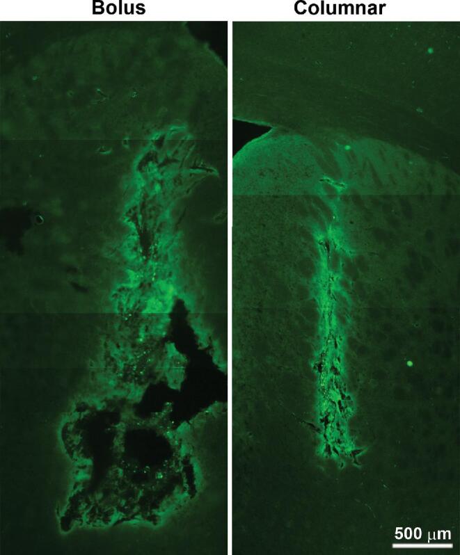
Objective: To describe a "columnar injection" technique designed to reduce damage to host tissue and result in a column of graft material with greater surface area to volume ratio than traditional injection techniques.

Methods: Using a clinically relevant model system of human embryonic stem cell-derived dopaminergic progenitors injected into athymic rat host brain, we describe a novel device that allows separate control of syringe barrel and plunger, permitting precise deposition of the contents into the cannula tract during withdrawal. Controls consist of contralateral injection using traditional techniques. Graft histology was examined at graft maturity.

Results: Bolus grafts were centered on the injection tract but were largely proximal to the "target" location. These grafts displayed a conspicuous peripheral distribution of cells, particularly of mature dopaminergic neurons. In contrast, column injections remained centered at the intended target, contained more evenly distributed cells, and had significantly more mature dopaminergic neurons.

Conclusion: We suggest that this columnar injection technique may allow better engraftment and development of intracerebral grafts, enhancing outcomes of cell therapy, compared to fixed-point injection techniques.

Keywords: Cell therapy; Dopaminergic neurons; Stereotaxy.


Figures

FIGURE 1.
FIGURE 1.
Columnar Injection device. A, Anterior view in unlocked position for cannula insertion. B, Anterior and posterior views in locked position for injection, showing the differential movement of the plunger and barrel. Lever arm has multiple detents allowing selection of several preset movement ratios between these two parts, thus determining volume injected per distance traveled.
FIGURE 2.
FIGURE 2.
Schematic of the bolus vs columnar injection principles. A, Bolus injection. Barrel and cannula are stationary. Plunger movement (black arrow) pushes out cell suspension (green) from syringe cannula tip at the target location. Most ejected material escapes proximally. B, Columnar injection. Differential between upward motion of these and of the plunger (black arrow) gives net downware pressure to express cell suspension (green) from cannula as it is withdrawn from the target. Green arrows indicate direction of movement of cell suspension. Black arrows indicate direction of movement of plunger. Arrow length is proportional to movement speed. Injection speed is the difference between barrel and plunger speeds. Initial stereotactic position is chosen so as to center the graft on the desired target.
FIGURE 3.
FIGURE 3.
Bolus vs columnar injection, in agarose. A1-5, A total of 25 μL of a solution of glycerol and Trypan Blue in saline was injected at 2 μL/min through a 22 g cannula with the tip at a fixed position (bolus injection). Images taken at successive time points show irregular extrusion of the material, most of it “refluxed” proximal to the cannula tip. B1-5, A total of 25 μL of an identical solution was injected via 22 g cannula using the column injection device with injection volume setting of 2 μL/mm and movement setting of 1mm/min at the starting depth of 12.5 mm. Arrows show the position of the cannula tip at different time points. Successive images show more even distribution of material as the cannula track is vacated.
FIGURE 4.
FIGURE 4.
Bolus vs columnar injection, short-term in Vivo results. Method as described in the text. Columnar injection is seen on the left, bolus on the right. Note the “rim” distribution of the bolus around a nonstaining central area, whereas the column appears more uniform. (scale bar indicates 500 μm). In standard bolus technique most of the graft lies proximal to the intended target. Columnar injection is initiated distal to the desired location of the center of the graft.
FIGURE 5.
FIGURE 5.
Bolus vs columnar injection, long-term (8 wk) in Vivo results. A and D, TH staining for bolus A and columnar D injections into the striatum, at low, medium (a’ and d’) and high (a” and d”) magnification. Boxes indicate areas enlarged in the frame below. Dense staining of dopaminergic neuropil originating in the host midbrain is seen, but TH+ cell bodies (a” and d”) are exclusively from the graft because no dopaminergic cell bodies are normally found in this region. B and E, hNuc staining showing overall distribution of grafted cells in bolus B and columnar E grafts. Note acellular areas in the bolus graft and more even distribution of cells in the columnar graft (b’ and e’). C and F, Double staining showing the distribution of TH+ and hNuc+ cells. In bolus grafts these cells were fewer in number and located mainly peripherally, whereas in the column, they are more evenly through the graft (c’, c”, f’, and f”). Arrows indicate location of individual TH+ cells. Scale bars in A-F represent 500 μm, and scale bars in a’-f’ and a”-f” represent 100 μm.
FIGURE 6.
FIGURE 6.
TH staining of bolus A and columnar B grafts at 8 wk, used for stereological cell counting. TH+ cell bodies in this location are exclusively from the graft. The stars indicate the intended final graft center using each technique. C, Histogram of cell counts of TH+ cell bodies in each graft, bolus vs column (n = 5). Total number of cells injected and injection volume was the same for each technique in each animal. Surviving TH+ cell numbers were significantly higher using the column technique (P < .003; 2-tailed ratio paired t-test).

References

    1. Backlund EO, Granberg PO, Hamberger Bet al.. Transplantation of adrenal medullary tissue to striatum in parkinsonism. First clinical trials. Proc Natl Acad Sci USA. 1985;62(2):1223-1232. - PubMed
    1. Lindvall O, Bjorklund A. Cell therapy in Parkinson disease. NeuroRx J Am Soc Experiment NeuroTher. 2004;1(4):382-393. - PMC - PubMed
    1. Bjorklund A, Lindvall O. Replacing dopamine neurons in parkinson disease: how did it happen? J Parkinson Dis. 2017;7(s1):S23-S33. - PMC - PubMed
    1. Stoker TB, Blair NF, Barker RA. Neural grafting for Parkinson disease: Challenges and prospects. Neural Regen Res. 2017;12(3):389-392. - PMC - PubMed
    1. Sonntag KC, Song B, Lee Net al.. Pluripotent stem cell-based therapy for Parkinson disease: Current status and future prospects. Prog Neurobiol. 2018;168(Sep):1-20. - PMC - PubMed

Publication types