

Towards clinical application of image mining: a systematic review on artificial intelligence and radiomics
- PMID: 31214791
- PMCID: PMC6879445
- DOI: 10.1007/s00259-019-04372-x
Towards clinical application of image mining: a systematic review on artificial intelligence and radiomics
Abstract
Purpose: The aim of this systematic review was to analyse literature on artificial intelligence (AI) and radiomics, including all medical imaging modalities, for oncological and non-oncological applications, in order to assess how far the image mining research stands from routine medical application. To do this, we applied a trial phases classification inspired from the drug development process.
Methods: Among the articles we considered for inclusion from PubMed were multimodality AI and radiomics investigations, with a validation analysis aimed at relevant clinical objectives. Quality assessment of selected papers was performed according to the QUADAS-2 criteria. We developed the phases classification criteria for image mining studies.
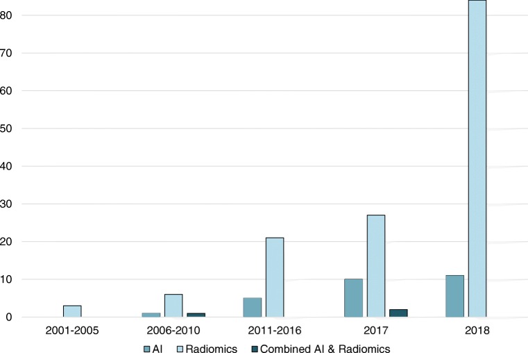
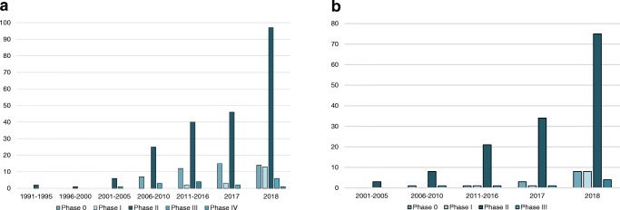
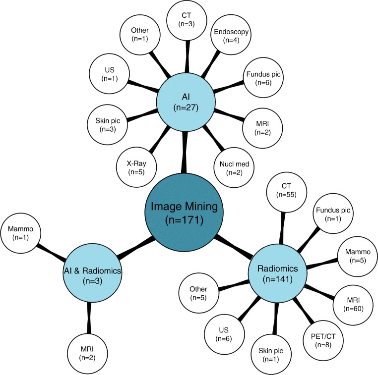
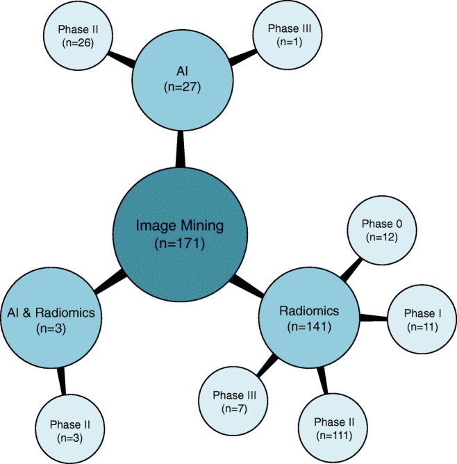
Results: Overall 34,626 articles were retrieved, 300 were selected applying the inclusion/exclusion criteria, and 171 high-quality papers (QUADAS-2 ≥ 7) were identified and analysed. In 27/171 (16%), 141/171 (82%), and 3/171 (2%) studies the development of an AI-based algorithm, radiomics model, and a combined radiomics/AI approach, respectively, was described. A total of 26/27(96%) and 1/27 (4%) AI studies were classified as phase II and III, respectively. Consequently, 13/141 (9%), 10/141 (7%), 111/141 (79%), and 7/141 (5%) radiomics studies were classified as phase 0, I, II, and III, respectively. All three radiomics/AI studies were categorised as phase II trials.
Conclusions: The results of the studies are promising but still not mature enough for image mining tools to be implemented in the clinical setting and be widely used. The transfer learning from the well-known drug development process, with some specific adaptations to the image mining discipline could represent the most effective way for radiomics and AI algorithms to become the standard of care tools.
Keywords: Artificial intelligence; Imaging; Radiomics; Systematic review; Texture analysis; Trial phases.
Conflict of interest statement
The authors declare no conflict of interest related to the present work.
Figures


Comment in
-
Reply to: "Lack of evidence and criteria to evaluate artificial intelligence and radiomics tools to be implemented in clinical settings".Eur J Nucl Med Mol Imaging. 2019 Dec;46(13):2814-2815. doi: 10.1007/s00259-019-04494-2. Eur J Nucl Med Mol Imaging. 2019. PMID: 31468183 No abstract available.
-
Lack of evidence and criteria to evaluate artificial intelligence and radiomics tools to be implemented in clinical settings.Eur J Nucl Med Mol Imaging. 2019 Dec;46(13):2812-2813. doi: 10.1007/s00259-019-04493-3. Epub 2019 Nov 6. Eur J Nucl Med Mol Imaging. 2019. PMID: 31696246 No abstract available.
References
-
- Bohannon J, Bohannon J. Fears of an AI pioneer. Science (80- ) 2015;349:252. - PubMed
-
- Lodwick GS, Keats TE, Dorst JP. The coding of roentgen images for computer analysis as applied to lung cancer. Radiology. 1963;81:185–200. - PubMed
-
- Haralick R, Shanmugam K, Dinstein I. Texture features for image classification. IEEE Trans Sys Man Cybern. 1973;SMC 3:610–621.
-
- Zorzela L, Loke Y, Ioannidis J, Golder S, Santaguida P, Altman D, et al. PRISMA harms checklist: improving harms reporting in systematic reviews. BMJ. 2016;352:i157. - PubMed
-
- Whiting PF, Rutjes AWS, Westwood ME, Mallett S, Deeks JJ, Reitsma JB, et al. QUADAS-2: strumento per valutare la qualità degli studi di accuratezza diagnostica. Evidence. 2016;8:e1000131.
Publication types
MeSH terms
LinkOut - more resources
Full Text Sources
