. 2019 Aug;110(8):2558-2572.
doi: 10.1111/cas.14103. Epub 2019 Jul 23.

Vasorin stimulates malignant progression and angiogenesis in glioma

Affiliations

Vasorin stimulates malignant progression and angiogenesis in glioma

Weiye Liang et al. Cancer Sci. 2019 Aug.

Abstract

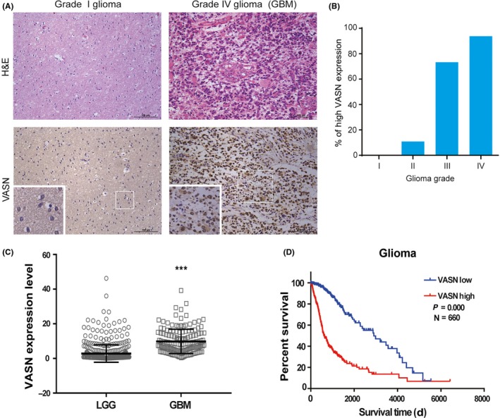
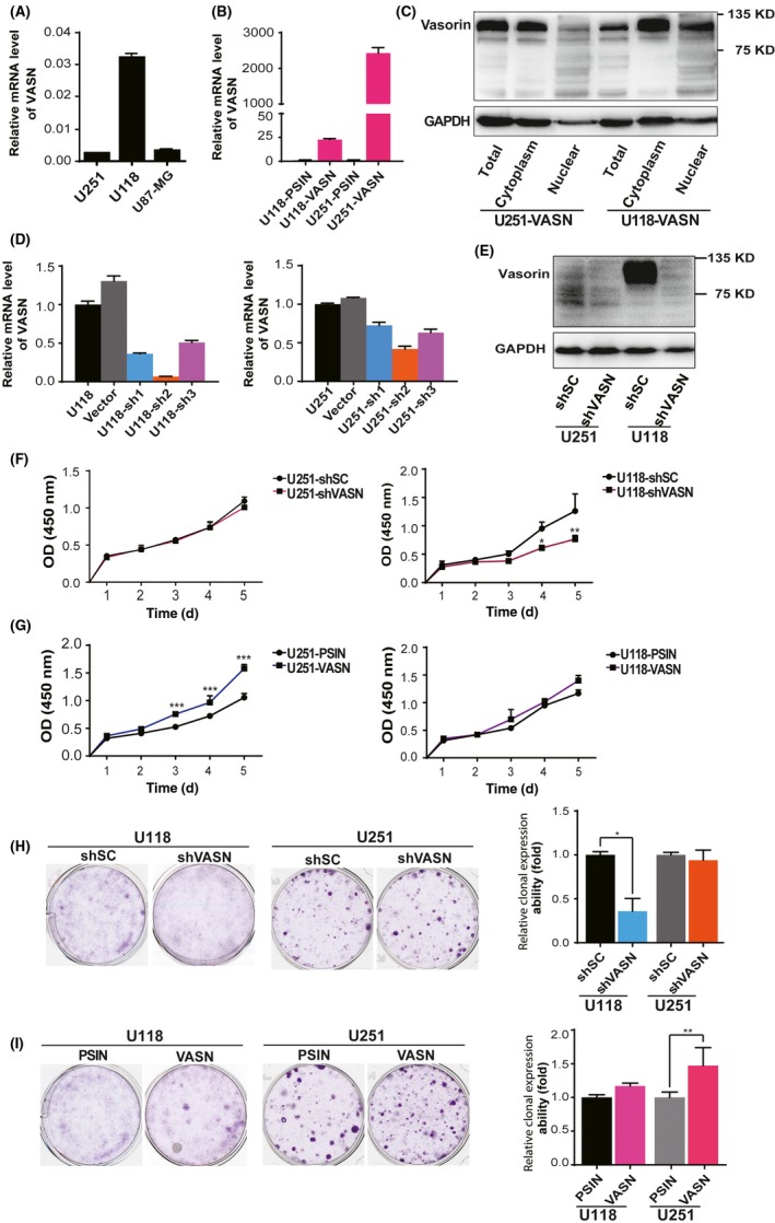
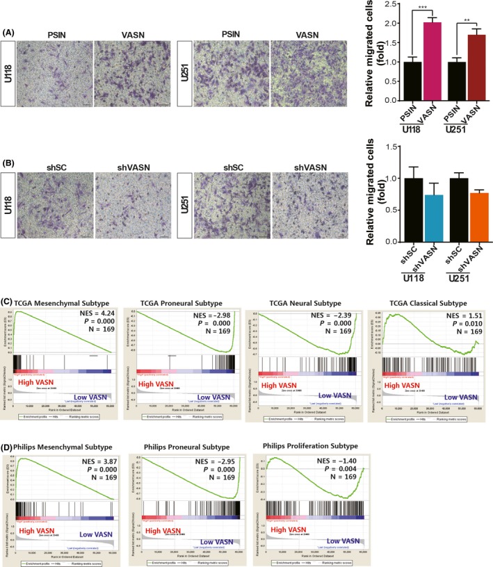
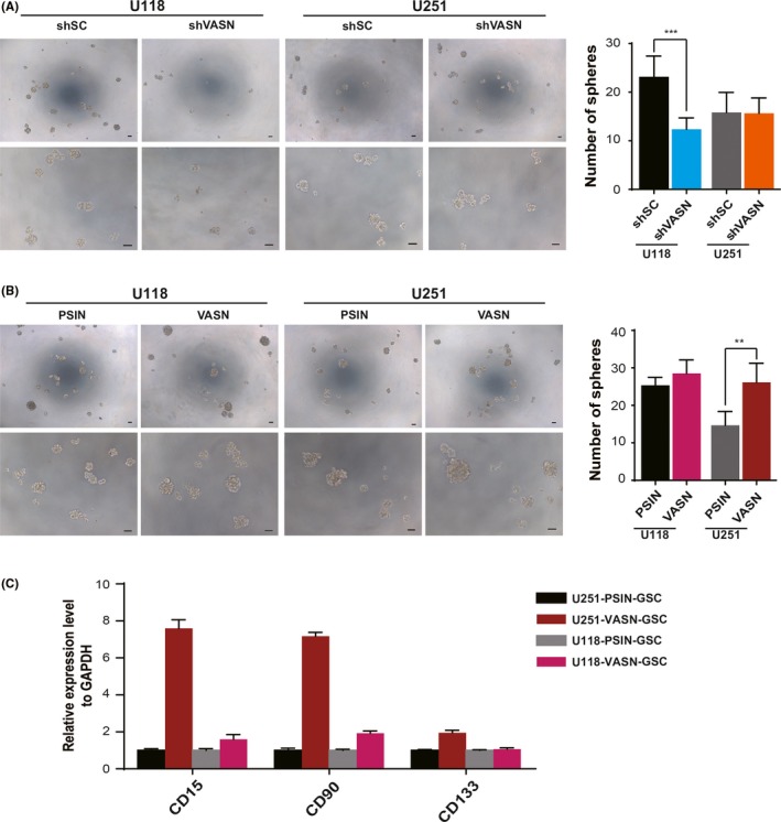
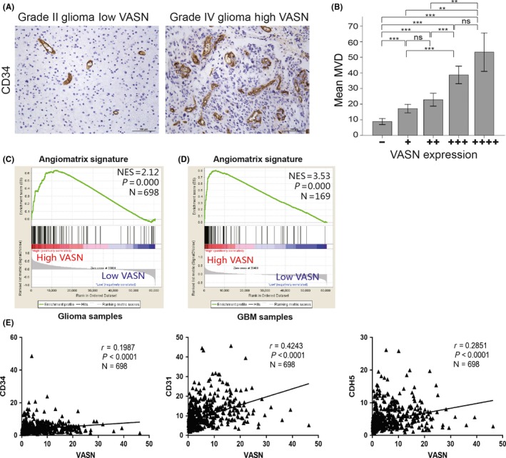
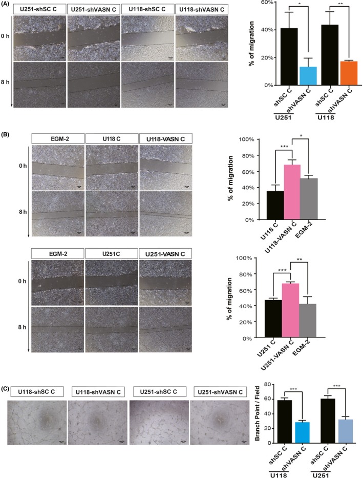
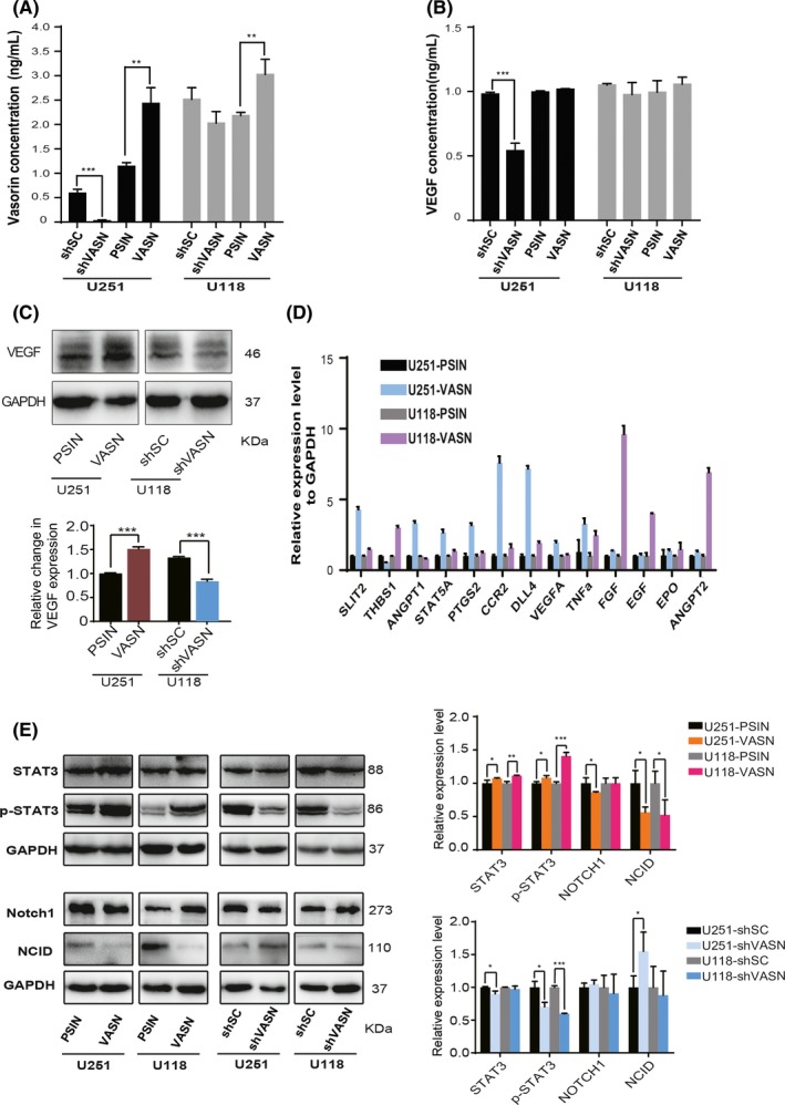
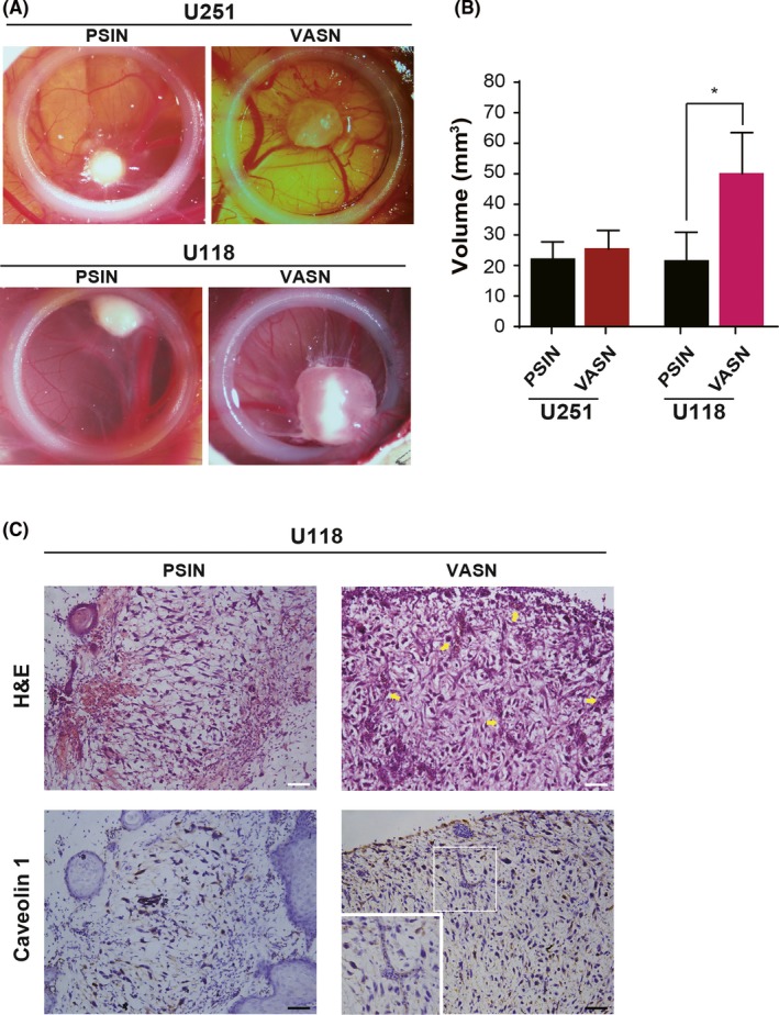
Glioma, the most common human primary brain tumor, is characterized by invasive capabilities and angiogenesis. Vasorin (VASN), a transmembrane protein, is reported to be associated with vascular injury repair and is overexpressed in some human tumors. However, its role in tumor progression and angiogenesis in glioma is unknown. In this study, VASN was shown to be overexpressed in high-grade gliomas, and the expression level correlated with tumor grade and microvessel density in glioma specimens. Glioma patients with high VASN expression had a shorter overall survival time. Knockdown of VASN in glioma cells by shRNA significantly inhibited the malignancy of glioma, including cell proliferation, colony formation, invasion, and sphere formation. Ectopic expression of VASN increased glioma progression in vitro. The expression of VASN correlated with the mesenchymal type of glioblastoma multiforme (GBM) subtyped by gene set enrichment analysis (GSEA). Our results showed that the concentration of VASN was increased in the conditioned medium (CM) from glioma cells with VASN overexpression, and the CM from glioma cells with knockdown or overexpressed VASN inhibited or promoted HUVEC migration and tubulogenesis in vitro, respectively. Glioma growth and angiogenesis were stimulated upon ectopic expression of VASN in vivo. The STAT3 and NOTCH pathways were found to be activated and inhibited by VASN overexpression. Our findings suggest that VASN stimulates tumor progression and angiogenesis in glioma, and, as such, represents a novel therapeutic target for glioma.

Keywords: tumor progression; STAT3; VASN; angiogenesis; glioma.


Conflict of interest statement

The authors have no conflict of interest to declare.

Figures

Figure 1
Figure 1
Vasorin (VASN) expression was elevated and correlates with poor prognosis in glioma. A, Representative H&E staining and immunohistochemical (IHC) staining images of VASN expression in glioma patients. B, The histogram illustrates the percentage of high VASN expression in gliomas across the 4 grades. C, VASN expression in low‐grade gliomas (LGG) and glioblastoma (GBM) from the TCGA diffuse glioma database. ***< 0.001. (D) Kaplan‐Meier survival analysis of glioma patients stratified by VASN expression. Median VASN expression was used for stratification into VASN‐high and VASN‐low tumors. Scale bar, 100 μm
Figure 2
Figure 2
Vasorin (VASN) expression promoted cell growth in glioma cells. A, Expression levels of VASN in glioma cell lines. B, The ectopic expression of the VASN gene was confirmed by quantitative real‐time PCR (qPCR). C, The ectopic expression and protein localization of the VASN was confirmed by western blot. The knockdown efficiency of VASN shRNA was confirmed by qPCR (D) and western blot (E). CCK‐8 assays were performed at different time points to detect the effect of knockdown (F) ectopic expression (G) of VASN on the growth of glioma cells. (H, I) Colony‐forming assays were performed on the same cell lines with either VASN knockdown or ectopic expression. The histograms illustrate the number of colonies counted. Data are presented as mean ± SD of 3 independent experiments. *P < 0.05, **P < 0.01. PSIN, negative control of ectopic expression of VASN; shSC, negative control of shRNA of VASN
Figure 3
Figure 3
Vasorin (VASN) promoted cell invasion and correlated with mesenchymal glioblastoma (GBM) gene signatures. Representative transwell invasion images and statistics for the number of counted glioma cells with VASN knockdown (A) and overexpression (B) that were able to migrate. GSEA was performed using the signatures for the TCGA GBM subtypes (C) or Phillips GBM subtypes (D). Patients were separated by high or low VASN mRNA expression. Scale bar, 50 μm
Figure 4
Figure 4
Vasorin (VASN) promoted tumor sphere formation in glioma cells. Representative images of sphere formation and statistics for the number of tumor spheres counted in glioma cells knockdown (A) and overexpression of VASN (B). C, The expression of cancer stem cell‐related genes (CD15, CD90 and CD133) was confirmed by quantitative real‐time PCR (qPCR). The data represent mean ± SD from 3 biological repeats. *P < 0.05, **P < 0.01, ***P < 0.001. Scale bar, 50 μm
Figure 5
Figure 5
Increased levels of vasorin (VASN) were associated with increased angiogenesis of glioma samples. A, CD34 staining for endothelial cells revealed increased tumor vascularization in high VASN expressing tissues; representative images are shown. B, The histogram illustrates microvessel density (MVD) among the 5 groups. Data represent the mean ± SD of 3 replicates. **P < 0.01, ***P < 0.001. Gene set enrichment analysis (GSEA) was performed using the AngioMatrix signature that is representative of glioblastoma (GBM) angiogenesis (C, D). Patients were separated by high or low VASN expression using TCGA glioma and GBM datasets. (E) VASN expression was correlated with markers of tumor vascularity using CD34, CD31 and CDH5. Scale bar, 100 μm
Figure 6
Figure 6
Vasorin (VASN)‐expressing tumor cells strongly promoted HUVEC migration and tube formation. Micrographs and histograms of the scratch wound‐healing assay of HUVEC grown in conditioned medium (CM) from glioma cell knockdown (A) and overexpression (B) of VASN. EGM‐2 served as a control. C, Micrographs of tube formation by HUVEC grown in CM from shVASN‐transfected glioma cells as indicated. Quantification of the number of branch points formed during the tube formation assay. Mean number of branching points per ×100 field ±SEM. *P < 0.05, **P < 0.01, ***P < 0.001. Scale bar, 100 μm
Figure 7
Figure 7
Vasorin (VASN) promoted glioma progression and angiogenesis by activating STAT3 and inhibiting NOTCH signaling. The concentrations of vasorin (A) and VEGF (B) in the conditioned medium (CM) from glioma cells with VASN knockdown or overexpression were detected by ELISA assay. C, Western blot analysis of VEGF in U118 cells knockdown and U251 cells overexpression of VASN. (D) qPCR analysis of angiogenic factors in glioma cells transfected with VASN. (E) Western blot analysis of STAT3, p‐STAT3, NOTCH1 and NICD in glioma cell knockdown or overexpression of VASN. *P < 0.05, **P < 0.01, ***P < 0.001
Figure 8
Figure 8
Transfection of vasorin (VASN) promoted tumor growth in vivo on chorioallantoic membranes (CAM). Glioma cells transfected with VASN were deposited on the CAM of fertilized eggs, and tumors were analyzed 4 days later. A, Tumor growth was assessed by biomicroscopy (×3 magnification). B, Tumor volume was quantified from the tumors obtained in CAM (8‐10 tumors/condition). *P < 0.05. C, H&E staining of the corresponding tumor sections is shown, and blood vessels are indicated by an arrow. Tumor sections were stained for caveolin 1 to mark endothelial cells. Numerous irregular and dilated blood vessels are visible in the tumor sections of gliomas overexpressing VASN. Scale bar, 50 μm

References

    1. Cavenee WK. High‐grade gliomas with chromosome 1p loss. J Neurosurg. 2000;92:1080‐1081. - PubMed
    1. Fine HA. How to get from here to there: tracking down invasive glioma cells. J Natl Cancer Inst. 2014;106:dju120. - PubMed
    1. Wong ML, Prawira A, Kaye AH, Hovens CM. Tumour angiogenesis: its mechanism and therapeutic implications in malignant gliomas. J Clin Neurosci. 2009;16:1119‐1130. - PubMed
    1. Boer JC, Walenkamp AM, den Dunnen WF. Recruitment of bone marrow derived cells during anti‐angiogenic therapy in GBM: the potential of combination strategies. Crit Rev Oncol Hematol. 2014;92:38‐48. - PubMed
    1. Carmeliet P, Jain RK. Principles and mechanisms of vessel normalization for cancer and other angiogenic diseases. Nat Rev Drug Discov. 2011;10:417‐427. - PubMed

MeSH terms