

A modified relief of unilateral ureteral obstruction model
- PMID: 31215300
- PMCID: PMC6586099
- DOI: 10.1080/0886022X.2019.1624263
A modified relief of unilateral ureteral obstruction model
Abstract
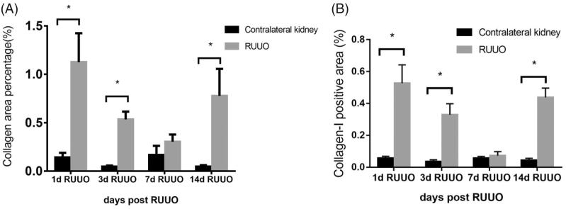
Objectives: To improve the mouse model of relief for unilateral ureteral obstruction (RUUO) and explore the pathological process of renal fibrosis after the obstruction was relieved. Methods: C57BL/6 mice in model group were randomly divided into RUUO group, improved RUUO group, and UUO group. After leaving Unilateral Ureteral Obstruction (UUO) for 3 days, the obstruction was released by reimplantation way in RUUO group and in reimplantation + catheter way in improved RUUO group. C57BL/6 mice in observation group were randomly divided into 1d RUUO group, 3d RUUO group, 7d RUUO group, and 14d RUUO group. Three days after UUO, the obstruction was released by reimplantation + catheter in four groups. We detected the renal volume, H&E, Masson staining, and immunohistochemistry of kidney pathology on the seventh day after RUUO in model group and on the 1st, 3rd, 7th, and 14th day after RUUO in observation group. Results: Comparing with mice in RUUO group, mice in improved RUUO group had lower renal volume, tubular damage score, and collagen area percentage. After the obstruction was relieved, the renal volume decreased gradually within 2 weeks. The tubular damage score in 7d RUUO group was lower than that in 1d RUUO and 3d RUUO group. However, the tubular damage score in 14d RUUO group was higher than that in 7d RUUO group. The tendency of collagen area percentage and α-SMA IOD value were consistent with the tubular damage score. Conclusions: Using the method of reimplantation + catheter, a reliable mice model of RUUO can be got. After RUUO, the de-obstructed kidneys are still in damage and fibrosis state.
Keywords: Relief of unilateral ureteral obstruction; catheter; pathology; renal fibrosis.
Figures


Similar articles
-
Regulatory T Cells Accelerate the Repair Process of Renal Fibrosis by Regulating Mononuclear Macrophages.Am J Med Sci. 2021 Jun;361(6):776-785. doi: 10.1016/j.amjms.2021.01.022. Epub 2021 Mar 2. Am J Med Sci. 2021. PMID: 33667434
-
Losartan accelerates the repair process of renal fibrosis in UUO mouse after the surgical recanalization by upregulating the expression of Tregs.Int Urol Nephrol. 2019 Nov;51(11):2073-2081. doi: 10.1007/s11255-019-02253-8. Epub 2019 Aug 10. Int Urol Nephrol. 2019. PMID: 31401712
-
Salt-sensitive hypertension after reversal of unilateral ureteral obstruction.Biochem Pharmacol. 2023 Apr;210:115438. doi: 10.1016/j.bcp.2023.115438. Epub 2023 Jan 27. Biochem Pharmacol. 2023. PMID: 36716827 Free PMC article.
-
The therapeutic approaches of renal recovery after relief of the unilateral ureteral obstruction: A comprehensive review.Iran J Basic Med Sci. 2020 Nov;23(11):1367-1373. doi: 10.22038/ijbms.2020.41984.9926. Iran J Basic Med Sci. 2020. PMID: 33235692 Free PMC article. Review.
-
Renal fibrosis progression following partial unilateral ureteral obstruction: mechanisms and therapeutic insights.World J Urol. 2025 Apr 17;43(1):229. doi: 10.1007/s00345-025-05580-x. World J Urol. 2025. PMID: 40244436 Free PMC article. Review.
Cited by
-
Renoprotective Effects of Luteolin: Therapeutic Potential for COVID-19-Associated Acute Kidney Injuries.Biomolecules. 2022 Oct 23;12(11):1544. doi: 10.3390/biom12111544. Biomolecules. 2022. PMID: 36358895 Free PMC article. Review.
-
Kidney-Draining Lymph Node Fibrosis Following Unilateral Ureteral Obstruction.Front Immunol. 2021 Dec 27;12:768412. doi: 10.3389/fimmu.2021.768412. eCollection 2021. Front Immunol. 2021. PMID: 35024041 Free PMC article.
-
Selective Detection of Nano-Sized Diagnostic Markers Using Au-ZnO Nanorod-Based Surface-Enhanced Raman Spectroscopy (SERS) in Ureteral Obstruction Models.Int J Nanomedicine. 2020 Oct 22;15:8121-8130. doi: 10.2147/IJN.S272500. eCollection 2020. Int J Nanomedicine. 2020. PMID: 33122904 Free PMC article.
-
Sulforaphane Restores Mitochondrial β-Oxidation and Reduces Renal Lipid Accumulation in a Model of Releasing Unilateral Ureteral Obstruction.Antioxidants (Basel). 2025 Feb 28;14(3):288. doi: 10.3390/antiox14030288. Antioxidants (Basel). 2025. PMID: 40227243 Free PMC article.
-
Improvement of the reversible unilateral ureteral obstruction model.Ren Fail. 2025 Dec;47(1):2454721. doi: 10.1080/0886022X.2025.2454721. Epub 2025 Mar 17. Ren Fail. 2025. PMID: 40097299 Free PMC article.
References
-
- Rabe M, Schaefer F. Non-transgenic mouse models of kidney disease. Nephron. 2016;133:53–61. - PubMed
-
- Ucero AC, Benito-Martin A, Izquierdo MC, et al. . Unilateral ureteral obstruction: beyond obstruction. Int Urol Nephrol. 2014;46:765–776. - PubMed
-
- Cochrane AL, Kett MM, Samuel CS, et al. . Renal structural and functional repair in a mouse model of reversal of ureteral obstruction. J Am Soc Nephrol JASN. 2005;16:3623–3630. - PubMed
-
- Chaabane W, Praddaude F, Buleon M, et al. . Renal functional decline and glomerulotubular injury are arrested but not restored by release of unilateral ureteral obstruction (UUO). Am J Physiol Renal Physiol. 2013;304:F432–F439. - PubMed
MeSH terms
LinkOut - more resources
Full Text Sources