Review
. 2019 Aug 1;122(2):563-571.
doi: 10.1152/jn.00200.2019. Epub 2019 Jun 19.

The rhythm of memory: how breathing shapes memory function

Affiliations
Review

The rhythm of memory: how breathing shapes memory function

Detlef H Heck et al. J Neurophysiol. .

Abstract

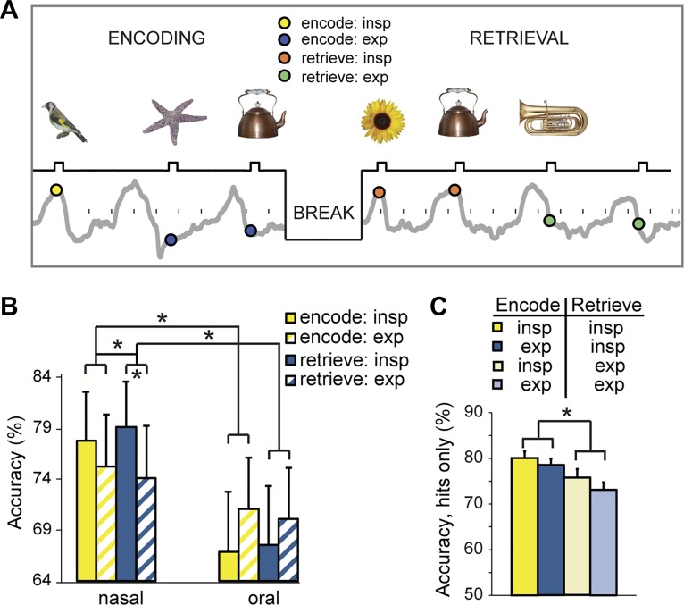
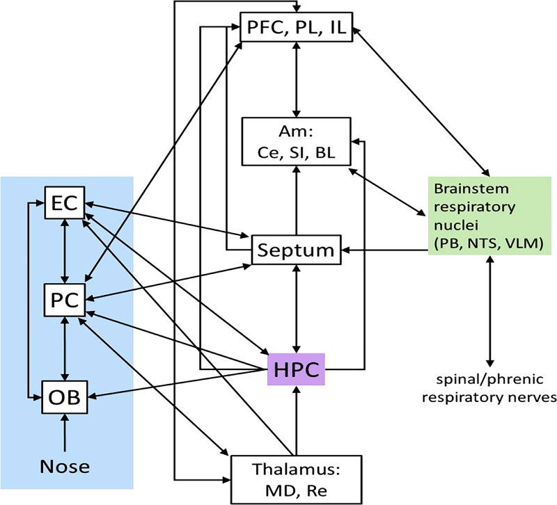
The mammalian olfactory bulb displays a prominent respiratory rhythm, which is linked to the sniff cycle and is driven by sensory input from olfactory receptors in the nasal sensory epithelium. In rats and mice, respiratory frequencies occupy the same band as the hippocampal θ-rhythm, which has been shown to be a key player in memory processes. Hippocampal and olfactory bulb rhythms were previously found to be uncorrelated except in specific odor-contingency learning circumstances. However, many recent electrophysiological studies in both rodents and humans reveal a surprising cycle-by-cycle influence of nasal respiration on neuronal activity throughout much of the cerebral cortex beyond the olfactory system, including the prefrontal cortex, hippocampus, and subcortical structures. In addition, respiratory phase has been shown to influence higher-frequency oscillations associated with cognitive functions, including attention and memory, such as the power of γ-rhythms and the timing of hippocampal sharp wave ripples. These new findings support respiration's role in cognitive function, which is supported by studies in human subjects, in which nasal respiration has been linked to memory processes. Here, we review recent reports from human and rodent experiments that link respiration to the modulation of memory function and the neurophysiological processes involved in memory in rodents and humans. We argue that respiratory influence on the neuronal activity of two key memory structures, the hippocampus and prefrontal cortex, provides a potential neuronal mechanism behind respiratory modulation of memory.

Keywords: cognition; cortical oscillations; memory; olfactory bulb; respiratory rhythm.


Conflict of interest statement

No conflicts of interest, financial or otherwise, are declared by the authors.

Figures

Fig. 1.
Fig. 1.
Respiratory phase modulates episodic memory performance. A: in a recognition memory task, subjects viewed a series of different visual objects that occurred at different times within the breathing cycle. Interstimulus interval: 3–6 s. After a 20-min break, subjects were presented with the pictures previously presented in the encoding session plus an equal number of new pictures. B: memory performance was more accurate during inspiration than during expiration, with effects more pronounced for nasal than oral breathing, both for encoding and retrieval. C: an analysis of all “hit” trials revealed that recognition memory was significantly enhanced for pictures that had appeared during the inspiratory (vs. expiratory) phase of retrieval, but it made no difference whether those same pictures had been encountered in the same phase during encoding. *P < 0.05 in all panels. [From Zelano et al. 2016.]
Fig. 2.
Fig. 2.
Hippocampal sharp-wave ripple (SWR) activity in relation to the respiratory cycle in mice. A: average local field potential (LFP) aligned on hippocampal SWRs (means ± SE). Data are aligned on the onset of ripple-activity (at time 0 s). B: time-frequency mapping of LFPs around CA1 ripples. Color represents normalized frequency power. C: polar plot reflecting the distribution of SWR events relative to respiratory phase. Red arrow represents the mean vector determined by circular statistics (Rayleigh test: n = 382; r = 0.14; z = 7.35; P = 0.02). 0° represents the end of expiration, and 180° corresponds to the end of inspiration. Concentric circles mark r values, as indicated in the lower half of the circle. [From Liu et al. 2017.]
Fig. 3.
Fig. 3.
Pathways by which respiratory drive may enter the cortical circuit. Left: olfactory system. Middle: regions connected with olfactory and respiratory systems, including the hippocampus. Right: respiratory brain stem connections (not detailed here). Not all connections are illustrated. Thalamic targets from the pyriform cortex are to the mediodorsal nucleus; thalamic efferents to the PC come from nucleus reuniens. Am, amygdala; BL, basolateral nucleus; Ce, central nucleus; EC, entorhinal cortex; HPC, hippocampus; IL, infralimbic; MD, mediodorsal nucleus; mPFC, medial prefrontal cortex; OB, olfactory bulb; PB, parabrachial nucleus; PC, pyriform cortex; PL, paralimbic; Re, nucleus reunions; SI, substantia innominata; NTS, nucleus tractus solitarius; VLM, ventrolateral medulla (Carlsen et al. 1982; Castle et al. 2005; Dobbins and Feldman 1994; Ellenberger and Feldman 1990; Ezure 2004; Gerrits and Holstege 1996; Hurley et al. 1991; Insausti et al. 2002; Künzle and Radtke-Schuller 2001; McKenna and Vertes 2004; Moga et al. 1990; Schwerdtfeger et al. 1990; Shipley and Adamek 1984; Van Groen et al. 1987; Van Groen and Wyss 1990; Vertes 2004; Wouterlood 1991).

Similar articles

Cited by

References

    1. Adhikari A, Topiwala MA, Gordon JA. Synchronized activity between the ventral hippocampus and the medial prefrontal cortex during anxiety. Neuron 65: 257–269, 2010. doi:10.1016/j.neuron.2009.12.002. - DOI - PMC - PubMed
    1. Adrian ED. The electrical activity of the mammalian olfactory bulb. Electroencephalogr Clin Neurophysiol 2: 377–388, 1950. doi:10.1016/0013-4694(50)90075-7. - DOI - PubMed
    1. Aristotle De Anima. ca. 350 BCE.
    1. Arshamian A, Iravani B, Majid A, Lundström JN. Respiration modulates olfactory memory consolidation in humans. J Neurosci 38: 10286–10294, 2018. doi:10.1523/JNEUROSCI.3360-17.2018. - DOI - PMC - PubMed
    1. Backus AR, Schoffelen JM, Szebényi S, Hanslmayr S, Doeller CF. Hippocampal-prefrontal theta oscillations support memory integration. Curr Biol 26: 450–457, 2016. doi:10.1016/j.cub.2015.12.048. - DOI - PubMed

Publication types