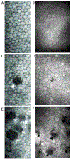
Imaging the Corneal Endothelium in Fuchs Corneal Endothelial Dystrophy
- PMID: 31215821
- PMCID: PMC6629500
- DOI: 10.1080/08820538.2019.1632355
Imaging the Corneal Endothelium in Fuchs Corneal Endothelial Dystrophy
Abstract
Fuchs endothelial corneal dystrophy (FECD) is characterized by the progressive degeneration of the corneal endothelium (CE). The purpose of this article is to review the diagnostic tools available to image and assess the CE in FECD. Slit-lamp biomicroscopy with specular reflection and retroillumination are important techniques to assess the CE. Objective diagnostic tests, such as retroillumination photographic analysis, specular microscopy, in vivo confocal microscopy (IVCM), and anterior segment optical coherence tomography, are valuable tools to evaluate the CE in FECD. Specular microscopy can be performed rapidly without touching the eye but requires a clear cornea with a smooth CE. In contrast, IVCM can image all layers of the cornea, even in advanced FECD. However, IVCM is contact-based and more technically challenging. It is important to select the appropriate objective diagnostic test to image and assess the CE in managing patients with FECD.
Keywords: Confocal; Fuchs; cornea; endothelium; specular.
Conflict of interest statement
Declaration of interest: None
Figures


Similar articles
-
Corneal endothelium features in Fuchs' Endothelial Corneal Dystrophy: A preliminary 3D anterior segment optical coherence tomography study.PLoS One. 2018 Nov 29;13(11):e0207891. doi: 10.1371/journal.pone.0207891. eCollection 2018. PLoS One. 2018. PMID: 30496218 Free PMC article.
-
[Imaging features of Fuchs endothelial corneal dystrophy].Zhonghua Yan Ke Za Zhi. 2020 Dec 11;56(12):938-943. doi: 10.3760/cma.j.cn112142-20200418-00228. Zhonghua Yan Ke Za Zhi. 2020. PMID: 33342121 Chinese.
-
In Vivo Confocal Microscopy Shows Alterations in Nerve Density and Dendritiform Cell Density in Fuchs' Endothelial Corneal Dystrophy.Am J Ophthalmol. 2018 Dec;196:136-144. doi: 10.1016/j.ajo.2018.08.040. Epub 2018 Sep 6. Am J Ophthalmol. 2018. PMID: 30194928 Free PMC article.
-
Dual corneal involvement by endothelial and epithelial corneal dystrophies in Steinert's disease: A case of triple dystrophy.Eur J Ophthalmol. 2021 Mar;31(2):NP23-NP26. doi: 10.1177/1120672119872374. Epub 2019 Sep 2. Eur J Ophthalmol. 2021. PMID: 31476892 Review.
-
The pathophysiology of Fuchs' endothelial dystrophy--a review of molecular and cellular insights.Exp Eye Res. 2015 Jan;130:97-105. doi: 10.1016/j.exer.2014.10.023. Epub 2014 Nov 1. Exp Eye Res. 2015. PMID: 25446318 Review.
Cited by
-
Imaging pathology in archived cornea with Fuchs' endothelial corneal dystrophy including tissue reprocessing for volume electron microscopy.Sci Rep. 2024 Dec 30;14(1):31786. doi: 10.1038/s41598-024-82888-5. Sci Rep. 2024. PMID: 39738318 Free PMC article.
-
Low-Cost, Smartphone-Based Specular Imaging and Automated Analysis of the Corneal Endothelium.Transl Vis Sci Technol. 2021 Apr 1;10(4):4. doi: 10.1167/tvst.10.4.4. Transl Vis Sci Technol. 2021. PMID: 34003981 Free PMC article.
-
Clinical Applications of Artificial Intelligence in Corneal Diseases.Vision (Basel). 2025 Aug 18;9(3):71. doi: 10.3390/vision9030071. Vision (Basel). 2025. PMID: 40843795 Free PMC article. Review.
-
Normal Corneal Thickness and Endothelial Cell Density in Rhesus Macaques (Macaca mulatta).Transl Vis Sci Technol. 2022 Sep 1;11(9):23. doi: 10.1167/tvst.11.9.23. Transl Vis Sci Technol. 2022. PMID: 36156731 Free PMC article.
-
Loss of Corneal Nerves and Corneal Haze in Patients with Fuchs' Endothelial Corneal Dystrophy with the Transcription Factor 4 Gene Trinucleotide Repeat Expansion.Ophthalmol Sci. 2022 Aug 27;3(1):100214. doi: 10.1016/j.xops.2022.100214. eCollection 2023 Mar. Ophthalmol Sci. 2022. PMID: 36275201 Free PMC article.
References
Publication types
MeSH terms
Grants and funding
LinkOut - more resources
Full Text Sources
