

Aptamer-based fluorometric determination of Salmonella Typhimurium using Fe3O4 magnetic separation and CdTe quantum dots
- PMID: 31216306
- PMCID: PMC6584018
- DOI: 10.1371/journal.pone.0218325
Aptamer-based fluorometric determination of Salmonella Typhimurium using Fe3O4 magnetic separation and CdTe quantum dots
Abstract
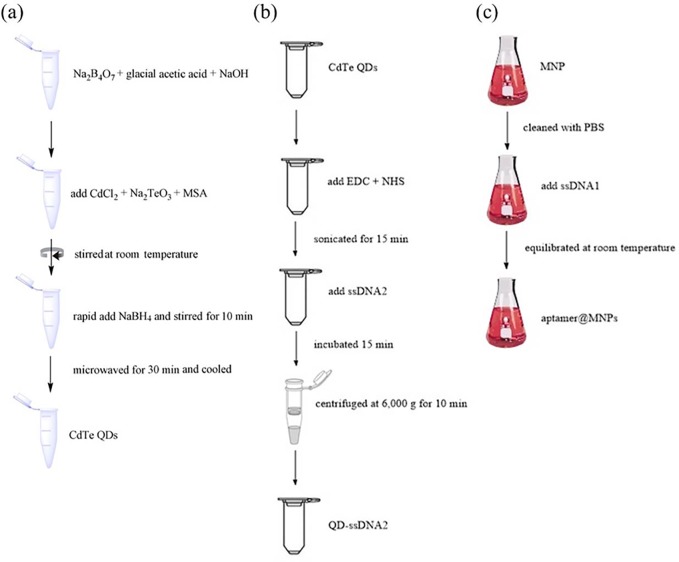
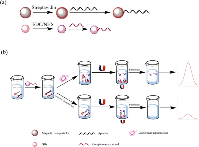
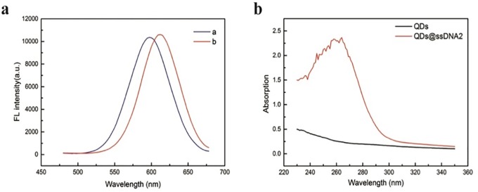
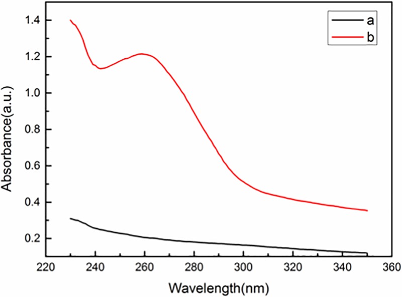
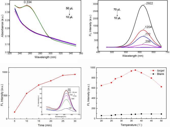
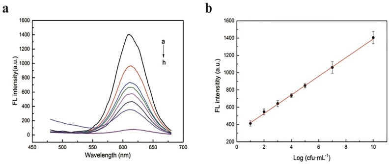
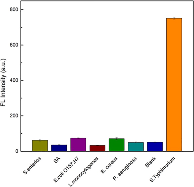
Based on the high sensitivity and stable fluorescence of CdTe quantum dots (QDs) in conjunction with a specific DNA aptamer, the authors describe an aptamer-based fluorescence assay for the determination of Salmonella Typhimurium. The fluorescence detection and quantification of S. Typhimurium is based on a magnetic separation system, a combination of aptamer-coated Fe3O4 magnetic particles (Apt-MNPs) and QD-labeled ssDNA2 (complementary strand of the aptamer). Apt-MNPs are employed for the specific capture of S. Typhimurium. CdTe QD-labeled ssDNA2 was used as a signaling probe. Simply, the as-prepared CdTe QD-labeled ssDNA2 was first incubated with the Apt-MNPs to form the aptamer-ssDNA2 duplex. After the addition of S. Typhimurium, they could specifically bind the DNA aptamer, leading to cleavage of the aptamer-ssDNA2 duplex, accompanied by the release of CdTe QD-labeled DNA. Thus, an increased fluorescence signal can be achieved after magnetic removal of the Apt-MNPs. The fluorescence of CdTe QDs (λexc/em = 327/612 nm) increases linearly in the concentration range of 10 to 1010 cfu•mL-1, and the limit of detection is determined to be 1 cfu•mL-1. The detection process can be performed within 2 h and is successfully applied to the analysis of spiked food samples with good recoveries from 90% to 105%.
Conflict of interest statement
The authors have declared that no competing interests exist.
Figures
Similar articles
-
Nucleic Acid-Based Nanobiosensor (NAB) Used for Salmonella Detection in Foods: A Systematic Review.Nanomaterials (Basel). 2022 Feb 28;12(5):821. doi: 10.3390/nano12050821. Nanomaterials (Basel). 2022. PMID: 35269310 Free PMC article. Review.
-
CdTe/CdSe quantum dot-based fluorescent aptasensor with hemin/G-quadruplex DNzyme for sensitive detection of lysozyme using rolling circle amplification and strand hybridization.Biosens Bioelectron. 2017 Jan 15;87:18-24. doi: 10.1016/j.bios.2016.08.003. Epub 2016 Aug 3. Biosens Bioelectron. 2017. PMID: 27504793
-
A semiconductor quantum dot-based ratiometric electrochemical aptasensor for the selective and reliable determination of aflatoxin B1.Analyst. 2019 Aug 5;144(16):4772-4780. doi: 10.1039/c9an00825j. Analyst. 2019. PMID: 31268094
-
Quantum dot-DNA aptamer conjugates coupled with capillary electrophoresis: A universal strategy for ratiometric detection of organophosphorus pesticides.Talanta. 2016;146:55-61. doi: 10.1016/j.talanta.2015.08.023. Epub 2015 Aug 14. Talanta. 2016. PMID: 26695234
-
Nanomaterials-based aptasensors: An efficient detection tool for heavy-metal and metalloid ions in environmental and biological samples.Environ Res. 2023 Dec 1;238(Pt 1):117170. doi: 10.1016/j.envres.2023.117170. Epub 2023 Sep 16. Environ Res. 2023. PMID: 37722582 Review.
Cited by
-
Enlarging the Toolbox Against Antimicrobial Resistance: Aptamers and CRISPR-Cas.Front Microbiol. 2021 Feb 19;12:606360. doi: 10.3389/fmicb.2021.606360. eCollection 2021. Front Microbiol. 2021. PMID: 33679633 Free PMC article. Review.
-
Nucleic Acid-Based Nanobiosensor (NAB) Used for Salmonella Detection in Foods: A Systematic Review.Nanomaterials (Basel). 2022 Feb 28;12(5):821. doi: 10.3390/nano12050821. Nanomaterials (Basel). 2022. PMID: 35269310 Free PMC article. Review.
-
Biosensors, modern technology for the detection of cancer-associated bacteria.Biotechnol Lett. 2022 Jun;44(5-6):683-701. doi: 10.1007/s10529-022-03257-8. Epub 2022 May 11. Biotechnol Lett. 2022. PMID: 35543825 Review.
-
Aptamers: precision tools for diagnosing and treating infectious diseases.Front Cell Infect Microbiol. 2024 Sep 25;14:1402932. doi: 10.3389/fcimb.2024.1402932. eCollection 2024. Front Cell Infect Microbiol. 2024. PMID: 39386170 Free PMC article. Review.
-
Oligonucleotide aptamers for pathogen detection and infectious disease control.Theranostics. 2021 Aug 27;11(18):9133-9161. doi: 10.7150/thno.61804. eCollection 2021. Theranostics. 2021. PMID: 34522231 Free PMC article. Review.
References
-
- Verma SK, Jha E, Sahoo B, Panda PK, Thirumurugan A, Parashar SKS, et al. Mechanistic insight into the rapid one-step facile biofabrication of antibacterial silver nanoparticles from bacterial release and their biogenicity and concentration-dependent in vitro cytotoxicity to colon cells. RSC Advances. 2017; 7(64): 40034–40045. 10.1039/C7RA05943D - DOI
-
- Brandt R, Petersen A, Brix S, Licht TR, Frøkiær H. Epithelial entry rather than the ensuing systemic immune response determines the pathogenicity of two Salmonella enterica serovar Typhimurium strains in a mouse model. Microbes and Infection. 2013; 15 (13):911–919. 10.1016/j.micinf.2013.08.004 - DOI - PubMed
Publication types
MeSH terms
Substances
LinkOut - more resources
Full Text Sources
Other Literature Sources
Medical
Molecular Biology Databases
