Review
. 2020 Jan;45(1):6-20.
doi: 10.1038/s41386-019-0444-2. Epub 2019 Jun 19.

Arousal and sleep circuits

Affiliations
Review

Arousal and sleep circuits

Barbara E Jones. Neuropsychopharmacology. 2020 Jan.

Abstract

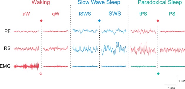
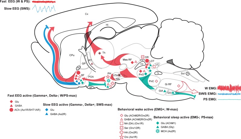
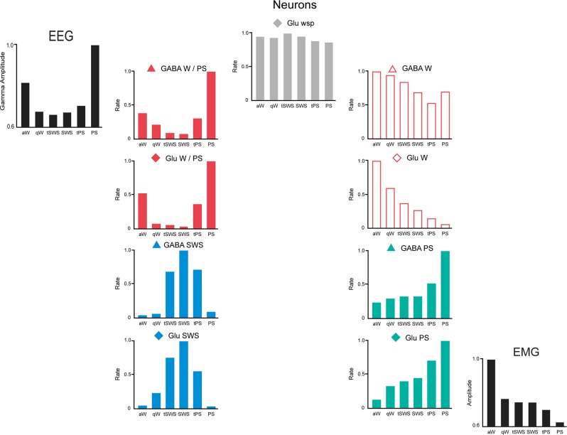
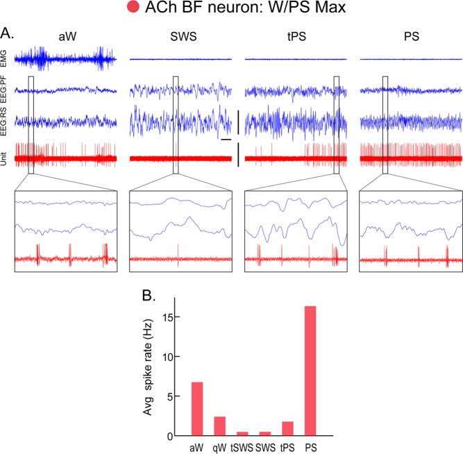
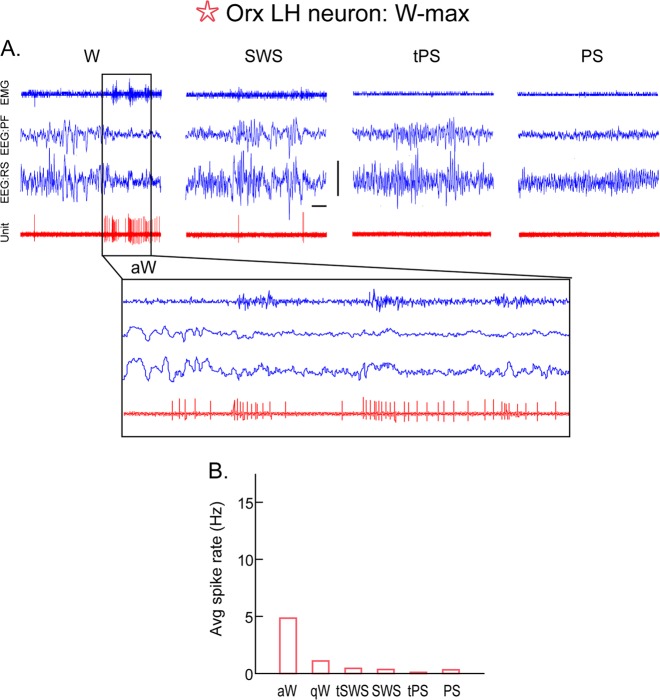
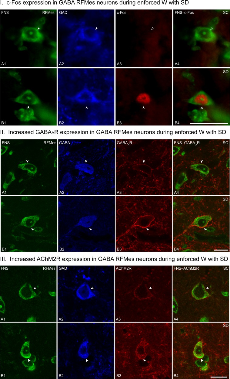
The principal neurons of the arousal and sleep circuits are comprised by glutamate and GABA neurons, which are distributed within the reticular core of the brain and, through local and distant projections and interactions, regulate cortical activity and behavior across wake-sleep states. These are in turn modulated by the neuromodulatory systems that are comprised by acetylcholine, noradrenaline, dopamine, serotonin, histamine, orexin (hypocretin), and melanin-concentrating hormone (MCH) neurons. Glutamate and GABA neurons are heterogeneous in their profiles of discharge, forming distinct functional cell types by selective or maximal discharge during (1) waking and paradoxical (REM) sleep, (2) during slow wave sleep, (3) during waking, or (4) during paradoxical (REM) sleep. The neuromodulatory systems are each homogeneous in their profile of discharge, the majority discharging maximally during waking and paradoxical sleep or during waking. Only MCH neurons discharge maximally during sleep. They each exert their modulatory influence upon other neurons through excitatory and inhibitory receptors thus effecting a concerted differential change in the functionally different cell groups. Both arousal and sleep circuit neurons are homeostatically regulated as a function of their activity in part through changes in receptors. The major pharmacological agents used for the treatment of wake and sleep disorders act upon GABA and neuromodulatory transmission.


Conflict of interest statement

The author declares no competing interests.

Figures

Fig. 1
Fig. 1
Sleep-wake states in the rat shown by EEG and EMG activity across the three major states, Waking (W), Slow Wave Sleep (SWS), and Paradoxical Sleep (PS) and their sub states (active, a, and quiet, q, W) or transitional (t) stages. Colors represent different activities of the EEG and EMG in the different states and the symbols the different neurons which discharge maximally during those states or activities, as represented in Figs. 2 and 3. PF, prefrontal cortex; RS, retrosplenial cortex
Fig. 2
Fig. 2
Sleep-wake state neural systems. Sagittal schematic view of the rat brain depicting neurons with their chemical neurotransmitters, pathways, and discharge profiles by which they influence cortical activity or behavior across the sleep/wake cycle. Waking (W) is characterized by fast (gamma, >30 Hz) activity on the cortical EEG (upper left) and high postural muscle tone on the neck EMG (lower right); slow wave sleep (SWS) by slow EEG (delta, <4 Hz) and low tone on the EMG; and paradoxical sleep (PS) by fast EEG and atonia on the EMG (Fig. 1). Neurons that are active during W (red symbols) include cells with ascending projections toward the cortex, which stimulate fast cortical activity (filled symbols), and cells with descending projections toward the spinal cord, which stimulate motor activity with postural muscle tone typical of behavioral W (open symbols). Those with predominantly ascending projections discharge in association with fast EEG activity (gamma+) and decrease or cease firing with delta activity (delta−) to be maximally active during W and PS (W/PS-max active, filled red symbols). (Those with similar average rates of discharge but different modes of discharge across states are red filled with gray.) They include glutamate (Glu), GABA, and acetylcholine (ACh) neurons. Those with more diffuse or descending projections discharge in association with behavioral arousal and EMG activity (EMG+) and decrease or cease firing with muscle atonia to be active during W and quiet during PS (EMG+, W-max active, open red symbols); they include Glu, GABA, noradrenaline (NA), serotonin (Ser), histamine (HA), and orexin (Orx or hypocretin) neurons. Neurons that are active during sleep include cells with ascending projections toward the cortex, which discharge maximally with slow wave activity during SWS (gamma−, delta+, SWS-max active, blue symbols) and those with descending projections toward the hypothalamus, brainstem, or spinal cord, which discharge maximally with muscle atonia during PS (EMG-, PS-max active, aqua symbols). They include GABA (with some glycine, Gly), Glu, and MCH neurons. Among these functionally distinguished cell types, certain have been shown to bear specific receptors to the neuromodulatory chemicals, including ACh inhibitory muscarinic type 2 receptors (AChM2R) on behavioral wake-active neurons, adrenergic excitatory (Aα1R) receptors on behavioral wake-active neurons, adrenergic inhibitory (Aα2R) receptors on slow EEG-active and behavioral sleep-active neurons, serotonergic (5-hydroxytryptamine) inhibitory (5HT1AR) receptors on fast EEG-active neurons and Orx excitatory (Orx1/2R) receptors on behavioral wake-active neurons. 7 g, genu 7th nerve; ac, anterior commissure; ACh, acetylcholine; BF, basal forebrain; CPu, caudate putamen; Cx, cortex; DA, dopamine; DR, dorsal raphe; EEG, electroencephalogram; EMG, electromyogram; GiA, gigantocellular, alpha part RF; Gi RF, gigantocellular RF; GiV, gigantocellular, ventral part RF; Glu, glutamate; Gly, glycine; GP, globus pallidus; HA, histamine; Hi, hippocampus; ic, internal capsule; LC, locus coeruleus nucleus; LDT, laterodorsal tegmental nucleus; PH, posterior hypothalamus; MCH, melanin-concentrating hormone; Mes RF, mesencephalic RF; NA, noradrenaline; opt, optic tract; Orx, orexin; PnC, pontine, caudal part RF; PnO, pontine, oral part RF; POA, preoptic area; PPT, pedunculopontine tegmental nucleus; PS, paradoxical sleep; RF, reticular formation; RT, reticular thalamic nucleus; s, solitary tract; scp, superior cerebellar peduncle; Ser, serotonin; SN, substantia nigra; Sol, solitary tract nucleus; SWS, slow wave sleep; Th, thalamus; VTA, ventral tegmental area; W, wake. (Modified with permission from Jones [199])
Fig. 3
Fig. 3
Discharge rates of principal cell types across sleep-wake stages. Normalized average rates of firing shown with normalized average gamma (30–60 Hz) EEG activity and EMG activity across sleep wake stages of active or attentive wake (aW), quiet wake (qW), transition to SWS (tSWS), slow wave sleep (SWS), transition to PS (tPS), and paradoxical sleep (PS). Rates were taken from one exemplary neuron for each cell type from neurons recorded in the basal forebrain. Cells were recorded and filled with Neurobiotin using the juxtacellular technique for subsequent immunohistochemical identification of their neurotransmitter as GABA or (putative or identified) glutamate (Glu). They were classified into one of four principal cell types, (W/PS-max, SWS-max, W-max, and PS-max active) or state (wsp, Wake-SWS-PS)-indifferent, by statistical analysis of their rates in aW, SWS, and PS. (Modified with permission from Hassani et al. [36])
Fig. 4
Fig. 4
Discharge of an acetylcholine (ACh) basal forebrain (BF) neuron across sleep-wake states. A Record of a neuron labeled by juxtacellular technique with Neurobiotin (Nb) and identified by immunohistochemistry for choline acetyltransferase (ChAT) as cholinergic in the magnocellular preoptic nucleus (MCPO) of the rat. As evident in 10 s traces (above), the unit fired during aW, virtually ceased firing during SWS, resumed firing during tPS, and discharged maximally during PS. As evident in expanded 0.5 s traces (below), the unit discharged in rhythmic bursts of spikes with theta EEG activity that was present intermittently during periods of aW, toward the end of tPS and continuously during PS. B Average discharge rate (Hz) across the sleep-wake states and transitions (t) of the same cell. Avg, average; aW, active wake; EEG, electroencephalogram; EMG, electromyogram; PF, prefrontal cortex; PS, paradoxical sleep; qW, quiet wake; RS, retrosplenial cortex; SWS, slow wave sleep; tPS, transition to PS; tSWS transition to SWS. Bar for horizontal scale: 1 s. Bar for vertical scales: 1 mV for EEG/EMG and 1.5 mV for Unit. (Reprinted with permission from Lee et al. [69])
Fig. 5
Fig. 5
Discharge of an orexin (Orx) lateral hypothalamus (LH) neuron across sleep-wake states. A Record of a neuron labeled by juxtacellular technique with Neurobiotin (Nb) and identified by immunohistochemistry for Orx in the rat. As evident in 10 s traces (above), the unit fired during W and was virtually silent during SWS, tPS, and PS. As evident in an expanded trace (of ~4 s, below), the unit discharged during active W (aW) and increased firing phasically in association with increases in muscle tone seen on the EMG. B Average discharge rate (Hz) across the sleep-wake states and transitions (t) of the same cell. Avg, average; aW, active wake; EEG, electroencephalogram; EMG, electromyogram; PF, prefrontal cortex; PS, paradoxical sleep; qW, quiet wake; RS, retrosplenial cortex; SWS, slow wave sleep; tPS, transition to PS; tSWS transition to SWS. Horizontal scale bar: 1 s. Vertical scale bar: 1 mV for EEG, 0.5 mV for EMG, and 2 mV for unit. (Reprinted with permission from Lee et al. [146])
Fig. 6
Fig. 6
GABA neurons in the mesencephalic reticular formation (RFMes) under conditions of sleep control (SC) and sleep deprivation (SD). I c-Fos in RFMes GABAergic neurons across groups. Fluorescent microscopic images show staining for Nissl with fluorescent Nissl stain (FNS, green, A1, B1), immunostaining for glutamic acid decarboxylase (GAD, blue, A2, B2, with positive staining indicated by filled arrowheads), and immunostaining for c-Fos (red, A3, B3, with positive staining indicated by filled arrowhead) along with dual staining for Nissl and c-Fos in merged images (green and red, A4, B4, with positive c-Fos staining indicated by filled arrowhead). Note that c-Fos immunostaining is prominent in the nucleus of a GABAergic neuron from an SD mouse (B3 and B4), whereas it is not apparent in images from SC mice (A3 and A4, indicated by open arrowheads). Scale bar: 20 μm. Image thickness: 500 nm in all panels. II GABAARs in RFMes GABAergic neurons across groups. Confocal microscopic images show all neurons stained for Nissl with FNS (green, A1, B1), the GABAergic neurons immunostained for GABA (blue, A2, B2, indicated by filled arrowheads), and for the GABAARs in single (red, A3, B3, indicated by filled arrowheads) and merged images (A4, B4, indicated by filled arrowheads). Note that in an SC mouse, the GABAAR immunofluorescence is minimally visible, whereas in an SD mouse, it is prominent and bright. In all cases, the immunostaining is relatively continuous though with nonuniform intensity along the plasma membrane of the GABA+ neurons. Scale bar: 20 μm. Image thickness: 500 nm in all panels. III AChM2Rs in RFMes GABAergic neurons across groups. Confocal microscopic images show all neurons stained for Nissl with FNS (green, A1, B1), the GABAergic neurons immunostained for GAD (blue, A2, B2, indicated by filled arrowheads), and for the AChM2Rs in single (red, A3, B3, indicated by filled arrowheads) and merged images (A4, B4, indicated by filled arrowheads). Note that in an SC mouse, the AChM2R immunofluorescence is minimally visible along the plasma membrane, whereas in an SD mouse, the AChM2R staining is bright and clearly visible along the full membrane of the GAD+ neuron. Scale bar: 20 μm. Image thickness: 500 nm in all panels. (Copied with permission from Toossi et al. [100])

References

    1. Maloney KJ, Cape EG, Gotman J, Jones BE. High-frequency gamma electroencephalogram activity in association with sleep-wake states and spontaneous behaviors in the rat. Neuroscience. 1997;76:541–55. - PubMed
    1. Moruzzi G, Magoun HW. Brain stem reticular formation and activation of the EEG. Electroencephalogr Clin Neurophysiol. 1949;1:455–73. - PubMed
    1. Lindsley DB, Schreiner LH, Knowles WB, Magoun HW. Behavioral and EEG changes following chronic brain stem lesions. Electroencephalogr Clin Neurophysiol. 1950;2:483–98. - PubMed
    1. Plum F, Posner JB. The diagnosis of stupor and coma. Philadelphia: Davis; 1980.
    1. Parvizi J, Damasio AR. Neuroanatomical correlates of brainstem coma. Brain. 2003;126:1524–36. - PubMed

Publication types