Case Reports
. 2019 Sep 2;216(9):2038-2056.
doi: 10.1084/jem.20181621. Epub 2019 Jun 19.

Severe influenza pneumonitis in children with inherited TLR3 deficiency

Affiliations
Case Reports

Severe influenza pneumonitis in children with inherited TLR3 deficiency

Hye Kyung Lim et al. J Exp Med. .

Abstract

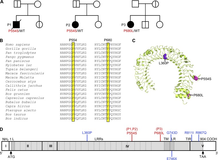
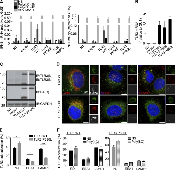
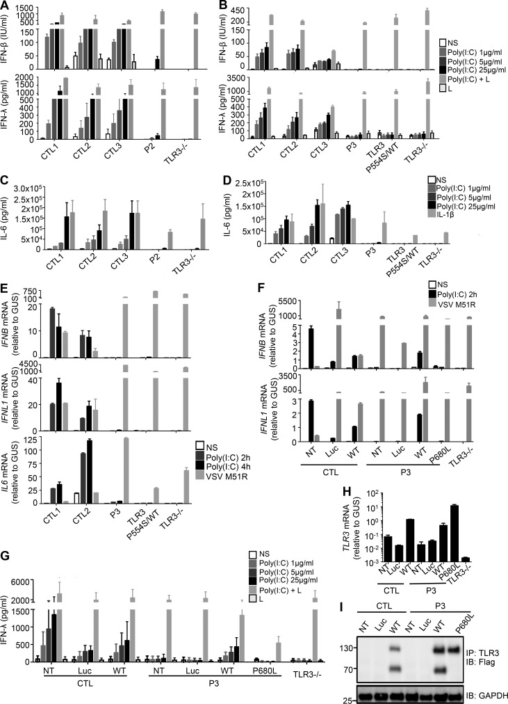
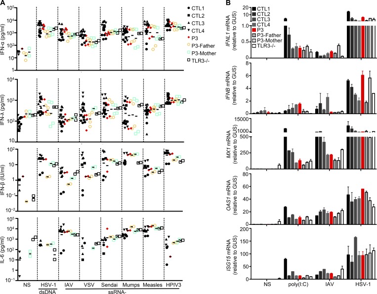
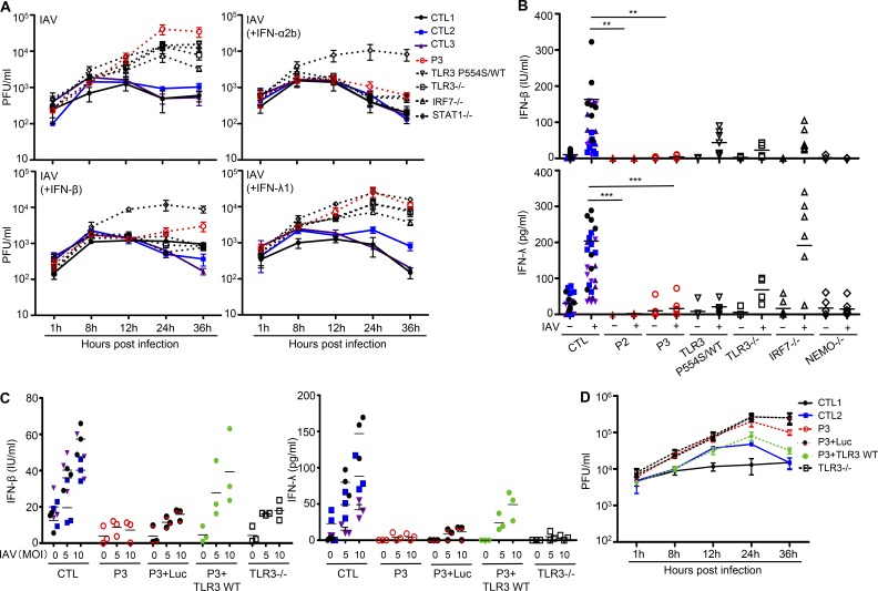
Autosomal recessive IRF7 and IRF9 deficiencies impair type I and III IFN immunity and underlie severe influenza pneumonitis. We report three unrelated children with influenza A virus (IAV) infection manifesting as acute respiratory distress syndrome (IAV-ARDS), heterozygous for rare TLR3 variants (P554S in two patients and P680L in the third) causing autosomal dominant (AD) TLR3 deficiency. AD TLR3 deficiency can underlie herpes simplex virus-1 (HSV-1) encephalitis (HSE) by impairing cortical neuron-intrinsic type I IFN immunity to HSV-1. TLR3-mutated leukocytes produce normal levels of IFNs in response to IAV. In contrast, TLR3-mutated fibroblasts produce lower levels of IFN-β and -λ, and display enhanced viral susceptibility, upon IAV infection. Moreover, the patients' iPSC-derived pulmonary epithelial cells (PECs) are susceptible to IAV. Treatment with IFN-α2b or IFN-λ1 rescues this phenotype. AD TLR3 deficiency may thus underlie IAV-ARDS by impairing TLR3-dependent, type I and/or III IFN-mediated, PEC-intrinsic immunity. Its clinical penetrance is incomplete for both IAV-ARDS and HSE, consistent with their typically sporadic nature.


Figures

Figure 1.
Figure 1.
Heterozygous TLR3 mutations in three unrelated IAV-ARDS patients. (A) Family pedigrees with TLR3 allele segregation. The black symbols indicate patients, and the bold vertical lines indicate healthy carriers of the mutant TLR3 allele. Healthy TLR3 WT relatives are indicated by white symbols. (B) Multiple sequence alignment across 19 vertebrate species. Both residues mutated in P1, P2, and P3 were highly conserved. (C) Predicted three-dimensional structure of the human TLR3 protein ectodomain. The residues affected by the two missense mutations found in IAV-ARDS patients are highlighted in magenta (P554S and P680L). The L360P mutation was previously identified in a patient with HSE (highlighted in violet). (D) Schematic diagram of the structure of the human TLR3 gene and protein, featuring the leader sequence (L), leucine-rich repeats (LRRs) of the ectodomain, transmembrane domain (TM), linker region (LR), and Toll/IL-1 receptor (TIR) domain. Roman numerals indicated the coding exons. Previously reported experimentally validated deleterious mutations found in HSE patients are shown in blue. The mutations found in the three children with severe influenza are shown in red (including the P554S mutation found in three previously reported and one unreported HSE patient).
Figure 2.
Figure 2.
Expression and function of the P680L mutant TLR3 allele. (A) RT-qPCR for IFNB and IFNL1 mRNA expression without stimulation (NS) or after 2 and 4 h of stimulation with 25 µg/ml poly(I:C) in P2.1 cells not transfected (NT) or stably transfected with empty vector, HA-tagged TLR3 WT, P554S, or P680L. VSV M51R, at a MOI of 1, was used as a positive stimulus for IFN induction via the TLR3-independent pathways. Mean values ± SD were calculated from two independent experiments, with biological duplicates in each experiment. (B) TLR3 mRNA levels were determined by RT-qPCR in P2.1 TLR3-deficient fibrosarcoma cells with or without transfection with empty vector, HA-tagged TLR3 WT, P554S, or P680L. Mean values ± SD were calculated from two independent experiments, with biological duplicates in each experiment. (C) TLR3 was detected on immunoblots. P2.1 cells not transfected or stably transfected with empty vector, HA-tagged TLR3 WT, or P680L were subjected to immunoprecipitation (IP) with anti-TLR3 antibody, and the immunoprecipitated protein was then immunoblotted (IB) with C-ter (C) HA antibody or N-ter (N) TLR3 antibody. GAPDH was used as a loading control for immunoblotting. Reproducible result from six independent experiments is shown. (D–F) Immunofluorescence imaging of P2.1 cells stably expressing HA-tagged WT or P680L. Intracellular distribution was assessed by colocalization (Coloc) with a subcellular marker: anti-PDI antibody for the ER, anti-EEA1 antibody for early endosomes, and anti-LAMP1 antibody for lysosomes. Cells were let un-treated (E) or incubated with 25 µg/ml poly(I:C) for 30 min (F). The images were analyzed with Imaris Coloc software, and plots were generated. About 200 cells were used for each analysis. Mean values ± SD were calculated from two independent experiments. *, P < 0.05; ****, P < 0.0001.
Figure 3.
Figure 3.
Impaired poly(I:C) responses in SV40-fibroblasts from patients heterozygous for TLR3 P554S or P680L, and rescue by WT TLR3. (A–D) Production of IFN-β, IFN-λ (A and B), and IL-6 (C and D) in SV40-fibroblasts from three healthy controls (CTL1/2/3), P2 (A and C), P3 (B and D), a TLR3 P554S/WT HSE patient, and a TLR3−/− HSE patient, 24 h after stimulation with 1, 5, or 25 µg/ml poly(I:C), or with 25 µg/ml poly(I:C) in the presence of lipofectamine (poly[I:C]+L; A and B), lipofectamine alone (L; A and B), or IL-1β (C and D), as assessed by ELISA. (E) IFNB, IFNL1, and IL6 mRNA levels in SV40-fibroblasts from two CTLs, P3, P554S/WT, and TLR3−/− patients, not stimulated (NS), or stimulated for 2 and 4 h with 25 µg/ml poly(I:C), or infected with VSV M51R at a MOI of 1 for 16 h. GUS was included for normalization. (F–I) Complementation of the impaired poly(I:C) response by introducing WT TLR3 into the patient’s fibroblasts. (F) IFNB, IFNL1 mRNA levels in SV40-fibroblasts from a healthy control (CTL) and P3, without plasmid transfection (NT) or after transfection with luciferase (Luc), Flag-tagged WT or P680L TLR3, and in fibroblasts from a TLR3−/− patient, not stimulated (NS), or stimulated for 2 h with 25 µg/ml poly(I:C), or infected with VSV M51R at a MOI of 1 for 16 h. GUS was included for normalization. (G) Production of IFN-λ, in the absence of stimulation, after 24 h of stimulation with 1, 5, or 25 µg/ml poly(I:C), and after stimulation with 25 µg/ml poly(I:C) in the presence of lipofectamine, or lipofectamine alone, as assessed by ELISA, in SV40-fibroblasts from a CTL and P3, without plasmid transfection or after transfection with Luc, Flag-tagged WT or P680L TLR3, and in fibroblasts from a TLR3−/− patient. (H) TLR3 mRNA levels were assessed by RT-qPCR. (I) TLR3 was detected on immunoblots following IP. Mean values ± SD were calculated from four (A–D and H) or two (E and F) independent experiments, with technical duplicates in each experiment. (G) Results from a single experiment with biological duplicates, representing three independent experiments. (I) Reproducible results from three independent experiments.
Figure 4.
Figure 4.
Normal IFN response to poly(I:C) and viruses in TLR3-mutated PBMCs. (A) PBMCs from four CTLs, P3, P3’s parents, and a TLR3−/− patient were infected with various types of viruses: double-stranded DNA (dsDNA; HSV-1) and single-stranded RNA (ssRNA; IAV strain pH1N1, VSV, Sendai virus, mumps virus, measles virus, HPIV3) viruses. The levels of IFN-α, -β, and -λ and IL-6 were measured by ELISA 24 h after infection. (B) The induction of IFNL1, IFNB, MX1, OAS1, and ISG15 mRNA was assessed by RT-qPCR in PBMCs from four CTLs, P3, P3’s parents, and a TLR3−/− patient. RNA was isolated from these cells after 8 h of poly(I:C) stimulation and 24 h of IAV pH1N1 and HSV-1 infection. Mean values ± SD were calculated from biological duplicates from two independent experiments.
Figure 5.
Figure 5.
Enhanced susceptibility to IAV in TLR3-mutated fibroblasts, and rescue by WT TLR3. (A) IAV replication, quantified by plaque assays, in SV40-fibroblasts from three CTLs, P3, P554S/WT, TLR3−/−, IRF7−/−, and STAT1−/− patients, 1, 8, 12, 24, and 36 h after infection at a MOI of 10. Cells were untreated or subjected to pretreatment with IFN-α2b, IFN-β, or IFN-λ for 16 h before infection. The data shown are representative of three independent experiments, with biological duplicates in each experiment. (B) Production of IFN-β and IFN-λ in the absence of infection or after 24 h of infection with IAV at a MOI of 1, in SV40-fibroblasts from seven CTLs, P2, P3, a TLR3 P554S/WT HSE patient, TLR3−/−, IRF7−/−, and NF-κB essential modulator (NEMO)–deficient patients, as assessed by ELISA. (C) Production of IFN-β and IFN-λ in the absence of infection or after 24 h of infection with IAV at a MOI of 5 or 10, in SV40-fibroblasts from a CTL and P3, without plasmid transfection or after transfection with Luc, Flag-tagged WT TLR3, and in fibroblasts from a TLR3−/− patient. (D) IAV replication, quantified by plaque assays, in SV40-fibroblasts from two CTLs and P3, without plasmid transfection or after transfection with Luc, Flag-tagged WT TLR3, and in fibroblasts from a TLR3−/− patient, 1, 8, 12, 24, and 36 h after infection at a MOI of 5. Mean values ± SD from five (B) or three (C and D) independent experiments are shown. Biological duplicates were tested in each experiment. **, P < 0.01; ***, P < 0.001.
Figure 6.
Figure 6.
Enhanced susceptibility to IAV in TLR3-mutated iPSC-derived lung epithelial cells. Lung epithelial cells were derived from the ES cells of a healthy control (CTL-RUES2, shown as black filled circles in the figure), iPSCs from two healthy controls (Sendai virus–reprogrammed iPSC CTL-SViPS, and mRNA-reprogrammed iPSC CTL-mRNA, shown as dark or light blue triangles, respectively), P3 (three iPSC clones from the same patient, shown with pink squares, yellow triangles, or red circles, respectively), TLR3−/−, IRF7−/−, STAT1−/−, and IL10RB−/− patients. The cells were untreated or subjected to 16 h of pretreatment with IFN-α2b or IFN-λ1, then infected with IAV at a MOI of 1 or 10 for 24 h. The cells were immunostained for influenza NP (green) and Nkx2.1 (red), and their nuclei were stained with DAPI (blue). (A) The percentage of Nkx2.1-positive cells was determined for the DAPI-positive cells. (B) Representative images of CTL and patient PECs, showing the immunostaining of NP, Nkx2.1, and DAPI, 24 h after infection with IAV. hES, human embryonic stem. (C) The percentage of influenza NP-positive cells was then determined for the Nkx2.1-positive cells. We analyzed ∼60,000 Nkx2.1 cells per cell line. Without IFN pretreatment, higher proportions of PECs derived from the iPSCs of TLR3 P680L/WT, TLR3−/−, IRF7−/−, STAT1−/−, and IL10RB−/− patients were positive for IAV NP, than of PECs from CTL-RUES2, iPSC CTL-SViPS, and iPSC CTL-mRNA. The data shown represent the mean values ± SD from three independent experiments. *, P < 0.05; **, P < 0.01; ***, P < 0.001.

References

    1. Alcaïs A., Quintana-Murci L., Thaler D.S., Schurr E., Abel L., and Casanova J.L.. 2010. Life-threatening infectious diseases of childhood: single-gene inborn errors of immunity? Ann. N. Y. Acad. Sci. 1214:18–33. 10.1111/j.1749-6632.2010.05834.x - DOI - PubMed
    1. Alexopoulou L., Holt A.C., Medzhitov R., and Flavell R.A.. 2001. Recognition of double-stranded RNA and activation of NF-kappaB by Toll-like receptor 3. Nature. 413:732–738. 10.1038/35099560 - DOI - PubMed
    1. Andersen L.L., Mørk N., Reinert L.S., Kofod-Olsen E., Narita R., Jørgensen S.E., Skipper K.A., Höning K., Gad H.H., Østergaard L., et al. . 2015. Functional IRF3 deficiency in a patient with herpes simplex encephalitis. J. Exp. Med. 212:1371–1379. 10.1084/jem.20142274 - DOI - PMC - PubMed
    1. Ank N., Iversen M.B., Bartholdy C., Staeheli P., Hartmann R., Jensen U.B., Dagnaes-Hansen F., Thomsen A.R., Chen Z., Haugen H., et al. . 2008. An important role for type III interferon (IFN-lambda/IL-28) in TLR-induced antiviral activity. J. Immunol. 180:2474–2485. 10.4049/jimmunol.180.4.2474 - DOI - PubMed
    1. Audry M., Ciancanelli M., Yang K., Cobat A., Chang H.H., Sancho-Shimizu V., Lorenzo L., Niehues T., Reichenbach J., Li X.X., et al. . 2011. NEMO is a key component of NF-κB- and IRF-3-dependent TLR3-mediated immunity to herpes simplex virus. J. Allergy Clin. Immunol. 128:610–7.e1: 4. 10.1016/j.jaci.2011.04.059 - DOI - PMC - PubMed

Publication types

MeSH terms