. 2019 Sep;9(9):1306-1323.
doi: 10.1158/2159-8290.CD-18-0083. Epub 2019 Jun 19.

PTEN Methylation by NSD2 Controls Cellular Sensitivity to DNA Damage

Affiliations

PTEN Methylation by NSD2 Controls Cellular Sensitivity to DNA Damage

Jinfang Zhang et al. Cancer Discov. 2019 Sep.

Abstract

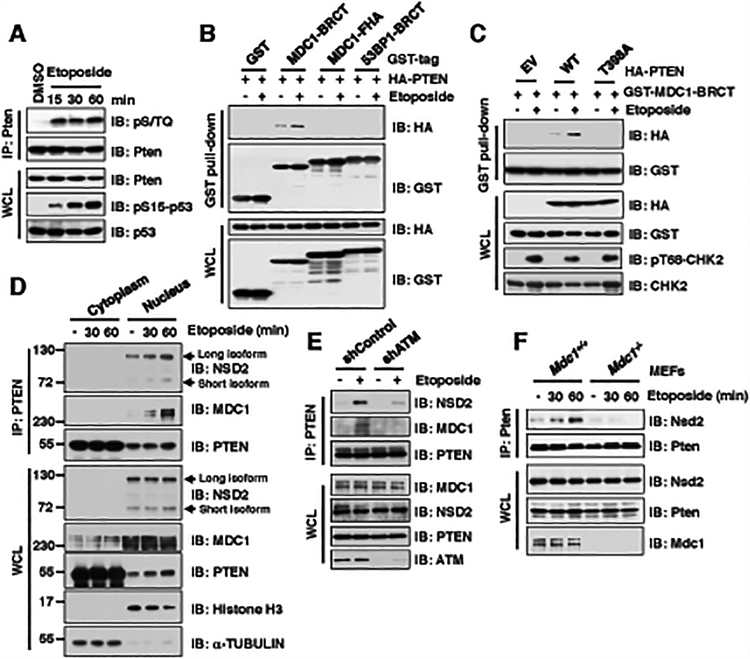
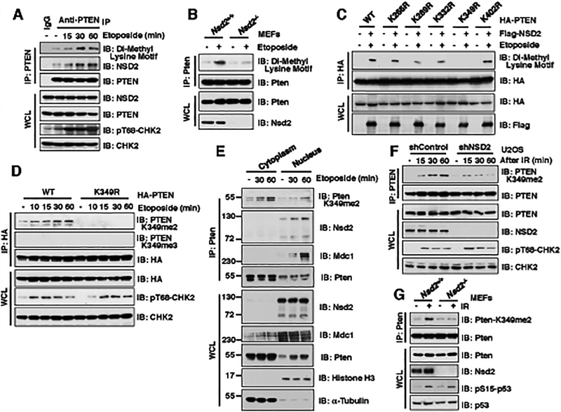
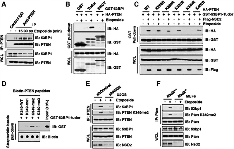
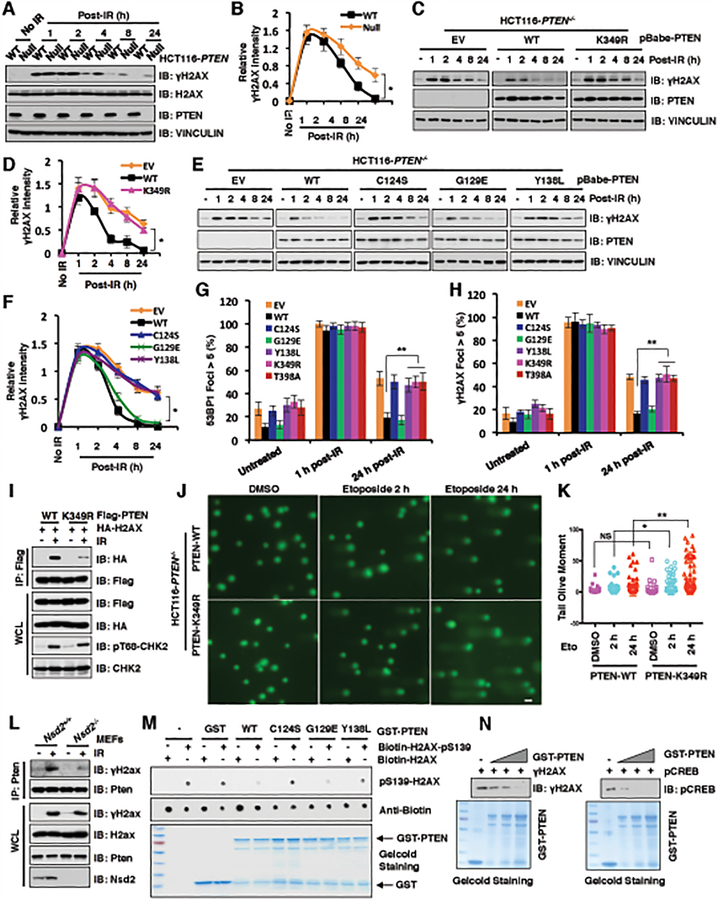
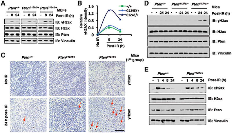
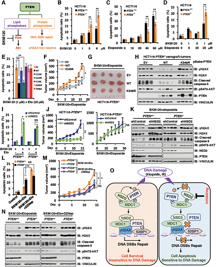
The function of PTEN in the cytoplasm largely depends on its lipid-phosphatase activity, though which it antagonizes the PI3K-AKT oncogenic pathway. However, molecular mechanisms underlying the role of PTEN in the nucleus remain largely elusive. Here, we report that DNA double-strand breaks (DSB) promote PTEN interaction with MDC1 upon ATM-dependent phosphorylation of T/S398-PTEN. Importantly, DNA DSBs enhance NSD2 (MMSET/WHSC1)-mediated dimethylation of PTEN at K349, which is recognized by the tudor domain of 53BP1 to recruit PTEN to DNA-damage sites, governing efficient repair of DSBs partly through dephosphorylation of γH2AX. Of note, inhibiting NSD2-mediated methylation of PTEN, either through expressing methylation-deficient PTEN mutants or through inhibiting NSD2, sensitizes cancer cells to combinatorial treatment with a PI3K inhibitor and DNA-damaging agents in both cell culture and in vivo xenograft models. Therefore, our study provides a novel molecular mechanism for PTEN regulation of DSB repair in a methylation- and protein phosphatase-dependent manner. SIGNIFICANCE: NSD2-mediated dimethylation of PTEN is recognized by the 53BP1 tudor domain to facilitate PTEN recruitment into DNA-damage sites, governing efficient repair of DNA DSBs. Importantly, inhibiting PTEN methylation sensitizes cancer cells to combinatorial treatment with a PI3K inhibitor combined with DNA-damaging agents in both cell culture and in vivo xenograft models.This article is highlighted in the In This Issue feature, p. 1143.


Conflict of interest statement

Conflict of interest: W.W. and P.P.P. are co-founder and stock holder of the Rekindle Therapeutics.

Figures

Figure 1.
Figure 1.. ATM-mediated phosphorylation of PTEN is required for its binding with the BRCT domain of MDC1 upon DNA damage signaling.
(A) Phosphorylation of Pten was detected using the phospho-(Ser/Thr) ATM/ATR substrate antibody (pS/TQ) upon etoposide treatment. Immunoblot (IB) analysis of anti-Pten immunoprecipitations (IPs) and whole cell lysates (WCL) derived from NIH3T3 cells treated with 30 μM etoposide as indicated time points before harvesting. (B) Etoposide treatment promoted PTEN interaction with MDC1 BRCT domain, but neither MDC1 FHA nor 53BP1 BRCT domain. IB analysis of GST pull-down and WCL derived from U2OS cells transfected with indicated constructs and treatment with/without 30 μM etoposide for 30 min before harvesting. (C) Etoposide treatment promoted wild type (WT), but not T398A mutant PTEN, interaction with MDC1 BRCT domain. IB analysis of GST pull-down and WCL derived from U2OS cells co-transfected with indicated constructs. 36 h after transfection, cells were treated with/without 30 μM etoposide for 30 min and harvested for IP assays. (D) PTEN, MDC1, and NSD2 formed a tertiary complex in the nucleus upon etoposide treatment. IB analysis of anti-PTEN IPs from cytoplasm or nucleus as well as WCL derived from U2OS cells treated with 30 μM etoposide as indicated time points before harvesting. (E) Depletion of ATM disrupted PTEN interaction with MDC1 and NSD2 upon etoposide treatment. IB analysis of anti-PTEN IPs and WCL derived from U2OS cell lines stably expressing shControl or shATM transfected with indicated constructs. 36 h after transfection, cells were treated with/without 30 μM etoposide for 30 min before harvesting. (F) Depletion of Mdc1 impaired the interaction between Pten and Nsd2. IB analysis of anti-Pten IPs and WCL derived from Mdc1+/+ and Mdc1−/− MEFs treated with 30 μM etoposide as indicated time points before harvesting.
Figure 2.
Figure 2.. DNA damage promotes NSD2-mediated di-methylation of PTEN at K349.
(A) Methylation of PTEN was detected using the Di-Methyl Lysine motif antibody. IB analysis of anti-PTEN IPs and WCL derived from U2OS cells treated with 30 μM etoposide as indicated time points before harvesting. (B) Depletion of Nsd2 impaired the di-methylation of Pten. IB analysis of anti-Pten IPs and WCL derived from Nsd2+/+ and Nsd2−/− MEFs treated with 30 μM etoposide for 30 min before harvesting. (C) K349 was identified as the major di-methylation site on PTEN. IB analysis of anti-HA IPs and WCL derived from 293T cells transfected with the indicated constructs and treated with/without 30 μM etoposide for 30 min before harvesting. (D) PTEN was detected by the specific K349 di-methylation (K349me2) antibody. IB analysis of anti-HA IPs and WCL derived from U2OS cells transfected with HA-PTEN WT or K349R mutant and treated with/without 30 μM etoposide at indicated time points. (E) The K349 di-methylation of Pten existed in both cytoplasm and nucleus. IB analysis of anti-Pten IPs from cytoplasm or nucleus as well as WCL derived from NIH3T3 cells treatment with 30 μM etoposide as indicated time points before harvesting. (F and G) NSD2 deficiency decreased the di-methylation of PTEN at K349. IB analysis of anti-PTEN IPs and WCL derived from U2OS cells stably expressing shNSD2 (F) or Nsd2−/− MEFs (G) that were treated with IR (5 Gy) at indicated time points before harvesting.
Figure 3.
Figure 3.. NSD2-mediated di-methylation of PTEN is recognized by the tudor domain of 53BP1.
(A) Etoposide treatment enhanced PTEN interaction with 53BP1. IB analysis of anti-PTEN IPs and WCL derived from U2OS cells treated with 30 μM etoposide as indicated time points before harvesting. (B and C) PTEN-K349R mutant disrupted its binding with 53BP1 upon etoposide treatment. IB analysis of GST pull-down and WCL derived from U2OS cells transfected with the indicated constructs. 36 h after transfection, cells were treated with/without 30 μM etoposide for 30 min and harvested for GST pull-down assays. (D) 53BP1 tudor domain had a high affinity with K349-me2-PTEN peptides. 1 μg of indicated biotin-labeled synthetic PTEN peptides were incubated with 250 ng purified recombinant GST-tagged 53BP1 tudor domain, respectively. Streptavidin beads were added to perform pull-down assays and precipitations were analyzed by IB. Dot blot assays were performed to show equal amount of biotinylated peptides was used for the pull-down assay. (E and F) Depletion of NSD2 disrupted PTEN interaction with 53BP1. IB analysis of anti-PTEN IPs and WCL derived from U2OS cells stably expressing shNSD2 (E) or Nsd2−/− MEFs (F) that were treated with/without 30 μM etoposide for 30 min before harvesting.
Figure 4.
Figure 4.. PTEN K349 di-methylation and protein phosphatase activity are required for efficient DSBs repair.
(A and B) PTEN deficiency elevated γH2AX after 4 h post-irradiation (IR) treatment. IB analysis of WCL derived from PTEN+/+ and PTEN−/− HCT116 cells after treatment with IR (5 Gy) as indicated time points (A). Quantification of protein intensity was performed using the ImageJ software (B). γH2AX immunoblot bands were normalized to VINCULIN, and then normalized to the control (no IR treatment). Data are represented as mean ± s.d., n = 3. *p < 0.05 (Student’s t-test). (C and D) PTEN wild type (WT), but not K349R mutant, rescued PTEN deficiency mediated high levels of γH2AX after 4 h post-IR treatment. IB analysis of WCL derived from PTEN−/− HCT116 cells introducing PTEN WT, K349R as well as EV, which were treated with IR (5 Gy) at indicated time points before harvesting (C). Quantification of protein intensity was performed using the ImageJ software (D). γH2AX immunoblot bands were normalized to VINCULIN, and then normalized to the control (no IR treatment). Data are represented as mean ± s.d., n = 3. *p < 0.05 (Student’s t-test). (E and F) PTEN protein phosphatase activity, but not lipid phosphatase activity, was required for regulating γH2AX levels. IB analysis of WCL derived from PTEN−/− HCT116 cells re-introducing PTEN WT, C124S, G129E, Y138L as well as EV, which were treated with IR (5 Gy) at indicated time points before harvesting (E). Quantification of protein intensity was performed using the ImageJ software (F). γH2AX immunoblot bands were normalized to VINCULIN, then normalized to the control (no IR treatment). Data are represented as mean ± s.d., n = 3. *p < 0.05 (Student’s t-test). (G and H) PTEN−/− HCT116 cells reconstituted with the indicated PTEN constructs were treated with IR (5 Gy) and immunostained at the indicated times with anti-53BP1 or anti-γH2AX. Quantification of 53BP1 (G) or γH2AX (H) foci positive cells (foci > 5 per cell) was performed by counting a total of 100 cells per sample, respectively. Data are represented as mean ± s.d., n = 3, and **p < 0.01 (Student’s t-test). (I) PTEN-K349R mutant decreased its interaction with H2AX. IB analysis of anti-Flag IPs and WCL derived from U2OS cells transfected with the indicated constructs. 36 h after transfection, cells were harvested for IP assays after treatment with IR (5 Gy) for 60 min. (J and K) PTEN-K349R mutation attenuated DNA damage repair. PTEN-WT or PTEN-K349R mutant expressed HCT116 cells were treated with DMSO or etoposide for 2h and 24h, respectively. (J) Images were captured following Comet assay; and (K) Tail olive moment values were calculated using Casplab software. Error bars represent standard error of the mean. **p<0.01, *p<0.05. n=80. Scale bar: 50 μM. (L) Nsd2 deficiency disrupted Pten interaction with γH2ax. IB analysis of anti-Pten IPs and WCL derived from Nsd2+/+ and Nsd2−/− MEFs treatment with/without IR (5 Gy) for 60 min before harvesting. (M and N) In vitro dephosphorylation assays were performed with bacterially purified recombinant GST-tagged PTEN WT and the indicated PTEN mutants including C124S, G129E, and Y138L incubating with indicated H2AX synthetic peptides (M) or purified γH2AX (N) from HCT116 cells after etoposide treatment, then analyzed by immunoblot analyses.
Figure 5.
Figure 5.. The protein phosphatase activity of PTEN is required for DSBs repair in vivo.
(A and B) PtenC124S/+ MEFs displayed higher levels of γH2ax at 24 h post γ-irradiation (IR) treatment compared with Pten+/+ and PtenG129E/+ MEFs. IB analysis of WCL derived from Pten+/+, PtenG129E/+ and PtenC124S/+ MEFs, which were treated with IR (5 Gy) at indicated time points before harvesting (A). Quantification of protein intensity was performed using the ImageJ software (B). γH2ax immunoblot bands were normalized to Vinculin, and then normalized to the control (no IR treatment). (C and D) Representative immunohistochemistry (IHC) analysis of spleen tissues derived from Pten+/+, PtenG129E/+ and PtenC124S/+ mice (1th group), which were treated with IR (3 Gy) and sacrificed at 24 h after irradiation (C). Scale bar, 50 μm. IB analysis of the sample was performed using indicated antibodies (D). Four mice each group. (E) PtenY138L/+ MEFs displayed higher levels of γH2ax at 8 or 24 h post IR treatment compared with Pten+/+ MEFs. IB analysis of WCL derived from Pten+/+ and PtenY138L/+ MEFs, which were treated with IR (5 Gy) at indicated time points before harvesting.
Figure 6.
Figure 6.. NSD2-mediated methylation of PTEN at K349 dictates cellular sensitivity to DNA-damaging agents.
(A) A schematic model to illustrate that PTEN has lipid phosphatase and protein phosphatase activity, which involves in PI3K/Akt signaling pathway in cytoplasm and DNA DSB repair/γH2AX pathway in nucleus, respectively. (B and C) PTEN+/+ and PTEN−/− HCT116 cells were treated with increased concentrations of BKM120 (B) for 72 h or etoposide (C) for 48 h, cells were harvested for cell apoptosis assays. Data are represented as mean ± s.d., n = 3, and **p < 0.01 (Student’s t-test). (D) PTEN+/+ and PTEN−/− HCT116 cells were pre-treated with 1 μM BKM120 for 24 h followed by additional etoposide (20 μM) treatment for 48 h, cells were harvested for cell apoptosis assays. Data are represented as mean ± s.d., n = 3, and **p < 0.01 (Student’s t-test). (E) PTEN−/− HCT116 cells reconstituted with the indicated PTEN constructs were pre-treated with 1 μM BKM120 for 24 h followed by additional etoposide (20 μM) treatment for 48 h. Cells were harvested for cell apoptosis assays. Data are represented as mean ± s.d., n = 3, and **p < 0.01 (Student’s t-test). (F-H) Tumor xenograft assays were performed by subcutaneous injection of PTEN−/− HCT116 cells stably expressing PTEN WT, K349R and empty vector (EV). Tumor growth rate in nude mice treated every other day with combination of etoposide (20 mg/kg) and BKM120 (25 mg/kg) was shown (F). Tumors were dissected and recorded after euthanizing the mice (G). IB analysis of the samples was performed using indicated antibodies (H). Four mice each group. *p < 0.05 (Student’s t-test). (I) PTEN+/+ and PTEN−/− HCT116 cells stably depleting NSD2 by shRNA (with shControl as a negative control) were pre-treated with 1 μM BKM120 for 24 h followed by additional etoposide (20 μM) treatment. 48 h post-treatment, cells were harvested for cell apoptosis assays. Data are represented as mean ± s.d., n = 3, and *p < 0.05 (Student’s t-test). (J and K) Tumor xenograft assays were performed by subcutaneous injection of PTEN+/+ and PTEN−/− HCT116 cells stably expressing shRNA against NSD2 or shControl as a negative control. Tumor growth rate in nude mice treated every other day with a combination of etoposide (20 mg/kg) and BKM120 (25 mg/kg) was shown in (J). Tumors were dissected after euthanizing the mice and were analyzed by IB with indicated antibodies (K). Statistical analysis of tumor volumes showed significant differences in mean tumor volumes between the shNSD2 and the shcontrol groups. Four mice each group. *p < 0.05, **p < 0.01, NS indicates no significant difference (Student’s t-test). (L) PTEN+/+ and PTEN−/− HCT116 cells were pre-treated with 1 μM BKM120 for 24 h followed by additional etoposide (20 μM) treatment. 48 h post-treatment, cells were harvested for cell apoptosis assays. Data are represented as mean ± s.d., n = 3. *p < 0.05, **p < 0.01, NS indicates no significant difference (Student’s t-test). (M and N) Tumor xenograft assays were performed by subcutaneously implanting PTEN+/+ and PTEN−/− HCT116 cells. Tumor growth rate in nude mice treated every other day with a combination of etoposide (20 mg/kg) and BKM120 (25 mg/kg) with DZNep (1 mg/kg) (or with vehicle as a negative control) was shown (M). Tumors were dissected after euthanizing the mice and were analyzed by IB using indicated antibodies (N). Four mice each group. *p < 0.05, NS indicates no significant difference (Student’s t-test). (O) A schematic model to show the molecular mechanism of NSD2-mediated methylation of PTEN involving in DNA damage repair. DNA damaging agents promote NSD2-mediated di-methylation of PTEN, which is recognized by the 53BP1 tudor domain to recruit PTEN on DNA damage sites and help DNA DSBs repair largely through dephosphorylating γH2AX (left panel). However, inhibiting NSD2 could suppress PTEN di-methylation at K349 and its recruitment on DNA damage sites, which subsequently leads to DSBs repair deficiency and sensitizes cancer cells to DNA damaging agents (right panel). P: phosphorylation; M: methylation.

Similar articles

Cited by

References

    1. Worby CA, Dixon JE. Pten. Annual review of biochemistry 2014;83:641–69 doi 10.1146/annurev-biochem-082411-113907. - DOI - PubMed
    1. Lee YR, Chen M, Pandolfi PP. The functions and regulation of the PTEN tumour suppressor: new modes and prospects. Nat Rev Mol Cell Biol 2018;19(9):547–62 doi 10.1038/s41580-018-0015-0. - DOI - PubMed
    1. Song MS, Salmena L, Pandolfi PP. The functions and regulation of the PTEN tumour suppressor. Nat Rev Mol Cell Biol 2012;13(5):283–96 doi 10.1038/nrm3330. - DOI - PubMed
    1. Ortega-Molina A, Serrano M. PTEN in cancer, metabolism, and aging. Trends in endocrinology and metabolism: TEM 2013;24(4):184–9 doi 10.1016/j.tem.2012.11.002. - DOI - PMC - PubMed
    1. Lee YR, Chen M, Lee JD, Zhang J, Lin SY, Fu TM, et al. Reactivation of PTEN tumor suppressor for cancer treatment through inhibition of a MYC-WWP1 inhibitory pathway. Science 2019;364(6441) doi 10.1126/science.aau0159. - DOI - PMC - PubMed

Publication types