. 2019 Jun 20;4(12):e128561.
doi: 10.1172/jci.insight.128561.

TRIOBP-5 sculpts stereocilia rootlets and stiffens supporting cells enabling hearing

Affiliations

TRIOBP-5 sculpts stereocilia rootlets and stiffens supporting cells enabling hearing

Tatsuya Katsuno et al. JCI Insight. .

Abstract

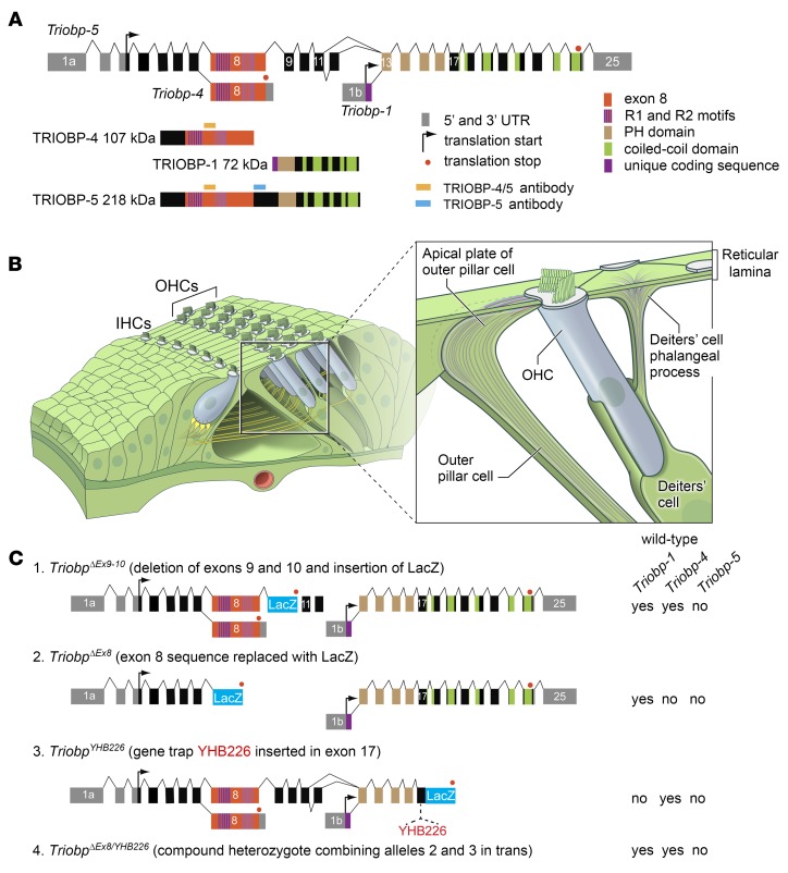
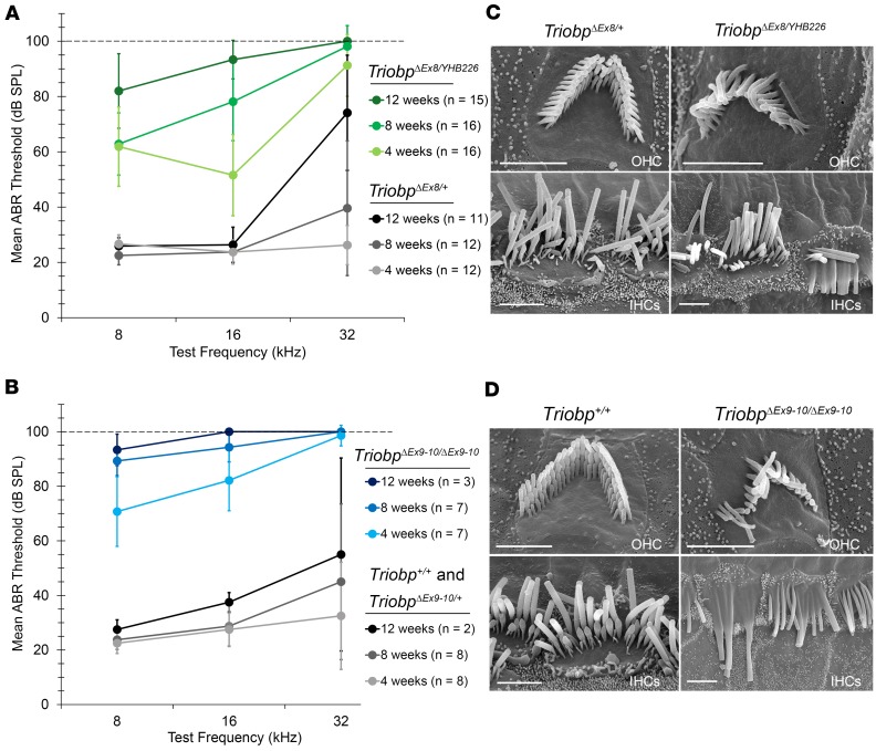
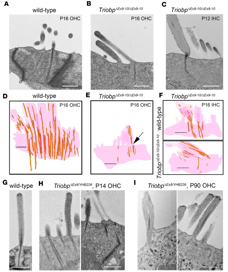
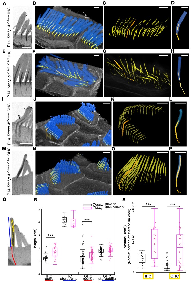
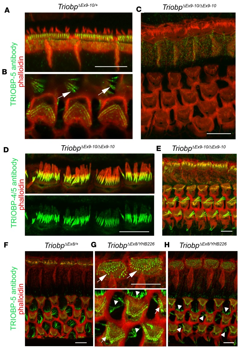
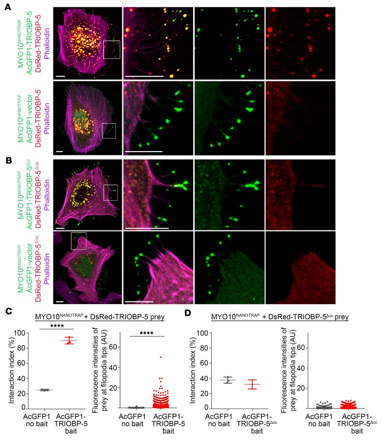
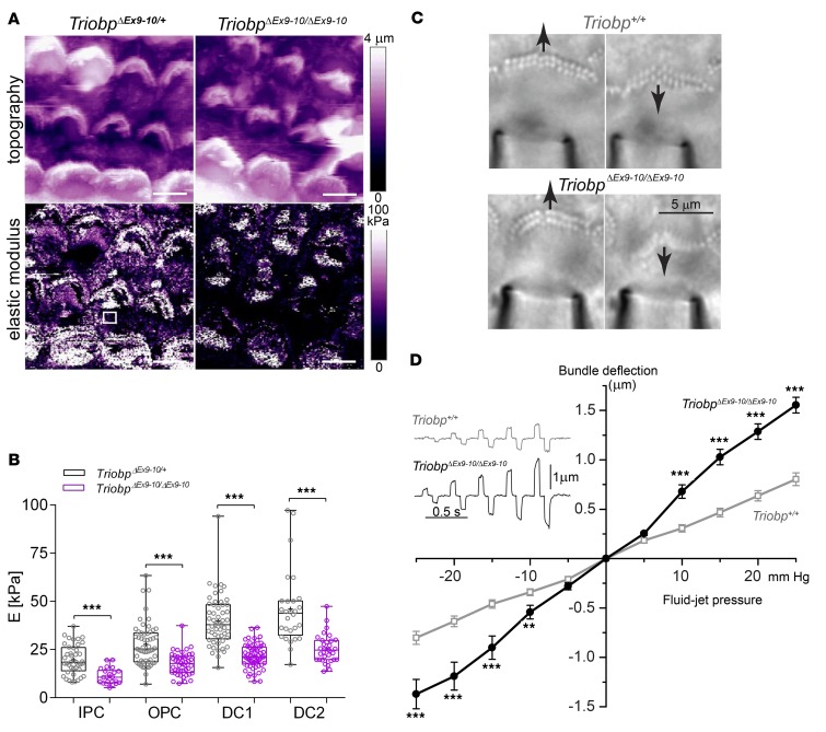
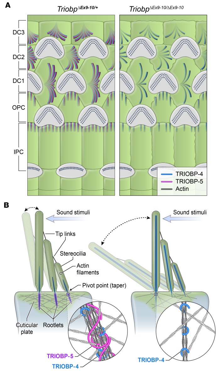
TRIOBP remodels the cytoskeleton by forming unusually dense F-actin bundles and is implicated in human cancer, schizophrenia, and deafness. Mutations ablating human and mouse TRIOBP-4 and TRIOBP-5 isoforms are associated with profound deafness, as inner ear mechanosensory hair cells degenerate after stereocilia rootlets fail to develop. However, the mechanisms regulating formation of stereocilia rootlets by each TRIOBP isoform remain unknown. Using 3 new Triobp mouse models, we report that TRIOBP-5 is essential for thickening bundles of F-actin in rootlets, establishing their mature dimensions and for stiffening supporting cells of the auditory sensory epithelium. The coiled-coil domains of this isoform are required for reinforcement and maintenance of stereocilia rootlets. A loss of TRIOBP-5 in mouse results in dysmorphic rootlets that are abnormally thin in the cuticular plate but have increased widths and lengths within stereocilia cores, and causes progressive deafness recapitulating the human phenotype. Our study extends the current understanding of TRIOBP isoform-specific functions necessary for life-long hearing, with implications for insight into other TRIOBPopathies.

Keywords: Cytoskeleton; Neuroscience.


Conflict of interest statement

Conflict of interest: The authors have declared that no conflict of interest exists.

Figures

Figure 1
Figure 1. Triobp gene structure, transcripts, protein isoforms, and mutant alleles.
(A) Alternative transcripts of mouse Triobp, the corresponding encoded protein isoforms, and their predicted domains. Locations of the epitopes for 2 antibodies (TRIOBP-4/5 and TRIOBP-5) are depicted with orange and light-blue rectangles, respectively. (B) Drawing of an organ of Corti sensory epithelium segment showing 1 row of inner (IHC) and 3 rows of outer (OHC) hair cells. Inset shows Deiters’ and outer pillar cells supporting an OHC. (C) Mutations of Triobp used to generate 2 genetically different TRIOBP-5–deficient mouse models. TriobpΔEx9-10/ΔEx9-10 has a LacZ cassette replacing exons 9 and 10 of Triobp-5 (schematic 1). The TriobpΔEx8 allele has a LacZ cassette replacing exon 8 (schematic 2) and the TriobpYHB226 allele has a trap cassette with a LacZ insertion in exon 17 (schematic 3). The combination in trans of these 2 Triobp mutations is a compound heterozygote designated TriobpΔEx8/YHB226. Expression of wild-type Triobp isoforms 1, 4, and 5 is indicated to the right of the schematics for each genotype.
Figure 2
Figure 2. Differential localization of TRIOBP isoforms in stereocilia rootlets revealed in the R26-EGFP-Triobp-4 transgenic reporter mouse.
(A) TRIOBP-5–specific antibody signal (red) observed predominantly in the rootlet segment inside the cuticular plate. See single channel images in Supplemental Figure 2. (B) Anti-GFP antibody recognized EGFP-TRIOBP-4 in the stereocilia rootlet segment that resides above the cuticular plate within the stereocilia core (green). A diffuse EGFP signal is present in the cuticular plate (green). (C) In Deiters’ cells located between OHCs, both TRIOBP-4 and TRIOBP-5 are present in F-actin bundles of obliquely ascending processes (arrows). Outer pillar cells show predominantly a TRIOBP-5 signal (red, arrowheads) close to junctions with OHCs. (D) P32 wild-type mouse (C57BL/6J) organ of Corti stained with TRIOBP-4/5 antibody. The inset shows representative IHC, in which TRIOBP-4/5 immunoreactivity (green) is prominent in stereocilia above the cuticular plate and a weaker signal is present in the rootlet segment within the cuticular plate. TRIOBP-4/5 immunoreactivity is also present in stereocilia of OHCs (inset and arrowheads) and in Deiters’ cells (arrows). (E and F) Helios gene gun–mediated transfections of P3 wild-type organ of Corti explants with plasmids expressing either AcGFP1-TRIOBP-5 (E) or AcGFP1-TRIOBP-4 cDNA (F) reveal targeting of AcGFP1-TRIOBP-5 (E) to stereocilia rootlets (green) within the cuticular plate. (F) AcGFP1-TRIOBP-4 predominantly targets the upper half of the rootlets within the stereocilia core, recapitulating the endogenous differential localization of TRIOBP-4 and TRIOBP-5. A barely detectable signal of AcGFP1-TRIOBP-4 was found in the lower half of rootlets within the cuticular plate. F-actin is visualized by blue phalloidin-405 in AC and rhodamine-phalloidin (red) staining in DF. All images are maximum intensity projections of confocal Z-stacks or their subsets (inserts). Scale bars: 5 μm (all panels).
Figure 3
Figure 3. Progressive hearing loss in TRIOBP-5deficient mice.
(A) Average auditory brainstem response (ABR) thresholds in TriobpΔEx8/+ heterozygous control mice (gray/black) and TriobpΔEx8/YHB226 compound heterozygous TRIOBP-5–deficient mice (shades of green). (B) Average ABR thresholds in Triobp+/+ (wild-type) and TriobpΔEx9-10/+ heterozygous controls (gray/black) and in TriobpΔEx9-10/ΔEx9-10 mice (shades of blue). ABRs were measured at 4, 8, and 12 weeks postnatally at frequencies of 8, 16, and 32 kHz. When no response was detected at a maximum stimulus level of 90 dB SPL, the threshold was assigned as 100 dB SPL (dashed line in A and B). Error bars indicate SD. (C) SEM images of P43 mouse IHC and OHC stereocilia bundles from normal-hearing TriobpΔEx8/+ heterozygotes (left) and from deaf TriobpΔEx8/YHB226 (right) compound heterozygotes. (D) SEM images of mouse P35 stereocilia bundles of wild-type and TriobpΔEx9-10/ΔEx9-10 OHCs and IHCs. Note fusion of stereocilia of mutant IHC and loss of stereocilia from longer row in OHC hair bundle. Scale bars: 2 μm in C and right panels in D, and 5 μm in D, left panel.
Figure 4
Figure 4. Structural abnormalities of stereocilia rootlets in TRIOBP-5–deficient mice.
(AC) TEM images of hair cell bundles (P12 and P16) showing representative rootlet morphology and (DF) 3D reconstructions of stereocilia rootlets from serial TEM sections (P16). wild-type (Triobp+/+) (A and D) and TriobpΔEx9-10/ΔEx9-10 OHCs (B and E), TriobpΔEx9-10/ΔEx9-10 IHC rootlet morphology (C), and 3D reconstruction of wild-type (Triobp+/+) and TriobpΔEx9-10/ΔEx9-10 IHC stereocilia and cuticular plates (F). (C) IHC stereocilium rootlet is dysmorphic, thin, and bent. (E) Arrow points to disrupted rootlet structure at stereocilia pivot points. (GI) TEM images of the OHC stereocilia in wild-type (Triobp+/+) at P16 (G), and TriobpΔEx8/YHB226 mouse at P14 (H) and P90 (I). Structural defects of rootlets at P14 include splayed bundles of F-actin or bent rootlets (H). Representative structural defects at P90 include fused stereocilia undergoing degeneration (I, left panel) and abnormal asymmetric localization of electron dense material within stereocilia F-actin cores (I, right panel). Scale bars: 500 nm.
Figure 5
Figure 5. FIB-SEM analyses of TriobpΔEx9-10/ΔEx9-10 hair cells show enlargement of rootlets within stereocilia cores and thinning of rootlets within the cuticular plate.
(AP) Reconstruction of stereocilia bundles and stereocilia rootlets from P14 TriobpEx9-10/ΔEx9-10 and TriobpEx9-10/+ IHCs and OHCs. (A, E, I, and M) Sagittal sections showing the morphology of rootlets reconstructed from FIB-SEM data sets. (B, F, J, and N) False-colored 3D reconstructions of hair bundles (blue) and rootlets (yellow and orange) were added to better visualize rootlet structure. (C, G, K, and O) Individual rootlets highlighted in orange show typical morphology for a particular genotype. (D, H, L, and P) Reconstruction of 1 stereocilium (blue) and rootlet selected in C, G, K, and O (orange) from hair cells in B, F, J, and N. The rootlet segments within stereocilia cores of TriobpEx9-10/ΔEx9-10 mice in EH and MP are longer and thicker compared with phenotypically wild-type controls in AD and IL, while TriobpEx9-10/ΔEx9-10 rootlets within the cuticular plate are abnormally thin. Scale bars: 1 μm. See also Supplemental Videos 1 and 2. (Q) Quantification of the shape of stereocilia and rootlets, rootlet length (red line), and stereocilium length (blue line) and volume of rootlet segment of stereocilia core (yellow) of the tallest row stereocilia of IHCs and OHCs. (R and S) Data in box-and-whisker plots are represented as mean ± SD. Data points represent length of rootlets within the cuticular plate (red) and within the stereocilia cores (blue) of IHCs or OHCs and volume of rootlets within stereocilia cores for Triobp-5ΔEx9-10/+ mice (black; n = 54, 17, 102, 98, 21 and 39) or Triobp-5ΔEx9-10/ΔEx9-10 mice (purple, n = 47, 17, 120, 96, 27, and 34) at P14. ***P < 0.001 compared with control by Mann-Whitney U test. There is a significant difference in rootlet length and rootlet volume in stereocilia cores when comparing Triobp-5ΔEx9-10/+ and Triobp-5ΔEx9-10/ΔEx9-10 mice, while there is no significant difference in stereocilia length.
Figure 6
Figure 6. Localization of TRIOBP isoforms in P14–P30 TRIOBP-5–deficient mice.
(A and B) TRIOBP-5 (green) is localized to the rootlet compartment within the cuticular plate of IHCs (A) and OHCs and in Deiters’ supporting cells (B, white arrows) of normal-hearing TriobpΔEx9-10/+ mice. (C) TRIOBP-5 was not detected in stereocilia rootlets and Deiters’ cells of TriobpΔEx9-10/ΔEx9-10 homozygous TRIOBP-5–deficient mice, confirming antibody specificity. (D) TRIOBP-4/5 antibody detects the TRIOBP-4 isoform in TriobpΔEx9-10/ΔEx9-10 IHC stereocilia rootlet compartment above the cuticular plate. (E) Immunoreactivity to TRIOBP-4/5 antibody is unchanged in Deiters’ cells and OHCs of TriobpΔEx9-10/ΔEx9-10 mice compared to immunoreactivity in phenotypically wild-type heterozygous (TriobpΔEx9-10/+) hair cells (Figure 2B). (FH) In contrast to the absence of a TRIOBP-5 signal in TriobpΔEx9-10/ΔEx9-10 mice, TRIOBP-5 antibody shows immunoreactivity in compound heterozygous TriobpΔEx8/YHB226 and control TriobpΔEx8/+ mice in the lower portion of stereocilia rootlets in both OHCs and IHCs (arrows in G) and in Deiters’ and outer pillar cells (arrowheads in G and H). Scale bars: 5 μm (all panels).
Figure 7
Figure 7. TRIOBP-5 homodimerization revealed by nanoscale pull-down assays.
(A) MYO10-HMM-Nanotrap (abbreviated MYO10NANOTRAP) specifically binds GFP derivatives, but not DsRed, and traffics GFP-tagged protein to filopodia tips. Representative image of a HeLa cell transfected with 3 expression vectors encoding MYO10NANOTRAP, AcGFP1-TRIOBP-5, and DsRed-TRIOBP-5 (upper panel), and a control HeLa cell transfected with the MYO10NANOTRAP, AcGFP1 vector, and DsRed-TRIOBP-5 (lower panel). AcGFP1-TRIOBP-5 and DsRed-TRIOBP-5 colocalize to filopodia tips (upper panel), while in the control, only the AcGFP1 vector localized at filopodia tips (lower panel). The complex of MYO10NANOTRAP and AcGFP1-TRIOBP-5 partnered with and transported DsRed-TRIOBP-5 to filopodia tips. The complex of MYO10NANOTRAP and AcGFP1 vector did not traffic DsRed-TRIOBP-5. These data indicate that TRIOBP-5 can homomultimerize in vivo. The second column shows enlarged images of the white-outlined areas from the first column. The third and fourth columns are the individual green and red channels of merged images in the second column. (B) Transfected HeLa cell with MYO10NANOTRAP, AcGFP1-TRIOBP-5Δcc, and DsRed-TRIOBP-5Δcc (upper panel), and control HeLa cell transfected with MYO10NANOTRAP, AcGFP1 vector, and DsRed-TRIOBP-5Δcc (lower panel). Triobp-5Δcc is deleted for sequence encoding the coiled-coil domains and 78 bp of upstream sequence. DsRed-TRIOBP-5Δcc was not trafficked to filopodia tips by the MYO10NANOTRAP and AcGFP1-TRIOBP-5Δcc, indicating that TRIOBP-5Δcc does not multimerize. (C) Left panel: Quantification of AcGFP1-TRIOBP-5 or AcGFP1 vector binding to DsRed-TRIOBP-5. When MYO10NANOTRAP, AcGFP1-TRIOBP-5, and DsRed-TRIOBP-5 were coexpressed, 90% ± 4.4% of filopodia (3 independent experiments) showed a statistically significant correlation between green and red fluorescence at filopodia tips (2-tailed t test). Right panel: Fluorescence intensities of DsRed-TRIOBP-5 at filopodia tips. The presence of AcGFP1-TRIOBP-5 caused a significant increase of fluorescence intensity of DsRed-TRIOBP-5 at the filopodia tips (Mann-Whitney U test). (D) Interaction index of either AcGFP1-TRIOBP-5Δcc or AcGFP1 vector with DsRed-TRIOBP-5Δcc (left panel). AcGFP1-TRIOBP-5Δcc does not bind to DsRed-TRIOBP-5Δcc. Fluorescence intensities of DsRed-TRIOBP-5Δcc at filopodia tips (right panel). AcGFP1-TRIOBP-5Δcc does not increase the intensity of DsRed-TRIOBP-5Δcc at filopodia tips, indistinguishable from the AcGFP1 vector control experiment. Scale bars: 10 μm. Data are mean ± SD. ****P < 0.0001.
Figure 8
Figure 8. TRIOBP-5 deficiency results in decreased supporting-cell stiffness and fragile hair cell stereocilia.
(A) Topographic image (top) and stiffness map (bottom) of live organ of Corti explants (P5, cochlear middle turn) from TriobpΔEx9-10/+ heterozygotes (left) and TriobpΔEx9-10/ΔEx9-10 mutant littermates (right) visualized by atomic force microscopy using PeakForce Tapping mode (PFT-AFM). Transverse stiffness (E, elastic Young’s modulus) of the reticular lamina measured in regions of interest (ROIs) overlaying supporting cells (for example, white square in TriobpΔEx9-10/ΔEx9-10 stiffness image). Scale bars: 5 μm. (B) Box-and-whisker plots of E values on apical surfaces of inner pillar (IPC), outer pillar (OPC), and Deiters’ row 1 (DC1) and 2 (DC2) cells. Bars show mean ± SD Asterisks indicate statistical significance between Triobp-5+/ΔEx9-10 and Triobp-5ΔEx9-10/ΔEx9-10 cells. ***P < 0.001 (2-tailed t test with Welch’s correction). n indicates the number of cells. Data points represent individual supporting cells for TriobpΔEx9-10/+ mice (gray) and TriobpΔEx9-10/ΔEx9-10 mice (purple), 5 and 4 mice, respectively. (C) Deflections of stereocilia bundles by a fluid-jet in wild-type (top) and TriobpΔEx9-10/ΔEx9-10 live IHCs (bottom). Responses to maximal pressure steps of ±25 mmHg are shown. See also Supplemental Video 3. The puff pipette is visible at the bottom of the panels. (D) Average displacements of stereocilia bundles as a function of fluid-jet pressure in wild-type (gray open symbols) and TriobpΔEx9-10/ΔEx9-10 (black solid symbols) IHCs at P8. Alternating positive and negative stimuli presented in increasing order. Examples of bundle deflections are shown in inset. Number of cells (number of mice): wild-type, n = 44 (6); TriobpΔEx9-10/ΔEx9-10 n = 49 (6). Data are shown as mean ± SEM. Asterisks indicate statistical significance of differences between wild-type and TriobpΔEx9-10/ΔEx9-10 mice. **P < 0.01; ***P < 0.001 (t test of independent variables).
Figure 9
Figure 9. Illustration of TRIOBP-4 and TRIOBP-5 distribution in inner ear supporting cells and working model of stereocilia pathology in TRIOBP-5–deficient mice.
(A) TRIOBP-4 and TRIOBP-5 are both present in Deiters’ (DC1, DC2, and DC3) and outer and inner pillar cells (OPC, IPC) of normal-hearing TriobpΔEx9-10/+ heterozygotes. Only TRIOBP-4 remains in the same cell types of deaf TriobpΔEx9-10/ΔEx9-10 mouse. (B) Schematic of wild-type stereocilia with normal rootlet architecture (left). TRIOBP-5–deficient stereocilia with abnormally thin rootlets in the cuticular plate (right) but abnormally thick rootlets in the F-actin core that extend aberrantly to tips of stereocilia (right). TRIOBP-5 (purple) and TRIOBP-4 (blue) wrap around rootlet F-actin (insets) and in wild-type are hypothesized to allow filaments to slide past one another during sound-induced stereocilia deflections. TRIOBP-5 recruits more F-actin to shape rootlets and generates thicker bundles due to oligomerization by its coiled-coil domains.

References

    1. Wang H, et al. Novel variants identified in multiple sclerosis patients from southern China. Front Neurol. 2018;9:582. - PMC - PubMed
    1. Bao J, Wang S, Gunther LK, Kitajiri S, Li C, Sakamoto T. The actin-bundling protein TRIOBP-4 and -5 promotes the motility of pancreatic cancer cells. Cancer Lett. 2015;356(2 Pt B):367–373. - PubMed
    1. Hoffmann TJ, Keats BJ, Yoshikawa N, Schaefer C, Risch N, Lustig LR. A large genome-wide association study of age-related hearing impairment using electronic health records. PLoS Genet. 2016;12(10):e1006371. doi: 10.1371/journal.pgen.1006371. - DOI - PMC - PubMed
    1. Pollak A, et al. Whole exome sequencing identifies TRIOBP pathogenic variants as a cause of post-lingual bilateral moderate-to-severe sensorineural hearing loss. BMC Med Genet. 2017;18(1):142. doi: 10.1186/s12881-017-0499-z. - DOI - PMC - PubMed
    1. Riazuddin S, et al. Mutations in TRIOBP, which encodes a putative cytoskeletal-organizing protein, are associated with nonsyndromic recessive deafness. Am J Hum Genet. 2006;78(1):137–143. doi: 10.1086/499164. - DOI - PMC - PubMed

Publication types