. 2019 Jun 20;4(12):e127167.
doi: 10.1172/jci.insight.127167.

Characterization of pathogenic monoclonal autoantibodies derived from muscle-specific kinase myasthenia gravis patients

Affiliations

Characterization of pathogenic monoclonal autoantibodies derived from muscle-specific kinase myasthenia gravis patients

Kazushiro Takata et al. JCI Insight. .

Abstract

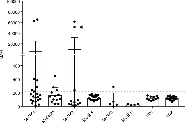
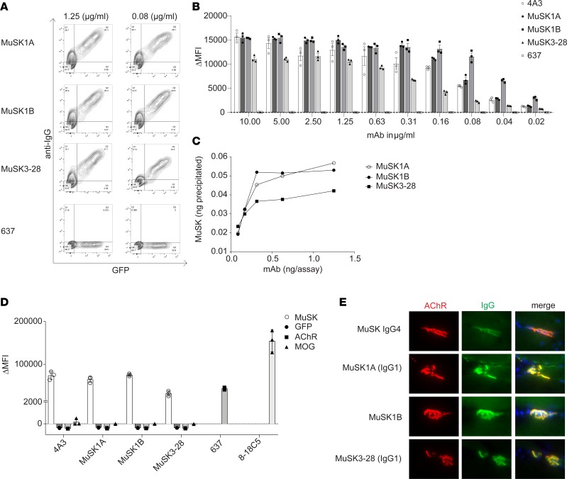
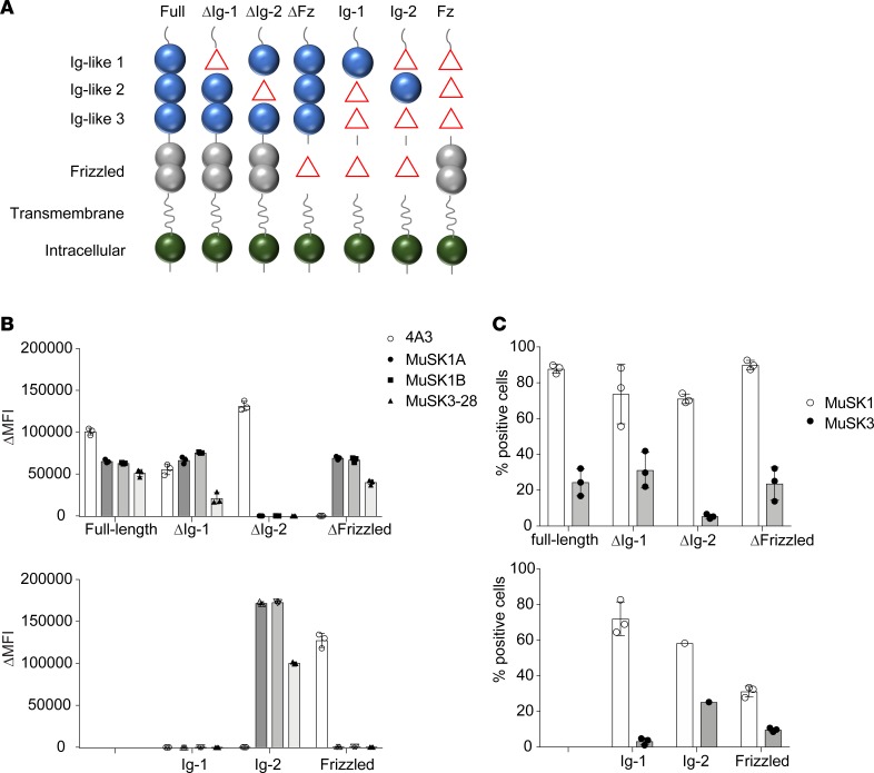
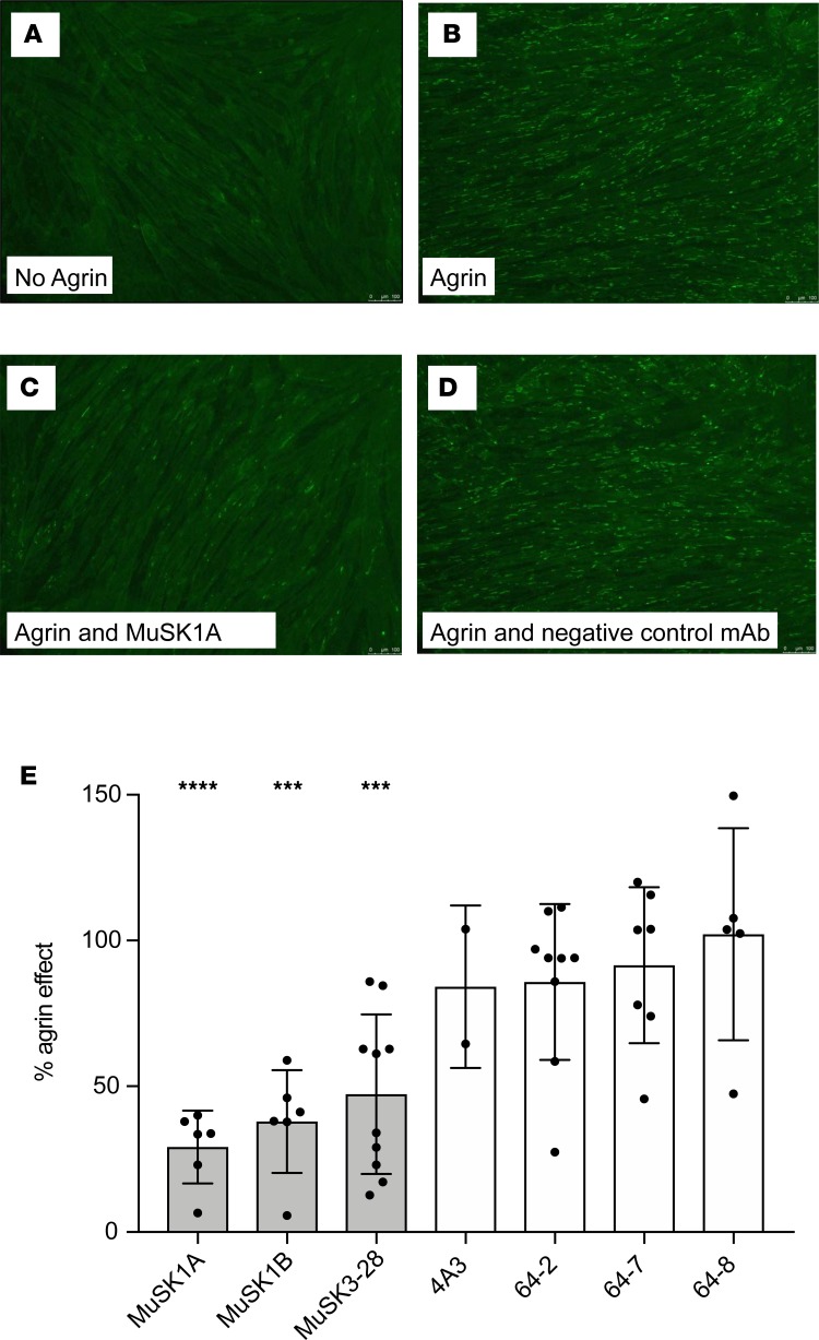
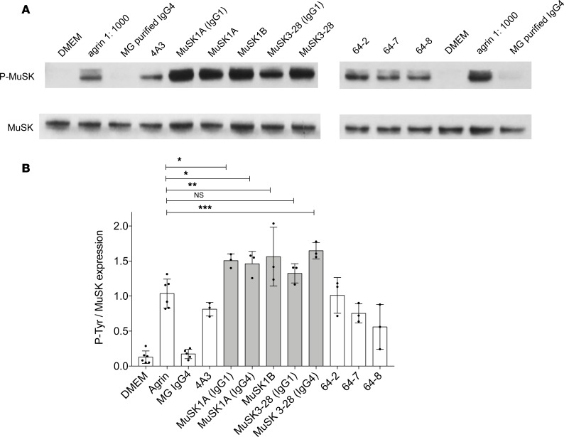
Myasthenia gravis (MG) is a chronic autoimmune disorder characterized by muscle weakness and caused by pathogenic autoantibodies that bind to membrane proteins at the neuromuscular junction. Most patients have autoantibodies against the acetylcholine receptor (AChR), but a subset of patients have autoantibodies against muscle-specific tyrosine kinase (MuSK) instead. MuSK is an essential component of the pathway responsible for synaptic differentiation, which is activated by nerve-released agrin. Through binding MuSK, serum-derived autoantibodies inhibit agrin-induced MuSK autophosphorylation, impair clustering of AChRs, and block neuromuscular transmission. We sought to establish individual MuSK autoantibody clones so that the autoimmune mechanisms could be better understood. We isolated MuSK autoantibody-expressing B cells from 6 MuSK MG patients using a fluorescently tagged MuSK antigen multimer, then generated a panel of human monoclonal autoantibodies (mAbs) from these cells. Here we focused on 3 highly specific mAbs that bound quantitatively to MuSK in solution, to MuSK-expressing HEK cells, and at mouse neuromuscular junctions, where they colocalized with AChRs. These 3 IgG isotype mAbs (2 IgG4 and 1 IgG3 subclass) recognized the Ig-like domain 2 of MuSK. The mAbs inhibited AChR clustering, but intriguingly, they enhanced rather than inhibited MuSK phosphorylation, which suggests an alternative mechanism for inhibiting AChR clustering.

Keywords: Autoimmune diseases; Autoimmunity; B cells; Immunology; Neuromuscular disease.


Conflict of interest statement

Conflict of interest: KCO has received research support from Ra Pharma and is a consultant and equity shareholder of Cabaletta Bio. KCO is the recipient of a sponsored research subaward from the University of Pennsylvania, the primary financial sponsor of which is Cabaletta Bio. ML and PMM have received research support from ArgenX. AV and the University of Oxford hold a patent (PCT/GB01/02661) for muscle-specific tyrosine kinase antibody tests, licensed to Athena Diagnostics, Massachusetts, USA. AV receives a proportion of royalties. RJN has received research support from Alexion Pharmaceuticals, Genentech, Grifols, and Ra Pharma.

Figures

Figure 1
Figure 1. Screening of human recombinant mAbs.
Recombinant mAbs were produced from single MuSK multimer-sorted B cells. Binding of these clones to MuSK-expressing cells was determined using a flow cytometry–based antibody-binding assay. Each data point represents the mean ΔMFI of each mAb tested at 1 μg/mL in triplicate. Bars represent the mean of means and error bars the SDs. The mAbs were derived from patients with MuSK MG and healthy donors (HDs): MuSK1 (n = 22), MuSK2a (n = 5), MuSK3 (n = 12), MuSK4 (n = 13), MuSK5 (n = 21), MuSK6 (n = 6), HD1 (n = 10), and HD2 (n = 19). A human recombinant MuSK mAb that we previously produced from single-cell–sorted plasmablasts (indicated with an arrow) was included with those tested from patient MuSK3. Values greater than the mean plus 4 SDs of the HD-derived mAbs (indicated by the horizontal dotted line) were considered positive.
Figure 2
Figure 2. Characterization of human MuSK mAb–binding properties.
Binding properties of mAbs MuSK1A, MuSK1B, and MuSK3-28 were tested in several in vitro antibody-binding assays. (A) Representative cell-based assay (CBA) flow cytometry plots are shown for 3 MuSK mAbs and a negative control (AChR-specific mAb 637). Binding was tested at both 1.25 and 0.08 μg/mL. The x axis represents GFP fluorescence intensity and, consequently, the fraction of transfected HEK cells. The y axis represents Alexa Fluor 647 fluorescence intensity, which corresponds to secondary anti–human IgG antibody binding and, consequently, primary antibody binding to MuSK. Hence, transfected cells are located in the right quadrants and transfected cells with MuSK autoantibody binding in the upper right quadrant. (B) Binding to MuSK was tested over a wide range of mAb concentrations in the CBA. Controls included the MuSK-specific humanized mAb 4A3 and AChR-specific mAb 637 tested with MuSK mAbs MuSK1A, MuSK1B, and MuSK3-28. Each data point represents a separate replicate within the same experiment. Bars represent means and error bars SDs. (C) A solution phase radioimmunoassay was used to measure MuSK binding over a range of mAb concentrations. Each data point represents a value within the same experiment. (D) Specificity of the mAbs was evaluated using CBAs that tested binding to HEK cells transfected with MuSK, GFP alone, AChR, or MOG. Positive controls included MuSK-specific humanized mAb 4A3, AChR-specific mAb 637, and MOG-specific 8-18C5. Each data point represents a separate replicate within the same experiment. Bars represent means and error bars SDs. (E) Immunofluorescent staining of mouse NMJs. Tibialis anterior muscles were cut longitudinally in cryosections and fixed with PFA. AChRs were stained with Alexa Fluor 648 α-bungarotoxin (shown in red) and DNA with Hoechst (shown in blue in the merged panels). The first row shows staining with polyclonal IgG4 from a patient with MuSK MG. Binding of mAbs (MuSK1A, MuSK1B, MuSK3-28) against MuSK (1.6 μg/mL for 1 hour) was detected with goat anti–human IgG Alexa Fluor 488 (IgG, shown in green). In AE the IgG4 subclass mAbs MuSK1A and MuSK3-28 were tested in their native IgG subclass unless indicated otherwise.
Figure 3
Figure 3. MuSK domain-binding results.
To map the human MuSK mAb epitopes, MuSK constructs that had particular domains deleted and full-length MuSK were each expressed in HEK cells and tested with the CBA. (A) The schematic illustrates the mutant forms of MuSK. For example, “ΔIg-1” includes only the Ig-like domains 2 and 3 and the frizzled-like (Fz-like) domain because the Ig-like domain 1 was deleted (shown as “Δ” in the schematic). Similarly, “Ig-1” includes only the Ig-like domain 1 because the Ig-like domains 2 and 3 and Fz-like domain were deleted (shown as “Δ” in the schematic). Binding of mAbs (MuSK1A, MuSK1B, MuSK3-28, and the positive control humanized MuSK mAb 4A3) to these mutant forms of MuSK was tested in our standardized flow cytometry CBA. Results for each (B) mAb or (C) serum specimen are shown. Serum was obtained from the same patients from whom the mAbs were derived. Each data point represents a separate replicate within the same experiment. Bars represent means and error bars SDs.
Figure 4
Figure 4. AChR-clustering assay in C2C12 mouse myotubes demonstrates pathogenic capacity of MuSK mAbs.
The presence of agrin in C2C12 myotube cultures leads to dense clustering of AChRs that can be readily visualized with fluorescent α-bungarotoxin and quantified. Pathogenic MuSK autoantibodies disrupt this clustering. Three different human MuSK-specific mAbs, the humanized murine control MuSK mAb 4A3, and 3 human non–MuSK-specific mAbs derived from AChR MG patient plasmablasts (plasmablasts 64-2, 64-7, and 64-8) were tested for their ability to disrupt the AChR clustering. Each mAb was added to the cultures at 1 μg/mL. (AD) Representative images (original magnification, ×100) from the clustering experiments are shown. (A) Cultured myotubes do not show AChR clustering until (B) agrin is added (bright spots reveal AChR clusters). (C) The mAb MuSK1A added at 1 μg/mL inhibits clustering (D), whereas a control mAb does not inhibit the formation of AChR clusters. (E) Clustering of AChR was quantified relative to the measured effect of agrin. Quantitative results are normalized to clustering induced by only agrin. Each data point represents the mean value from an independent experiment. Bars represent the mean of means and error bars the SDs. Multiple-comparisons ANOVA (against the pooled results for the 3 human non–MuSK-specific mAbs), Dunnett’s test; *P < 0.05, **P < 0.01, ***P < 0.001, and ****P < 0.0001, shown only when significant.
Figure 5
Figure 5. MuSK mAbs can amplify agrin-induced tyrosine phosphorylation.
(A) Immunoblots showing phosphotyrosine bands and related MuSK expression in C2C12 murine myotubes that were incubated with agrin in the presence of MuSK MG serum-derived IgG4 or recombinant MuSK/control mAbs. 4A3 is a humanized murine MuSK mAb; MuSK1A, MuSK1B, and MuSK3-28 are human MuSK mAbs from patients with MuSK MG; and 64-2, 64-7, and 64-8 are non–MuSK-binding human mAbs derived from AChR MG patient plasmablasts. IgG4 subclass mAbs MuSK1A and MuSK3-28 were expressed in vectors reflecting the native subclass and as IgG1 (as indicated). (B) Normalized densitometry analysis results from the MuSK phosphorylation immunoblots are plotted. Each data point represents an independent experiment. Bars represent means and error bars SDs. Phosphorylation of MuSK was determined by normalizing to MuSK expression, detected by a commercial anti-MuSK antibody after stripping the blot, and the ratio of phosphotyrosine MuSK/MuSK is plotted. Multiple-comparisons ANOVA (versus agrin), Dunnett’s test; ns P > 0.05, *P < 0.05, **P < 0.01, and ***P < 0.001, shown for MuSK mAbs versus agrin comparisons.

Similar articles

Cited by

References

    1. Vincent A. Unravelling the pathogenesis of myasthenia gravis. Nat Rev Immunol. 2002;2(10):797–804. doi: 10.1038/nri916. - DOI - PubMed
    1. Gilhus NE. Myasthenia gravis. N Engl J Med. 2016;375(26):2570–2581. doi: 10.1056/NEJMra1602678. - DOI - PubMed
    1. Yi JS, Guptill JT, Stathopoulos P, Nowak RJ, O’Connor KC. B cells in the pathophysiology of myasthenia gravis. Muscle Nerve. 2018;57(2):172–184. doi: 10.1002/mus.25973. - DOI - PMC - PubMed
    1. Vincent A, Beeson D, Lang B. Molecular targets for autoimmune and genetic disorders of neuromuscular transmission. Eur J Biochem. 2000;267(23):6717–6728. doi: 10.1046/j.1432-1033.2000.01785.x. - DOI - PubMed
    1. Lindstrom JM, Engel AG, Seybold ME, Lennon VA, Lambert EH. Pathological mechanisms in experimental autoimmune myasthenia gravis. II. Passive transfer of experimental autoimmune myasthenia gravis in rats with anti-acetylcholine receptor antibodies. J Exp Med. 1976;144(3):739–753. doi: 10.1084/jem.144.3.739. - DOI - PMC - PubMed

Publication types