

Hemangioma of the umbilical cord with associated amnionic inclusion cyst: two uncommon entities occurring simultaneously
- PMID: 31217617
- PMCID: PMC8138541
- DOI: 10.32074/1591-951X-26-17
Hemangioma of the umbilical cord with associated amnionic inclusion cyst: two uncommon entities occurring simultaneously
Erratum in
-
Hemangioma of the umbilical cord with associated amnionic inclusion cyst: two uncommon entities occurring simultaneously.Pathologica. 2019 Jun;111(2):86. doi: 10.32074/1591-951X-26-17-EC. Pathologica. 2019. PMID: 31596275 Free PMC article.
Abstract
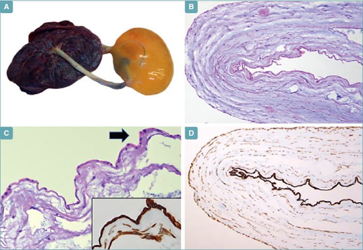
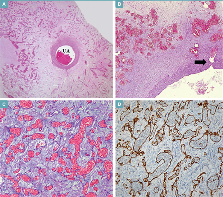
Umbilical cord hemangioma is an uncommon benign vascular neoplasm arising from the free segment of the umbilical cord, distinct from placental and fetal insertion, and is thought to originate from endothelial cells of the umbilical vessels. Cystic changes in the umbilical cord rarely occur as a consequence of the damage to the amnionic surface of the cord caused by the presence of the hemangioma. Until now, a total of 8 cases of umbilical cord hemangioma associated with cystic changes in the umbilical cord have been reported in the literature, however, among these cases, only one showed an associated cyst derived from inclusion of the amniotic epithelium, and the remaining seven cases consisted of hemangiomas with associated pseudocyst of the umbilical cord. We herein report a case of umbilical cord hemangioma with an associated amnionic epithelial inclusion cyst. Clinicopathological features and differential diagnostic considerations are also discussed.
Keywords: Amnionic epithelium; Cyst; Hemangioma; Plancenta; Umbilical cord.
Copyright © 2019 Società Italiana di Anatomia Patologica e Citopatologia Diagnostica, Divisione Italiana della International Academy of Pathology.
Conflict of interest statement
None declared.
Figures
References
-
- Ghidini A, Romero R, Eisen RN, et al. . Umbilical cord hemangioma. Prenatal identification and review of the literature. J Ultrasound Med 1990;9:297-300. - PubMed
-
- Heifetz SA, Rueda-Pedraza ME. Hemangiomas of the umbilical cord. Pediatr Pathol 1983;1:385-98. - PubMed
-
- Nguyen M, Addicott B, Chu J, et al. . Congenital cyst of the umbilical cord. Fetal Pediatr Pathol 2016;5:1-4. - PubMed
-
- Yavner DL, Redline RW. Angiomyxoma of the umbilical cord with massive cystic degeneration of Wharton’s jelly. Arch Pathol Lab Med 1989;113:935-7. - PubMed
Publication types
MeSH terms
LinkOut - more resources
Full Text Sources
Medical
