. 2019 May 14:2019:5406468.
doi: 10.1155/2019/5406468. eCollection 2019.

Spermidine Prevents Heart Injury in Neonatal Rats Exposed to Intrauterine Hypoxia by Inhibiting Oxidative Stress and Mitochondrial Fragmentation

Affiliations

Spermidine Prevents Heart Injury in Neonatal Rats Exposed to Intrauterine Hypoxia by Inhibiting Oxidative Stress and Mitochondrial Fragmentation

Nannan Chai et al. Oxid Med Cell Longev. .

Erratum in

Abstract

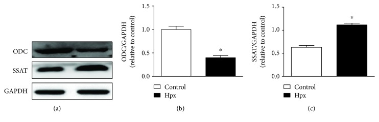
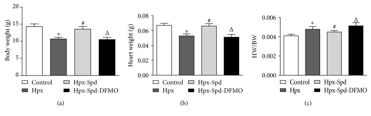
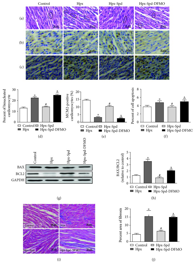
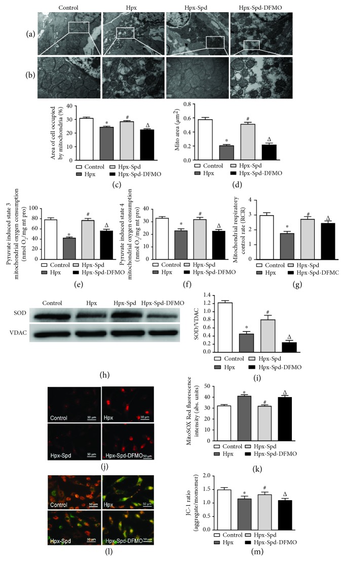
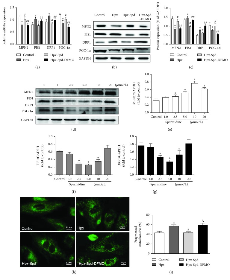
Intrauterine hypoxia (IUH) is a common intrauterine dysplasia that can cause programming of the offspring cardiovascular system. In this study, we hypothesized that placental treatment with spermidine (SPD) can prevent heart injury in neonatal offspring exposed to IUH. Pregnant rats were exposed to 21% O2 or 10% O2 (hypoxia) for 7 days prior to term or were exposed to hypoxia and intraperitoneally administered SPD or SPD+difluromethylornithine (DFMO) on gestational days 15-21. Seven-day-old offspring were then sacrificed to assess several parameters. Our results demonstrated that IUH led to decreased myocardial ornithine decarboxylase (ODC) and increased spermidine/spermine N1-acetyltransferase (SSAT) expression in the offspring. IUH also resulted in decreased offspring body weight, heart weight, cardiomyocyte proliferation, and antioxidant capacity and increased cardiomyocyte apoptosis and fibrosis. Furthermore, IUH caused mitochondrial structure abnormality, dysfunction, and decreased biogenesis and led to a fission/fusion imbalance in offspring hearts. In vitro, hypoxia induced mitochondrial ROS accumulation, decreased membrane potential, and increased fragmentation. Notably, all hypoxia-induced changes analyzed in this study were prevented by SPD. Thus, in utero SPD treatment is a potential strategy for preventing IUH-induced neonatal cardiac injury.


Figures

Figure 1
Figure 1
Polyamine metabolism in the neonatal rat hearts. Protein levels of ornithine decarboxylase (ODC) and spermidine/spermine acetyltransferase (SSAT) in cardiac tissue collected from neonatal offspring were determined by western blotting. Data are shown as the mean ± SEM; n = 4 per group. P < 0.05 versus control. Hpx: hypoxia.
Figure 2
Figure 2
Evaluation of neonatal offspring and heart characteristics. (a) Body weight (BW), (b) heart weight (HW), and (c) BW/HW ratio of neonatal rats. Data are shown as the mean ± SEM; n = 8 per group. P < 0.05 versus control, # P < 0.05 versus the Hpx group, and P < 0.05 versus the Hpx-Spd group. Hpx: hypoxia; Hpx-Spd: hypoxia and SPD treatment; Hpx-Spd-DFMO: hypoxia and SPD+DFMO treatment.
Figure 3
Figure 3
Observation of myocardial morphological structure, cell proliferation, apoptosis, and fibrosis in offspring rats. (a) Representative left ventricle sections stained by HE in the normal (control), hypoxia (Hpx), SPD-treated (Hpx-Spd), and SPD+polyamine synthesis inhibitor- (Hpx-Spd-DMFO-) treated groups. (b) Representative immunohistochemical staining for protein expression and localization of MCM2-positive cardiomyocytes. (c) Brown-stained nuclei indicate TUNEL-positive cells. (d) Evaluation of the percentage of binucleated cardiomyocytes (n = 6). (e) Evaluation of the percentage of MCM2-positive cells (n = 10). (f) The percentage of TUNEL-positive nuclei in different groups (n = 8). (g) BAX and BCL2 protein expression detected by western blotting. (h) Quantification of the BAX and BCL2 protein level ratio (n = 4). (i) Representative Masson's trichrome staining in ventricle sections in each group. (j) Evaluation of interstitial fibrotic areas in ventricle sections in each group (n = 8). Data are shown as the mean ± SEM. P < 0.05 versus control, # P < 0.05 versus the Hpx group, and P < 0.05 versus the Hpx-Spd group.
Figure 4
Figure 4
Effects of SPD on mitochondrial quantity and quality, mitochondrial function, and MnSOD expression in the myocardium of neonatal offspring and on mitochondrial ROS production and membrane potential in primary cardiomyocytes (NRCMs). (a) TEM of myocardial ultrastructure of normal (control), hypoxia (Hpx), SPD-treated (Hpx-Spd), and SPD+polyamine synthesis inhibitor- (Hpx-Spd-DMFO-) treated groups (magnification, 10,000x). (b) Representative TEM images showing ultrastructural changes in the myocardial mitochondria of each group (magnification, 30,000x). (c, d) Quantification of the area of cells occupied by mitochondria (%) and the quantitative mitochondrial area (n = 10). (e–g) Mitochondrial function was evaluated based on mitochondrial state 3 (e) and state 4 (f) oxygen consumption and the respiratory control rate (RCR) (g) in cardiac mitochondria isolated from 7-day-old offspring hearts (n = 10). (h) The expression ratio of SOD proteins in isolated mitochondria detected by western blotting. (i) Quantification of the protein levels (n = 4). (j) Detection of superoxide production in myocardial mitochondria of NRCMs by the MitoSOX Red fluorescent probe. (k) Statistical quantification of the average fluorescence intensity of MitoSOX Red (n = 6). (l) Detection of mitochondrial membrane potential (ΔΨm) of NRCMs by the JC-1 fluorescent probe. (m) Statistical analysis of the ratio of red to green fluorescence (n = 6). Data are shown as the mean ± SEM. P < 0.05 versus control, # P < 0.05 versus the Hpx group, and P < 0.05 versus the Hpx-Spd group.
Figure 5
Figure 5
Effects of SPD on mitochondrial biogenesis and fission/fusion dynamics in the offspring myocardium and NRCMs exposed to hypoxia. (a) mRNA expression levels of myocardial MFN2, FIS1, DRP1, and PGC-1α were determined by qRT-PCR; GAPDH was used as an internal control (n = 6). (b) Protein levels of MFN2, FIS1, DRP1, and PGC-1α in the offspring myocardium collected from the control, Hpx, Hpx-Spd, and Hpx-Spd-DFMO groups were assessed by western blotting. (c) Quantification of MFN2, FIS1, DRP1, and PGC-1α protein levels normalized to GAPDH in each group (n = 4). Data are shown as the mean ± SEM. P < 0.05 versus control, # P < 0.05 versus the Hpx group, and P < 0.05 versus the Hpx-Spd group. (d) Western blotting showed a dose-dependent effect of SPD (0, 2.5, 5, 10, and 20 μmol) on the protein levels of MFN2, FIS1, and DRP1 in NRCMs after exposure to hypoxia. Quantitative analysis of (e) MFN2, (f) FIS1, and (g) DRP1 protein levels normalized to that of GAPDH. Data are shown as the mean ± SEM. P < 0.05 versus the control group (0 μmol/L SPD treatment; n = 6). (h) Detection of fused and fragmented mitochondria in NRCMs by MitoTracker Green staining (magnification, 400x). (i) The percentage of fragmented mitochondria. n = 6 for each group. Data are shown as the mean ± SEM. P < 0.05 versus the control group, # P < 0.05 versus the Hpx group, and P < 0.05 versus the Hpx-Spd group.

References

    1. Giussani D. A., Bennet L., Sferruzzi-Perri A. N., Vaughan O. R., Fowden A. L. Hypoxia, fetal and neonatal physiology: 100 years on from Sir Joseph Barcroft. The Journal of Physiology. 2016;594(5):1105–1111. doi: 10.1113/JP272000. - DOI - PMC - PubMed
    1. Giussani D. A., Davidge S. T. Developmental programming of cardiovascular disease by prenatal hypoxia. Journal of Developmental Origins of Health and Disease. 2013;4(5):328–337. doi: 10.1017/S204017441300010X. - DOI - PubMed
    1. Zhang P., Ke J., Li Y., et al. Long-term exposure to high altitude hypoxia during pregnancy increases fetal heart susceptibility to ischemia/reperfusion injury and cardiac dysfunction. International Journal of Cardiology. 2019;274(274):7–15. doi: 10.1016/j.ijcard.2018.07.046. - DOI - PMC - PubMed
    1. Brain K. L., Allison B. J., Niu Y., et al. Intervention against hypertension in the next generation programmed by developmental hypoxia. PLOS Biology. 2019;17(1, article e2006552) doi: 10.1371/journal.pbio.2006552. - DOI - PMC - PubMed
    1. Rodríguez-Rodríguez P., Ramiro-Cortijo D., Reyes-Hernández C. G., López de Pablo A. L., González M. C., Arribas S. M. Implication of oxidative stress in fetal programming of cardiovascular disease. Frontiers in Physiology. 2018;9, article 602 doi: 10.3389/fphys.2018.00602. - DOI - PMC - PubMed