. 2019 May 15;11(5):2668-2682.
eCollection 2019.

Berberine prevents non-alcoholic steatohepatitis-derived hepatocellular carcinoma by inhibiting inflammation and angiogenesis in mice

Affiliations

Berberine prevents non-alcoholic steatohepatitis-derived hepatocellular carcinoma by inhibiting inflammation and angiogenesis in mice

Yan Luo et al. Am J Transl Res. .

Abstract

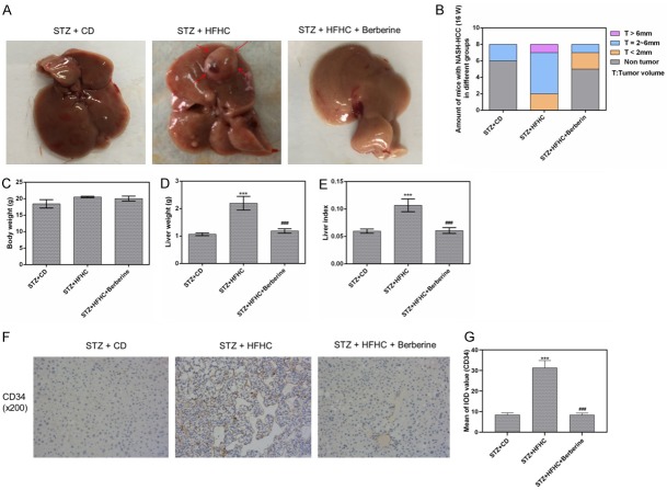
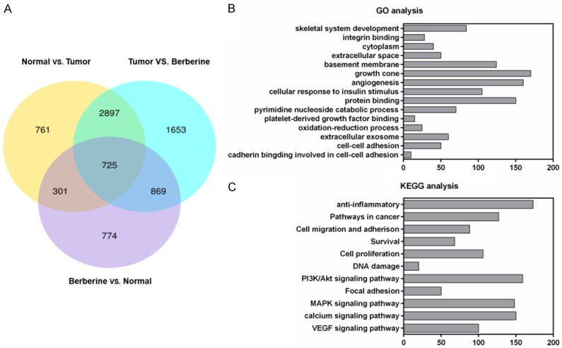
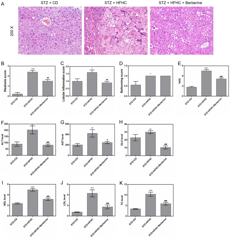
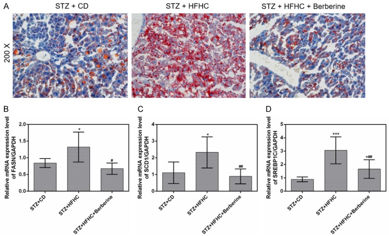
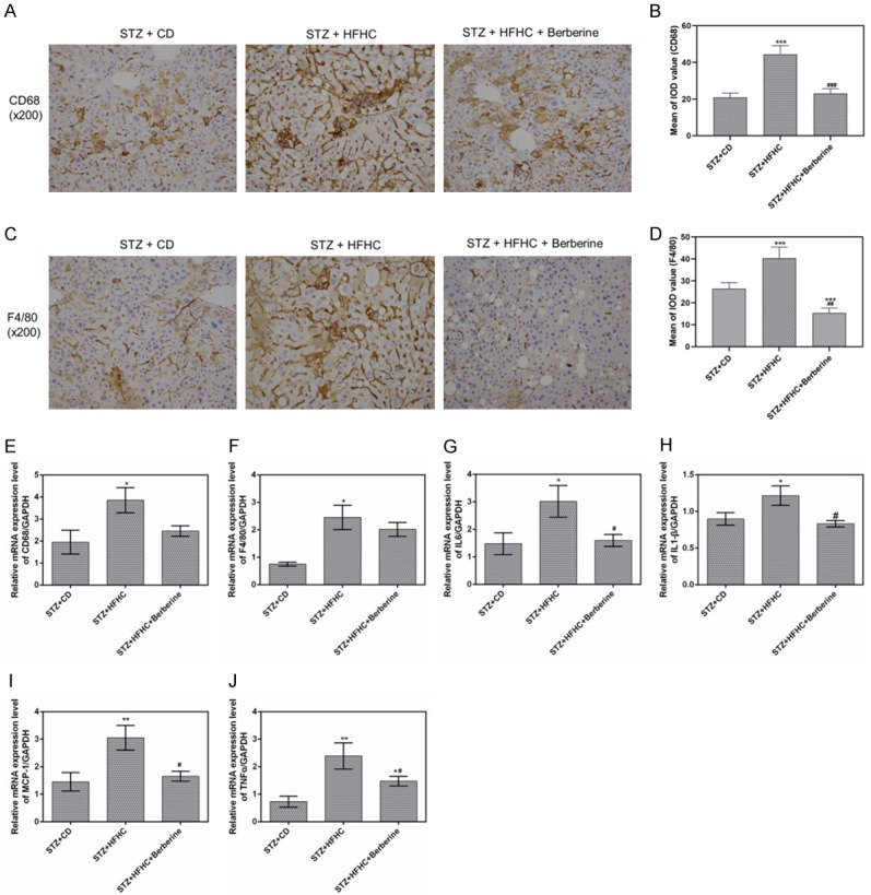
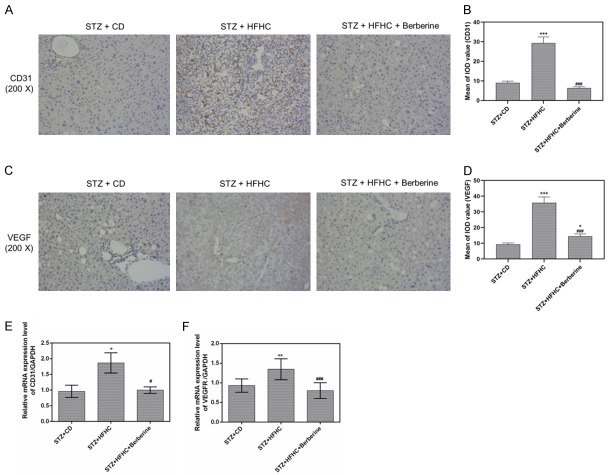
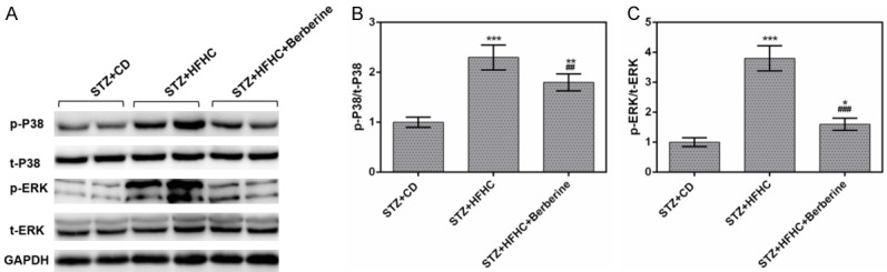
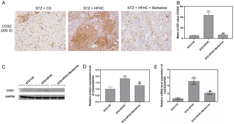
Hepatocellular carcinoma (HCC) is one of the most malignant and poor prognosis tumors, which was increasingly caused by nonalcoholic fatty liver disease/nonalcoholic steatohepatitis (NAFLD/NASH) in western countries. In this study, we aimed to investigate the mechanism and therapeutic prospect of berberine in the treatment of NASH-HCC mice. Combination of STZ injection and high fat and high-cholesterol diet (HFHC) was used to establish NASH-HCC model. The effect of berberine intervention is studied from histology, biochemistry and molecular level. Our results showed that administration of berberine to NASH-HCC mice reduced the incidence of tumors and mitigated NASH. Berberine significantly reduced the levels of alanine aminotransferase (ALT), aspartate aminotransferase (AST), glucose (GLU), high-density lipoprotein (HDL), low-density lipoprotein (LDL) and total cholesterol (TC). Transcriptome sequencing and bioinformatics analysis identified numberous genes and various pathways may participate in the favorite effect of berberine. Specifically, berberine suppressed the expressions of genes related to lipogenesis, inflammation, fibrosis and angiogenesis. Moreover, our results showed that berberine suppressed phosphorylation of p38MAPK and ERK as well as COX2 expression significantly. This suggested berberine achieved its biological functions mainly by regulating inflammation and angiogenesis genes involving p38MAPK/ERK-COX2 pathways. This study demonstrated the anti-tumor effects of berberine and its possible mechanism, providing a potential drug for treating NASH-HCC.

Keywords: Berberine; HCC; NASH; angiogenesis; inflammation.


Conflict of interest statement

None.

Figures

Figure 1
Figure 1
Berberine attenuated the tumorigenesis and reduced liver index. (A) The representative livers from mice with different treatments. The arrows represent the HCC. (B) The numbers and size of tumors in mice in the three groups. The body weight (C) and the liver weight (D) and the liver index (E). (F and G) The expression of HCC biomarker CD34 was measured by immunohistochemistry. Data are presented as the mean ± standard deviation. ***P < 0.001 versus STZ + CD group, ###P < 0.001 versus STZ + HFHC group.
Figure 2
Figure 2
Effects and pathways involved in berberine-treated NASH-HCC mice by bioinformatics analysis. (A) Venn diagrams revealed the differently expressed genes in the liver tissues of nontumor, tumor and berberine groups. GO function analysis (B) and KEGG pathway enrichment analysis (C) for 725 differently expressed genes.
Figure 3
Figure 3
Berberine alleviated the liver steatohepatitis and biochemical profiles. (A) Liver sections in the three groups were stained with H&E. (B) Steatosis scores, (C) lobular inflammation scores, (D) ballooning scores and (E) NAS for the three experimental groups. The serum levels of ALT (F), AST (G), GLU (H), HDL (I), LDL (J) and TC (K) were detected using a Hitachi automatic analyser 7600. Data are presented as the mean ± standard deviation. *P < 0.05, **P < 0.01, ***P < 0.001 versus STZ + CD group, #P < 0.05, ##P < 0.01, ###P < 0.001 versus STZ + HFHC group.
Figure 4
Figure 4
Berberine suppressed the lipid accumulation in liver. (A) Liver sections in the three groups were stained with Oil Red O. The mRNA expressions of FASN (B), SCD1 (C) and SREBP1c (D) in livers were measured by qPCR. Data are presented as the mean ± standard deviation. GAPDH was used as a load control. *P < 0.05, ***P < 0.001 versus STZ + CD group, #P < 0.05, ##P < 0.01 versus STZ + HFHC group.
Figure 5
Figure 5
Berberine exerted the anti-inflammatory effect in mice. (A and B) The protein levels of CD68 were measured by immunohistochemistry, (C and D) The protein levels of F4/80 were measured by immunohistochemistry. The mRNA expressions of CD68 (E), F4/80 (F), IL-6 (G), IL-1β (H), MCP-1 (I) and TNF-α (J) in livers were measured by qPCR. Data are presented as the mean ± standard deviation. GAPDH was used as a load control. *P < 0.05, **P < 0.01, ***P < 0.001 versus STZ + CD group, #P < 0.05, ##P < 0.01, ###P < 0.001 versus STZ + HFHC group.
Figure 6
Figure 6
Berberine inhibited the liver fibrosis. (A) Liver sections in the three groups were stained with Masson staining. The mRNA expressions of Col1A1 (B), α-SMA (C) and TIMP-1 (D) in livers were measured by qPCR. Data are presented as the mean ± standard deviation. GAPDH was used as a load control. *P < 0.05, **P < 0.01, ***P < 0.001 versus STZ + CD group, #P < 0.05, ##P < 0.01 versus STZ + HFHC group.
Figure 7
Figure 7
Berberine prevented the angiogenesis in mice. A and B. CD31 immunohistochemistry showed the MVD (intensive brownish staining) in the livers. C and D. VEGF immunohistochemistry showed the MVD (intensive brownish staining) in the livers. E and F. The mRNA expression of CD31 and VEGFR were determined by qPCR analysis. Data are presented as the mean ± standard deviation. GAPDH was used as a load control. *P < 0.05, **P < 0.01, ***P < 0.001 versus STZ + CD group, #P < 0.05, ###P < 0.001 versus STZ + HFHC group.
Figure 8
Figure 8
Berberine inactivated the phosphorylation of p38MAPK and ERK. (A) The protein levels of p-p38MAPK, p38MAPK, p-ERK and ERK were measured by performing Western blot. The ratios of p-p38MAPK/p38MAPK (B) and p-ERK/ERK (C) were significantly lowered by berberine. Data are presented as the mean ± standard deviation. GAPDH was used as a load control. *P < 0.05, **P < 0.01, ***P < 0.001 versus STZ + CD group, ##P < 0.01, ###P < 0.001 versus STZ + HFHC group.
Figure 9
Figure 9
Berberine inhibited COX2 expression in NASH-HCC mice. A and B. The protein level of COX2 was measured by immunohistochemistry. C and D. The protein level of COX2 in livers was measured by performing Western blot. E. The mRNA level of COX2 in livers was measured by qPCR. Data are presented as the mean ± standard deviation. GAPDH was used as a load control. ***P < 0.001 versus STZ + CD group, ##P < 0.01, ###P < 0.001 versus STZ + HFHC group.

References

    1. Ferlay J, Soerjomataram I, Dikshit R, Eser S, Mathers C, Rebelo M, Parkin DM, Forman D, Bray F. Cancer incidence and mortality worldwide: sources, methods and major patterns in GLOBOCAN 2012. Int J Cancer. 2015;136:E359–386. - PubMed
    1. Younossi ZM, Koenig AB, Abdelatif D, Fazel Y, Henry L, Wymer M. Global epidemiology of nonalcoholic fatty liver disease-Meta-analytic assessment of prevalence, incidence, and outcomes. Hepatology. 2016;64:73–84. - PubMed
    1. Dyson J, Jaques B, Chattopadyhay D, Lochan R, Graham J, Das D, Aslam T, Patanwala I, Gaggar S, Cole M, Sumpter K, Stewart S, Rose J, Hudson M, Manas D, Reeves HL. Hepatocellular cancer: the impact of obesity, type 2 diabetes and a multidisciplinary team. J Hepatol. 2014;60:110–117. - PubMed
    1. Leung C, Yeoh SW, Patrick D, Ket S, Marion K, Gow P, Angus PW. Characteristics of hepatocellular carcinoma in cirrhotic and non-cirrhotic non-alcoholic fatty liver disease. World J Gastroenterol. 2015;21:1189–1196. - PMC - PubMed
    1. Chang CH, Huang WY, Lai CH, Hsu YM, Yao YH, Chen TY, Wu JY, Peng SF, Lin YH. Development of novel nanoparticles shelled with heparin for berberine delivery to treat helicobacter pylori. Acta Biomater. 2011;7:593–603. - PubMed

LinkOut - more resources