. 2019 May 15;11(5):2784-2800.
eCollection 2019.

FAT4-USP51 complex regulates the proliferation and invasion of endometrial cancer via Hippo pathway

Affiliations

FAT4-USP51 complex regulates the proliferation and invasion of endometrial cancer via Hippo pathway

Xiaoxia Che et al. Am J Transl Res. .

Abstract

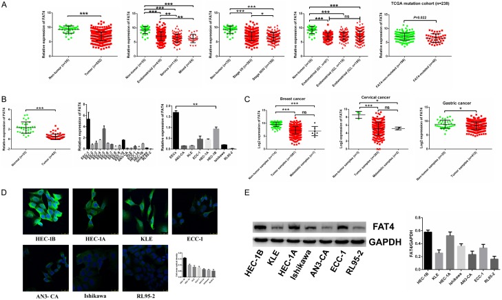
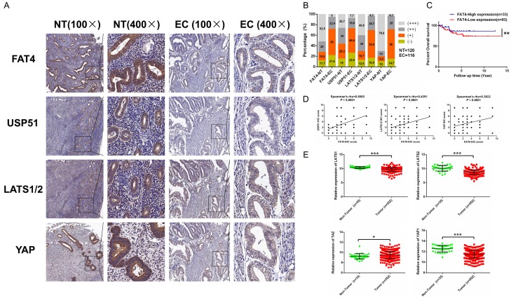
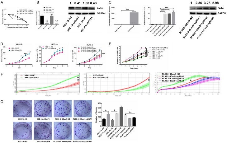
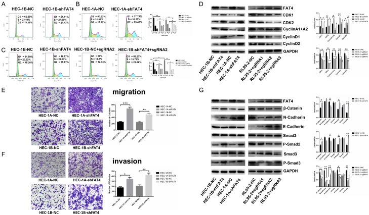
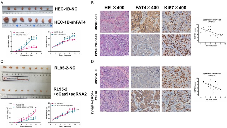
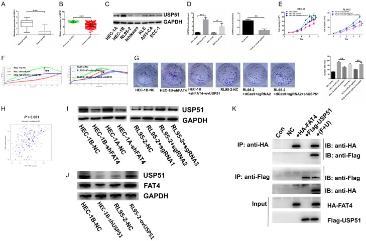
Recent studies have identified FAT tumour suppressor homologue 4 (FAT4), an essential component of adherents junctions, involved in several cancers. However, its role in endometrial cancer (EC) remains unclear. In this study, we first analyzed the association between FAT4 expression and tumour stage, tumour type, and patient prognosis in 552 tumour samples and 35 non-tumour samples from The Cancer Genome Atlas (TCGA) database. The association of decreased FAT4 expression with advanced signature (lymph node metastasis, lymphovascular invasion and muscular infiltration) in EC patients was also confirmed by our own dataset. Stable FAT4 Knockdown promoted EC cell lines proliferation and invasion. FAT4 overexpression inhibited the parental cell phenotype. FAT4 silencing resulted in decreased phosphorylation of the LATS1/2 and YAP while increased YAP nuclear translocation which was associated with the promotion of proliferation and invasion. PCR array analysis of the negative control and shFAT4 HEC-1B cell lines revealed that the deubiquitinating enzyme USP51 was a FAT4 interacting target gene. Ablating USP51 by shRNA decreased cellular FAT4 protein level while overexpression of USP51 increased FAT4 protein level. Coimmunoprecipitation confirmed the direct binding of FAT4 and USP51 which was essential for FAT4's function in EC. The growth inhibitory effect of FAT4 was also attenuated by USP51 down-regulation. In conclusion, suppression of FAT4 by inactivation of deubiquitinating enzyme USP51 promoted proliferation and invasion of EC cells via inhibiting Hippo pathway.

Keywords: FAT4; Hippo pathway; USP51; endometrial cancer.


Conflict of interest statement

None.

Figures

Figure 1
Figure 1
The frequent down-regulation of FAT4 in human EC in the Fudan cohort and in the TCGA database. A: FAT4 mRNA expression was significantly decreased in 552 endometrial cancer (EC) samples compared with their non-tumorous samples. FAT4 downregulated mRNA expression was associated with advanced stage (*P<0.05, t test) and histological type (**P<0.01, t test) while no association with grade. Analysis of TCGA mutation cohort, the mRNA expression of FAT4 in FAT4-mutated group was relative lower compared with FAT4-non-mutated group (P = 0.022, t test). B: FAT4 mRNA expression was significantly decreased in 65 EC samples compared with 33 normal samples in the Fudan cohort (***P<0.0001, t test). FAT4 mRNA expression was also decreased in original normal epithelial endometrial cells extracted from 10 patients during hysteroscopy compared with 7 EC cell lines (***P<0.0001, t test). C: FAT4 expression was also strikingly downregulated in multiple tumour types available in the TCGA (*P<0.05, ***P<0.0001, t test). D, E: FAT4 protein expression in 7 EC cell lines was examined by immunofluorescence and Western blot. FAT4 protein expression was relatively high in the HEC-1A and HEC-1B cell lines and was low in the RL95-2 cell line.
Figure 2
Figure 2
FAT4 protein expression in EC tissues. A: Immunohistochemical staining of FAT4, USP51, LATS1/2 and YAP in EC tissues, noncancerous tissues and normal tissues. Original magnification, × 100 and × 400, scale bar = 100 μm and 50 μm, respectively. B: Proportions of samples negative (- or +) or positive (++ or +++) for FAT4, USP51, LATS1/2 and YAP among 116 EC tissues (EC) and 120 normal tissues (NT). C: EC patients with low expression of FAT4 exhibited worse overall survival compared with patients with high expression of FAT4 (**P<0.01, Log rank test). Kaplan-Meier method was used to draw the cumulative survival curves. D: The correlations of the scores for the four molecules, FAT4, USP51, LATS1/2 and YAP, were measured by Pearson’s coefficient. E: The core components of the Hippo signalling pathway, including LATS1, LATS2, TAZ and YAP. LATS1, LATS2 and YAP mRNA expression was downregulated in 552 tumour samples compared with 35 noncancerous samples, as revealed by the TCGA RNA-Seq analysis, while TAZ showed the opposite trend (*P<0.05, ***P<0.0001, t test).
Figure 3
Figure 3
Silencing FAT4 promoted EC proliferation, and overexpressing FAT4 suppressed EC proliferation. A: IC50 values of puromycin in HEC-1A, HEC-1B and RL95-2 cells. B: Knockdown (shFAT4) efficiency of FAT4 at the mRNA and protein levels in HEC-1A and HEC-1B cells. The data are presented mean ± SD, t-test, *P<0.05, **P<0.01. C: Efficiency of upregulating dCas9, FAT4 mRNA and FAT4 protein levels in RL95-2 cells. The data are presented as mean ± SD, t-test, ***P<0.001. D-F: Knockdown (shFAT4) of FAT4 in both the HEC-1B and HEC-1A cell lines significantly promoted EC cell proliferation compared with the negative (non-target) control (NC); upregulating (sgRNA) FAT4 in RL95-2 cells significantly inhibited EC cell proliferation compared with NC. The data are presented mean ± SD, t-test, *P<0.05, **P<0.01. G: EC cell colony formation was significantly increased by knockdown (shFAT4) of FAT4 in both HEC-1B and HEC-1A cells and significantly decreased upon upregulating (sgRNA) FAT4 in the RL95-2 cell line, The data are presented mean ± SD, t-test, *P<0.05, **P<0.01.
Figure 4
Figure 4
FAT4 inhibited EC cell cycle progression, cell invasion and altered cell cycle-associated protein markers and invasion-associated protein markers in the HEC-1A, HEC-1B and RL95-2 cell lines. A, B: Flow cytometry assay showed a decrease in the percentage of cells in G1 phase and a concomitant increase in that in G2 phase, while the percentage in S phase remained stable in FAT4-silenced HEC-1A and HEC-1B cells. The data are presented as mean ± SD, t-test, *P<0.05, **P<0.01. C: Flow cytometry assay revealed that upregulating FAT4 in HEC-1B-NC cells increased the G1 phase population and that upregulating FAT4 in HEC-1B-shFAT4 cells rescued the increase in proliferation upon FAT4 silencing. The data are presented mean ± SD, t-test, *P<0.05, **P<0.01. D: Western blotting showed that CDK1, CDK2, cyclin A1+A2, cyclin D1 and cyclin D2 expression was increased in HEC-1A-shFAT4 and HEC-1B-shFAT4 cells compared with HEC-1A-NC and HEC-1B-NC cells. The expression of these markers was decreased in RL95-2-sgRNA cells with upregulated FAT4 expression compared with RL95-2 negative control cells, The data are presented as the mean ± SD, t test. *P<0.05, **P<0.01. E, F: Knockdown of FAT4 in both the HEC-1A and HEC-1B cell lines significantly promoted EC cell migration and invasion, as shown by adhesion and transwell cell migration and invasion assays. The data are reported as the average cell count in five random areas in each transwell membrane. Each sample was tested in triplicate. **P<0.01, ***P<0.001, t test. G: FAT4 modulated the expression of migration-associated markers. HEC-1A and HEC-1B cells were infected with NC or shFAT4, and the expression of β-catenin, N-cadherin, P-Smad2 and P-Smad3 was significantly decreased, E-cadherin expression increased, and Smad2 and Smad3 showed stable expression. RL95-2 cells were infected with NC or sgRNA1-3, and the expression of β-catenin, N-cadherin, P-Smad2 and P-Smad3 was significantly upregulated, E-cadherin was downregulated, and Smad2 and Smad3 were stably expressed. GAPDH served as a loading control. The data are presented as the mean ± SD, t test. *P<0.05, **P<0.01.
Figure 5
Figure 5
FAT4 inhibited EC carcinogenicity in vivo. A: HEC-1B cells were tranfected with either NC or shFAT4 and injected into BALB/c nude mice subcutaneously (0.2 mL, 1 × 10⁶ cells). Tumor size and weight were monitored every 3 days. Silenced FAT4 significantly promoted EC tumor growth, the data are mean ± SD, *P<0.05, t test. C: RL95-2 cells were tranfected with either NC or sgRNA2 and injected into BALB/c nude mice subcutaneously (0.2 mL, 1 × 10⁶ cells). Tumor size and weight were monitored every 3 days. Upregulated FAT4 significantly inhibited EC tumor growth, the data are mean ± SD, *P<0.05, t test. B and D: IHC staining for FAT4 and Ki67 in sections of transplanted tumors. *P<0.05, t test. Person’s correlation.
Figure 6
Figure 6
Altered FAT4 levels regulated the Hippo signalling pathway and the PCR-array analysis of HEC-1B-NC and HEC-1B-shFAT4 including 70 common ubiquitin and deubiquitin enzymes. A: The mRNA expression of the core modulators of the Hippo pathway (LATS1, LATS2, MST1, MST2, TAZ and YAP) in HEC-1A and HEC-1B cells infected with either NC or shFAT4 and in RL95-2 cells infected with NC or sgRNA, The data are presented as the mean ± SD, t test. *P<0.05, **P<0.01. B: LATS1, P-LATS1, LATS2, P-LATS2, YAP and P-YAP protein expression in HEC-1A and HEC-1B cells infected with either NC or shFAT4 and in RL95-2 cells infected with NC or sgRNA, The data are presented as the mean ± SD, t test. *P<0.05, **P<0.01. C: FAT4 repression increased the nuclear accumulation of YAP. Cytoplasmic P-YAP expression decreased, nuclear YAP levels increased, and cytoplasmic total YAP levels remained stable in FAT4-silenced cells compared with NC cells (HEC-1A and HEC-1B). GAPDH and histone 3 were used as loading controls. The data are presented as the mean ± SD, t test. *P<0.05, **P<0.01. D, E: Cluster analysis and volcano plot of the difference genes regulated by FAT4, red point represents upregualted genes and green point downregulated genes; F: Validating the significantly regulated genes (USP45, USP46, USP51, TRAF2, RNF19A, IRAK1 and RNF181) by FAT4 in HEC-1A and HEC-1B infected with either NC or shFAT4. The data are presented as the mean ± SD, t test. *P<0.05, **P<0.01.
Figure 7
Figure 7
Suppression of USP51 contributed to promoting tumorigenic function of FAT4 and FAT4 directly connected with USP51 to regulate mutual expression. A: mRNA expression of USP51 downregulated in EC tissues compared with normal tissues in Fudan cohort (***P<0.001, t test); B: mRNA expression of USP51 decreased in EC tissues compared with non-cancerous tissues in TCGA database (***P<0.001, t test); C: Protein expression of USP51 in 7 EC cell lines; D: To overexpress USP51 in HEC-1B-shFAT4 and HEC-1A-shFAT4 cell lines and knockdown USP51 in RL95-2+dCas9+sgRNA2 cell line (**P<0.01, ***P<0.001, t test); E-G: Increasing USP51 rescued the promoting proliferation function of silenced-FAT4 in HEC-1B cell line while HEC-1A presented no significance. Decreasing USP51 rescued the inhibiting proliferation function of upregulated-FAT4 in RL95-2 cell line (*P<0.05, **P<0.01, One-way ANOVA); H: mRNA expression of FAT4 and USP51 association based on TCGA database (Person’s correlation = 0.467); I: Validating the protein expression of USP51 in HEC-1A and HEC-1B infected with either NC or shFAT4 and RL95-2 infected with either NC or sgRNA. GAPDH was a loading control; J: Detection the protein expression of USP51 and FAT4 in HEC-1B cells transfected either with NC or shUSP51; RL95-2 cells transfected either with NC or ovUSP51 plasmid. K: CoIP assay was used to test the direct connection between FAT4 and USP51 in HEK293T cell line.

Similar articles

Cited by

References

    1. Chen W, Zheng R, Baade PD, Zhang S, Zeng H, Bray F, Jemal A, Yu XQ, He J. Cancer statistics in China, 2015. CA Cancer J Clin. 2016;66:115–32. - PubMed
    1. Siegel RL, Miller KD, Jemal A. Cancer statistics, 2018. CA Cancer J Clin. 2018;68:7–30. - PubMed
    1. Bokhman JV. Two pathogenetic types of endometrial carcinoma. Gynecol Oncol. 1983;15:10–7. - PubMed
    1. Setiawan VW, Yang HP, Pike MC, McCann SE, Yu H, Xiang YB, Wolk A, Wentzensen N, Weiss NS, Webb PM, van den Brandt PA, van de Vijver K, Thompson PJ, Strom BL, Spurdle AB, Soslow RA, Shu XO, Schairer C, Sacerdote C, Rohan TE, Robien K, Risch HA, Ricceri F, Rebbeck TR, Rastogi R, Prescott J, Polidoro S, Park Y, Olson SH, Moysich KB, Miller AB, McCullough ML, Matsuno RK, Magliocco AM, Lurie G, Lu L, Lissowska J, Liang X, Lacey JJ, Kolonel LN, Henderson BE, Hankinson SE, Hakansson N, Goodman MT, Gaudet MM, Garcia-Closas M, Friedenreich CM, Freudenheim JL, Doherty J, De Vivo I, Courneya KS, Cook LS, Chen C, Cerhan JR, Cai H, Brinton LA, Bernstein L, Anderson KE, Anton-Culver H, Schouten LJ, Horn-Ross PL. Type I and II endometrial cancers: have they different risk factors? J Clin Oncol. 2013;31:2607–2618. - PMC - PubMed
    1. Hoeng JC, Ivanov NV, Hodor P, Xia M, Wei N, Blevins R, Gerhold D, Borodovsky M, Liu Y. Identification of new human cadherin genes using a combination of protein motif search and gene finding methods. J Mol Biol. 2004;337:307–317. - PubMed

LinkOut - more resources