

Melatonin improves therapeutic potential of mesenchymal stem cells-derived exosomes against renal ischemia-reperfusion injury in rats
- PMID: 31217862
- PMCID: PMC6556638
Melatonin improves therapeutic potential of mesenchymal stem cells-derived exosomes against renal ischemia-reperfusion injury in rats
Abstract
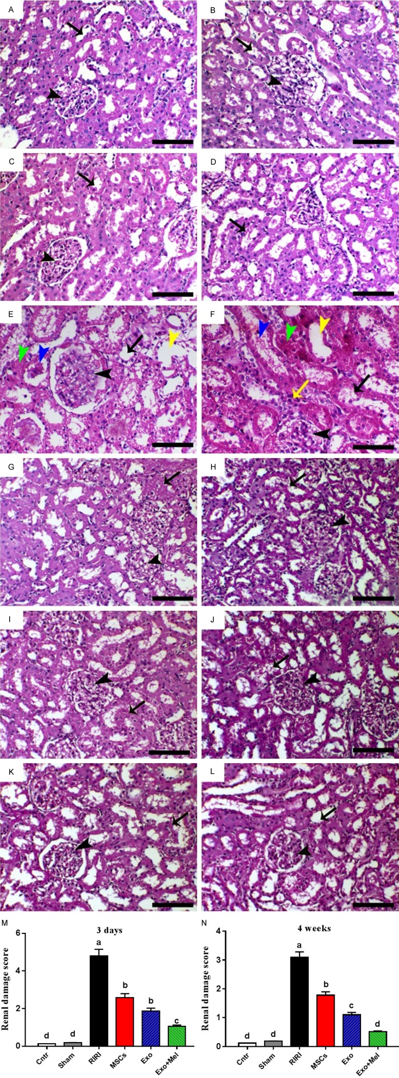
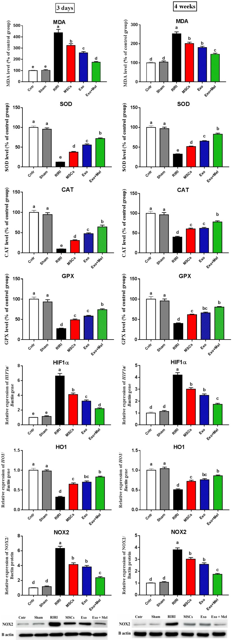
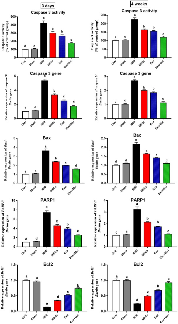
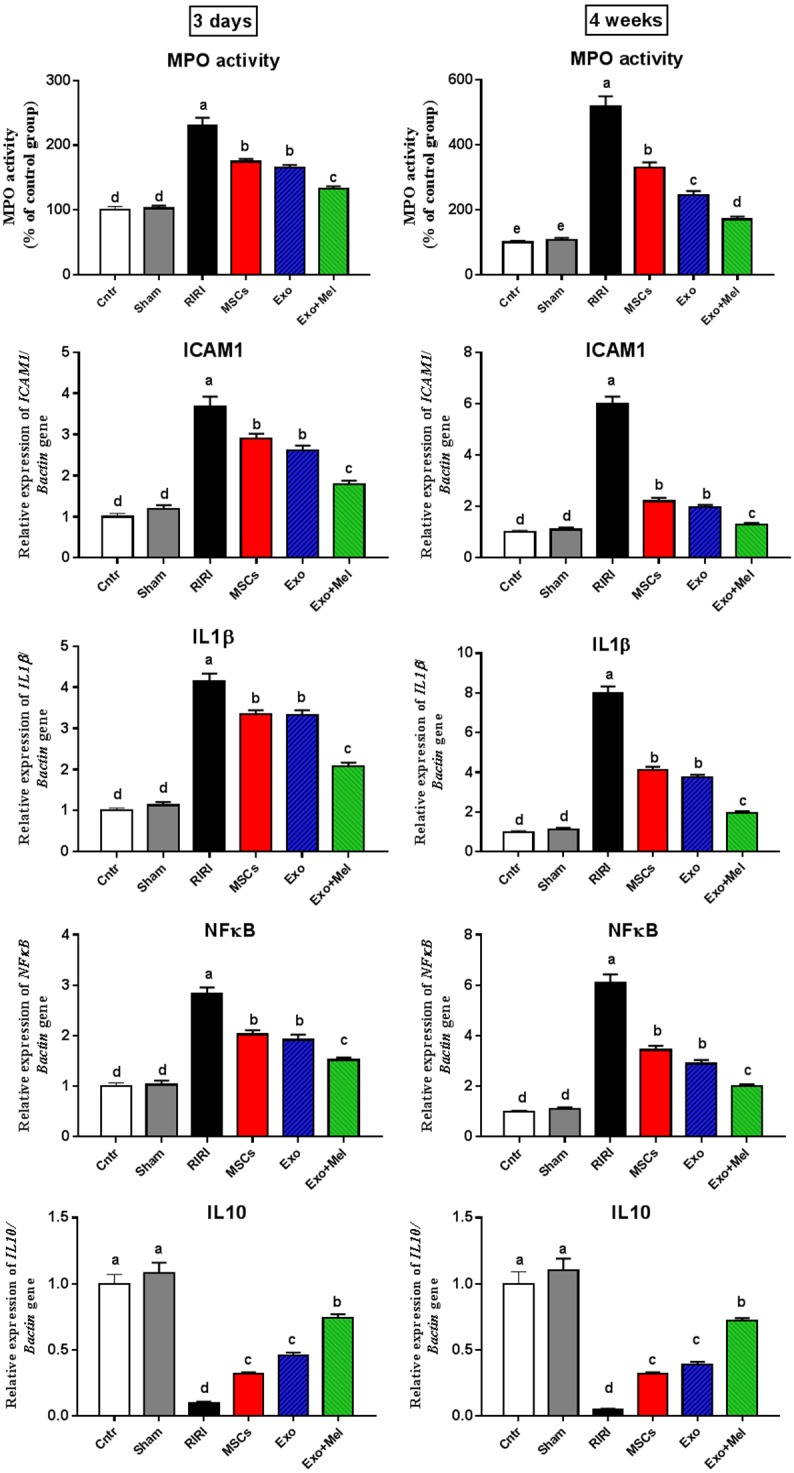
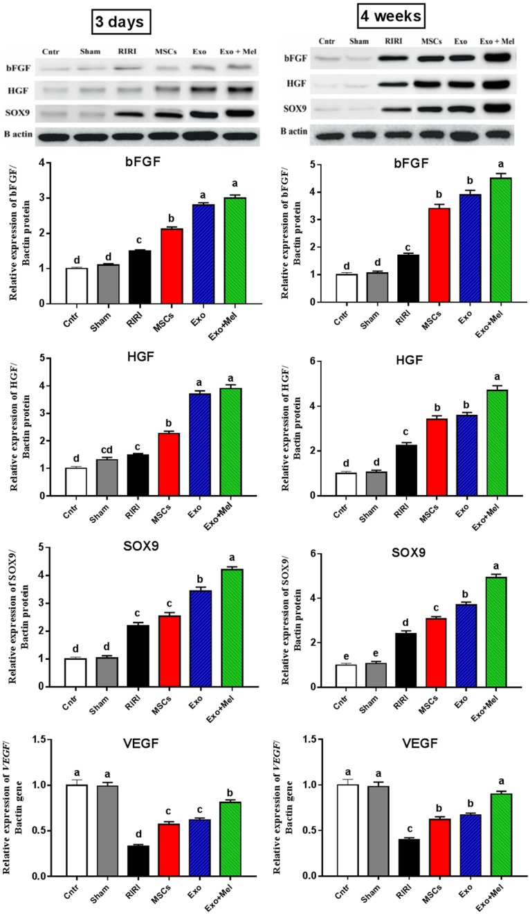
Renal ischemia-reperfusion injury (RIRI) is one of the main causes for acute kidney injury (AKI). Many previous attempts failed to adopt a suitable treatment regimen for AKI. Recently, combined melatonin (Mel) and mesenchymal stem cell (MSC)-derived exosomes (Exo) therapy gave a promising therapeutic option for acute liver ischemic injury, however this treatment approach has not been tested against RIRI yet. This study tested the hypothesis that administration of exosomes derived from MSCs preconditioned with Mel gave best protection against RIRI as compared to therapy by MSCs or exosomes derived from non-preconditioned MSCs. Female adult rats (n = 60) equally divided into control group, sham group, RIRI group (induced by bilateral renal arteries clamping), RIRI + MSCs group (1 × 10⁶ bone marrow derived MSCs), RIRI + Exo group (250 μg Exo derived from no-preconditioned MSCs), and RIRI + Mel + Exo group (250 μg Exo derived from Mel preconditioned MSCs). MSCs or Exo was bilaterally injected once in each renal artery during reperfusion. The obtained results revealed notable improvement in RIRI following all treatment (MSCs, Exo, and Exo + Mel) with best improvement in Exo + Mel group as evidenced by: 1) decreased kidney injury histopathological score; 2) reduced blood levels of kidney damage markers [blood urea nitrogen (BUN) and creatinine]; 3) declined oxidative stress status (MDA level, HIF1α gene, and NOX2 protein); 4) increased anti-oxidant status (HO1 gene, and SOD, CAT, GPX activities); 5) declined apoptosis (caspase 3 activity and mRNA, and PARP1, Bax genes), 6) induced anti-apoptotic effect (Bcl2 gene); 7) inhibition of inflammation (decreased MPO activity and ICAM1, IL1B, NFkB genes and increased IL10 genes); 8) improved regeneration (bFGF, HGF and SOX9 proteins); and 9) enhanced angiogenesis (VEGF gene). These data indicate that treatment with exosomes derived from MSCs preconditioned with melatonin gave best protective effect against renal ischemia-reperfusion injury as compared to therapy by non-preconditioned MSCs or their exosomes.
Keywords: Melatonin; exosomes; mesenchymal stem cells; renal ischemia.
Conflict of interest statement
None.
Figures
References
-
- Williams P, Lopez H, Britt D, Chan C, Ezrin A, Hottendorf R. Characterization of renal ischemia-reperfusion injury in rats. J Pharmacol Toxicol Methods. 1997;37:1–7. - PubMed
-
- Morigi M, Imberti B, Zoja C, Corna D, Tomasoni S, Abbate M, Rottoli D, Angioletti S, Benigni A, Perico N, Alison M, Remuzzi G. Mesenchymal stem cells are renotropic, helping to repair the kidney and improve function in acute renal failure. J Am Soc Nephrol. 2004;15:1794–804. - PubMed
-
- Togel F, Weiss K, Yang Y, Hu Z, Zhang P, Westenfelder C. Vasculotropic, paracrine actions of infused mesenchymal stem cells are important to the recovery from acute kidney injury. Am J Physiol Renal Physiol. 2007;292:F1626–35. - PubMed
LinkOut - more resources
Full Text Sources
Other Literature Sources
Medical
Research Materials
Miscellaneous