

Overexpression of MEF2D contributes to oncogenic malignancy and chemotherapeutic resistance in ovarian carcinoma
- PMID: 31218100
- PMCID: PMC6556600
Overexpression of MEF2D contributes to oncogenic malignancy and chemotherapeutic resistance in ovarian carcinoma
Abstract
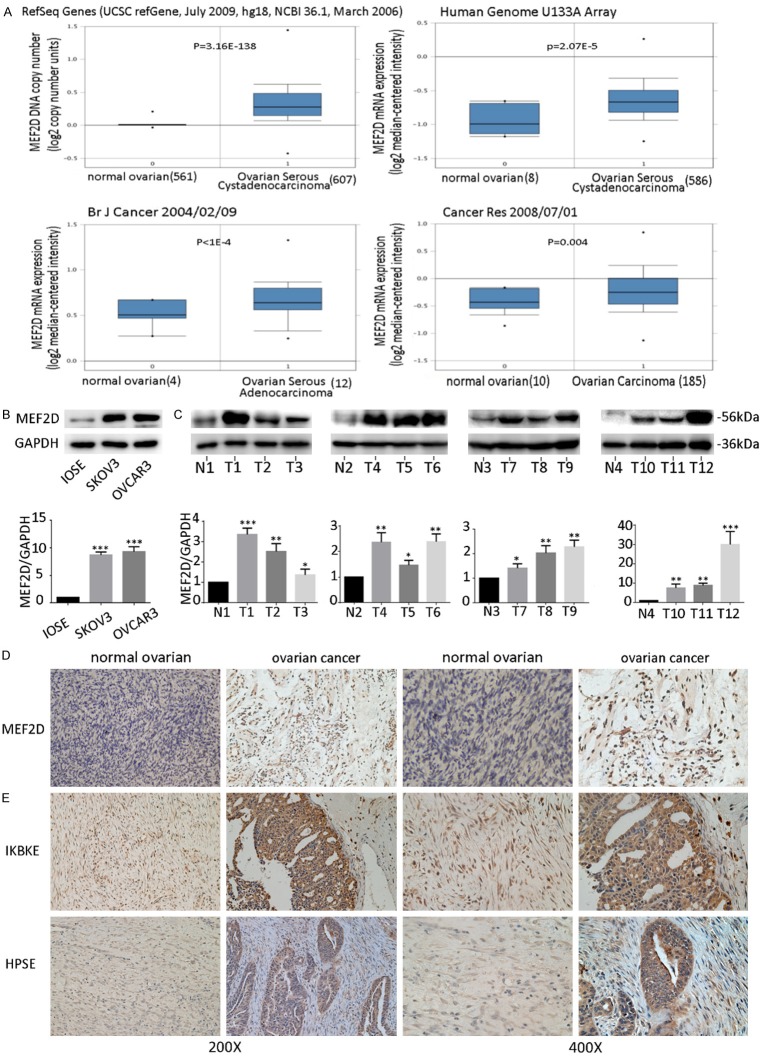
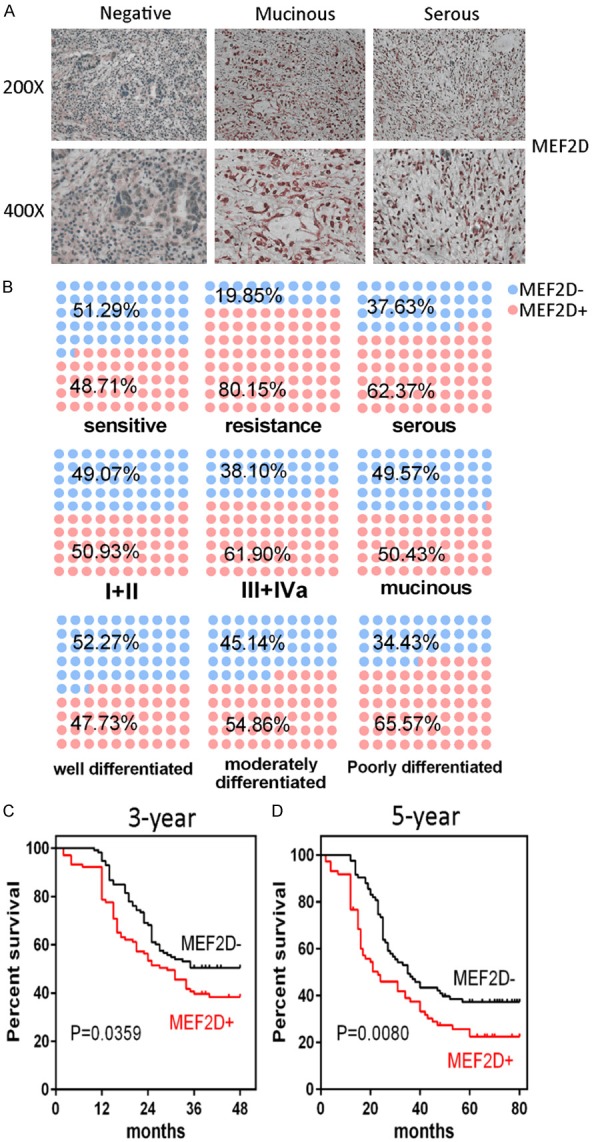
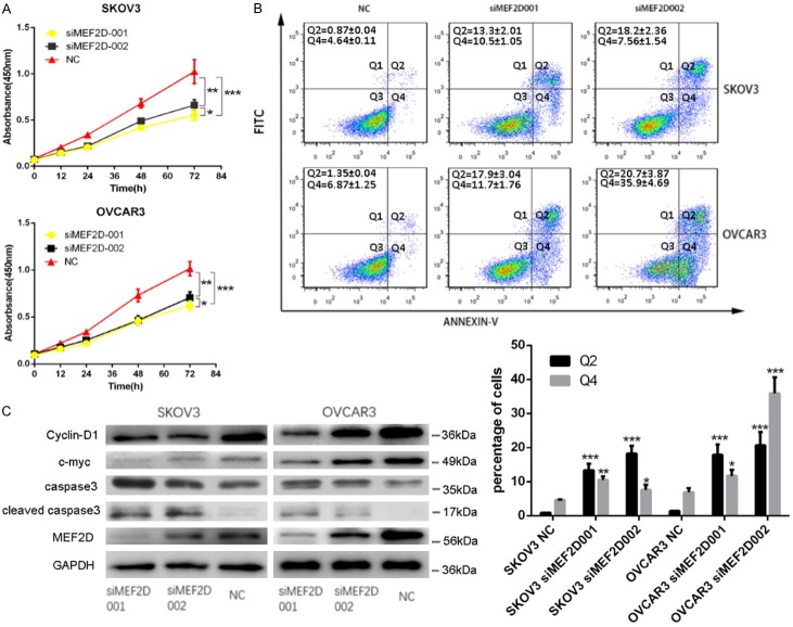
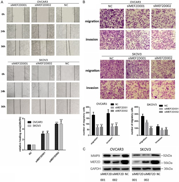
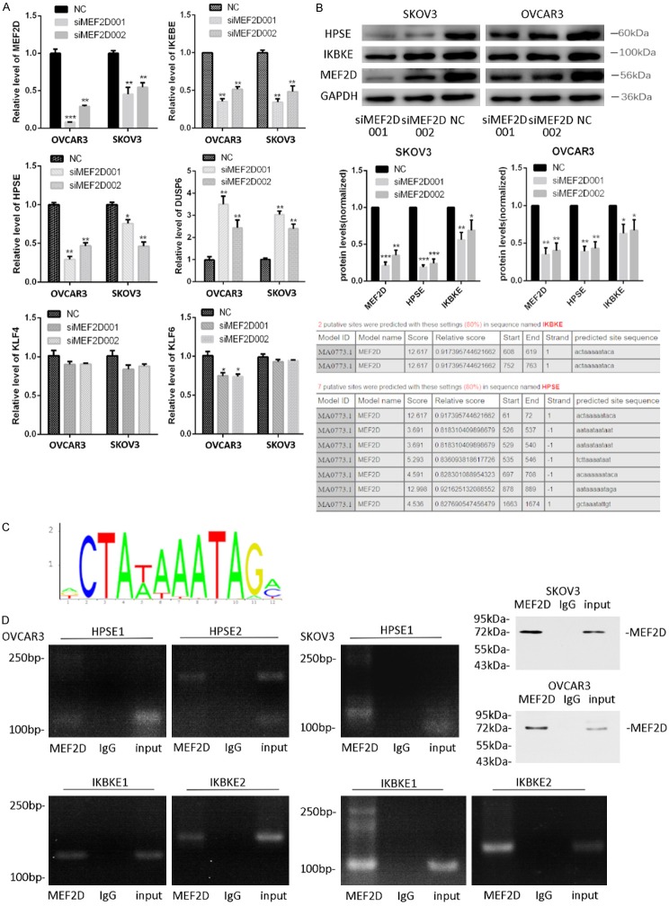
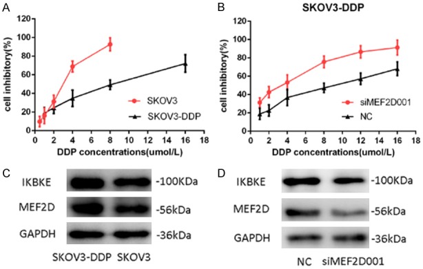
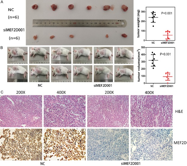
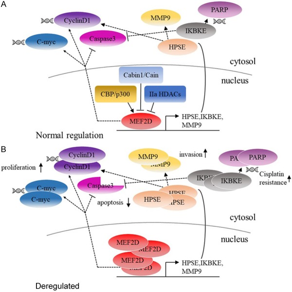
The transcription factor MEF2 promotes survival in various cell types and a number of studies indicate that abnormal regulation of MEF2 is linked to oncogenicity in several carcinomas. We have found that MEF2D, a member of the MEF2 family, is upregulated in Ovarian Cancer (OC). Immunohistochemistry analysis of tumor sections of 402 OC patients revealed that MEF2D is significantly elevated at the protein level. We have also found that the expression level of MEF2D is associated with cisplatin-resistance and poor prognosis by a retrospective analysis. Furthermore, Downregulation of MEF2D by siRNA reduces proliferation and invasiveness of OC cells SKOV3 and OVCAR3, induces apoptosis in vitro, and abolishes OVCAR3 tumorigenicity in xenograft model. Mechanistic study via ChIP analysis identified two of MEF2D-targeted genes, HPSE and IKBKE, which are associated with tumor invasion and chemotherapy-resistance, in accord with MEF2D expression in OC. Remarkably, knock-down of MEF2D invariably lead to the downregulation of IKBKE and reversed cisplatin (DDP)-resistance in cisplatin-resistant cells SKOV3-DDP. Our results suggest that MEF2D promotes malignant biological behaviors and cisplatin-resistance in OC and establish MEF2D as a new therapeutic target in OC treatment.
Keywords: HPSE; IKBKE; MEF2D; Ovarian cancer (OC); cisplatin-resistant.
Conflict of interest statement
None.
Figures
Similar articles
-
Myocyte enhancer factor 2D promotes tumorigenicity in malignant glioma cells.Tumour Biol. 2016 Jan;37(1):601-10. doi: 10.1007/s13277-015-3791-6. Epub 2015 Aug 4. Tumour Biol. 2016. PMID: 26234765
-
Overexpression of the transcription factor MEF2D in hepatocellular carcinoma sustains malignant character by suppressing G2-M transition genes.Cancer Res. 2014 Mar 1;74(5):1452-62. doi: 10.1158/0008-5472.CAN-13-2171. Epub 2014 Jan 3. Cancer Res. 2014. PMID: 24390737
-
Long non-coding RNA Linc00312 modulates the sensitivity of ovarian cancer to cisplatin via the Bcl-2/Caspase-3 signaling pathway.Biosci Trends. 2018 Jul 17;12(3):309-316. doi: 10.5582/bst.2018.01052. Epub 2018 Jun 28. Biosci Trends. 2018. PMID: 29952351
-
Long non-coding RNA H19 mediates ovarian cancer cell cisplatin-resistance and migration during EMT.Int J Clin Exp Pathol. 2019 Jul 1;12(7):2506-2515. eCollection 2019. Int J Clin Exp Pathol. 2019. PMID: 31934077 Free PMC article.
-
Soluble CD40 ligands sensitize the epithelial ovarian cancer cells to cisplatin treatment.Biomed Pharmacother. 2016 Apr;79:166-75. doi: 10.1016/j.biopha.2016.01.006. Epub 2016 Feb 26. Biomed Pharmacother. 2016. PMID: 27044825
Cited by
-
Comprehensive Analysis of the Potential Immune-Related Biomarker Transporter Associated With Antigen Processing 1 That Inhibits Metastasis and Invasion of Ovarian Cancer Cells.Front Mol Biosci. 2021 Dec 10;8:763958. doi: 10.3389/fmolb.2021.763958. eCollection 2021. Front Mol Biosci. 2021. PMID: 34957213 Free PMC article.
-
Efficacy and Safety of Anlotinib Combined with PD-1 Blockades for Patients with Previously Treated Epithelial Ovarian Cancer: A Retrospective Study.Int J Gen Med. 2022 Apr 12;15:3977-3989. doi: 10.2147/IJGM.S352536. eCollection 2022. Int J Gen Med. 2022. PMID: 35440872 Free PMC article.
-
MEF2D Functions as a Tumor Suppressor in Breast Cancer.Int J Mol Sci. 2024 May 10;25(10):5207. doi: 10.3390/ijms25105207. Int J Mol Sci. 2024. PMID: 38791246 Free PMC article.
-
Comprehensive Analysis of the Tumor Microenvironment and Ferroptosis-Related Genes Predict Prognosis with Ovarian Cancer.Front Genet. 2021 Nov 15;12:774400. doi: 10.3389/fgene.2021.774400. eCollection 2021. Front Genet. 2021. PMID: 34868262 Free PMC article.
-
A Comprehensive Bioinformatic Analysis Based on Functional Studies of MEF-2 Family in NSCLC.Yale J Biol Med. 2025 Jun 30;98(2):117-134. doi: 10.59249/PMMF2985. eCollection 2025 Jun. Yale J Biol Med. 2025. PMID: 40589937 Free PMC article.
References
-
- Board PDQCGE. PDQ cancer information summaries. Bethesda (MD): National Cancer Institute (US); 2002. Genetics of breast and gynecologic cancers (PDQ(R)): health professional version. - PubMed
-
- Gupta KK, Gupta VK, Naumann RW. Ovarian cancer: screening and future directions. Int J Gynecol Cancer. 2019;29:195–200. - PubMed
-
- Colombo N, Peiretti M, Parma G, Lapresa M, Mancari R, Carinelli S, Sessa C, Castiglione M. Newly diagnosed and relapsed epithelial ovarian carcinoma: ESMO clinical practice guidelines for diagnosis, treatment and follow-up. Ann Oncol. 2010;21(Suppl 5):v23–30. - PubMed
-
- Hall TR, Dizon DS. Neoadjuvant chemotherapy for advanced epithelial ovarian cancer. Clin Adv Hematol Oncol. 2016;14:262–268. - PubMed
LinkOut - more resources
Full Text Sources