. 2019 May 1;9(5):959-974.
eCollection 2019.

Type Iγ phosphatidylinositol phosphate kinase dependent cell migration and invasion are dispensable for tumor metastasis

Affiliations

Type Iγ phosphatidylinositol phosphate kinase dependent cell migration and invasion are dispensable for tumor metastasis

Shiheng Li et al. Am J Cancer Res. .

Abstract

Type Iγ phosphatidylinositol phosphate kinase (PIPKIγ) has been associated with poor prognosis in breast cancer patients by promoting metastasis. Among the six alternative-splicing isoforms of PIPKIγ, PIPKIγ_i2 specifically targets to focal adhesions and regulates focal adhesion turnover, thus was proposed responsible for tumor metastasis. In the present study, we specifically depleted PIPKIγ_i2 from mouse triple negative breast cancer (TNBC) 4T1 cells and analyzed their behaviors. As expected, PIPKIγ_i2-depleted 4T1 cells exhibited reduced proliferation, migration, and invasion in vitro at a comparable level as pan-PIPKIγ depleted cells. However, PIPKIγ_i2 depletion had no effect on metastasis and progression of 4T1 tumors in vivo. PIPKIγ_i2-depleted tumors showed similar levels of growth, hypoxia state, macrophage infiltration, and angiogenesis as parental tumors, although the pan-PIPKIγ depletion led to substantial inhibition on these aspects. Further investigation revealed that depleting PIPKIγ_i2 alone, unlike depleting all PIPKIγ isoforms, had no effect on PD-L1 expression, the status of the epithelial-to-mesenchymal transition, and the anchorage-independent growth of 4T1 cells. In human TNBC MDA-MB-231 cells, we obtained similar results. Thus, while PIPKIγ_i2 indeed is required for cell migration and invasion, these characteristics are not decisive for metastasis. Other PIPKIγ isoform(s) that regulate the expression of key factors to support cell survival under stresses is more critical for the malignant progression of TNBCs.

Keywords: PIPKIγ; cell migration; metastasis; the epithelial-to-mesenchymal transition; triple negative breast cancer.


Conflict of interest statement

None.

Figures

Figure 1
Figure 1
Knockdown of pan-PIPKIγ and PIPKg_i2 reduce the proliferation and migration of 4T1 cells in vitro to a similar level. (A) The expression of all PIPKIγ isoforms or PIPKIγ_i2 alone was silenced by stably expressing short hairpin RNAs (shRNAs) targeting a common mRNA sequence shared by all PIPKIγ isoforms (shIγ) or a PIPKIγ_i2 specific sequence (shIγ_i2), respectively. Cells stably expressing a non-specific shRNA (shNC) were used as control. The knockdown efficiency of both shRNAs was confirmed by immunoblotting using indicated antibodies. (B) MTT assay was applied to 4T1 cells expressing shNC, shIγ or shIγ_i2 in 96-well plates. Triplicates were set for each time point. (C) 4T1 cells expressing shNC, shIγ or shIγ_i2 were subjected to immunoblotting to determine the levels of phosphorylated AKT (pAKT), total AKT, phosphorylated ERK1/2 (pERK1/2), and total ERK1/2 proteins. b-actin was blotted in same cell lysates for loading control. (D) 4T1 cells expressing shNC, shIγ or shIγ_i2 were subjected to wound healing assay. Representative images were taken at 0 and 12 hours after the wound was created. (E) Motility of cells descripbed in (D) was calculated by measuring the width of the wound. Migration Index = (width0h-width12h)/width0h. (F) EGF-induced directional migration was measured using Boyden chamber. Cells migrated to the reverse side of the membrane were stained with DAPI, counted under microscope, and normalized against non-treated samples. Duplicates were used for each condition. (E and F) results from three independent experiments were statistically analyzed and plotted. *P < 0.05; **P < 0.01; ***P < 0.001.
Figure 2
Figure 2
Loss of PIPKIγ_i2 alone exhibited similar effect on inhibiting the in vitro invasion of 4T1 cells as depletion of all PIPKIγ isoforms. (A) 4T1 cells stably expressing control shRNA (shNC), pan-PIPKIγ shRNA (shIγ), or PIPKIγ_i2 shRNA (shIγ_i2) were subjected to in vitro invasion assay using Matrigel-coated Transwell invasion chamber. Duplicates were applied to each experimental sampel and the invasion index was calculated following the manufacturer’s introduction. (B) MMP9 expression in 4T1 cells described in (A) was analyzed by immunoblotting using indicated antibodies. (C) Representative images of Matrix degradation performed by each group of 4T1 cell. (D) The percentage of cells showing matrix degradation (% of total counted cells), relative area of degraded matrix on slide (% of control group), and particles per degraded cell were quantified under microscope (total counted cells = 200). (A and D) results from three independent experiments were statistically analyzed and plotted. *P < 0.05; **P < 0.01.
Figure 3
Figure 3
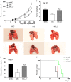
Depletion of PIPKIγ_i2 alone failed to suppress the growth and lung metastasis of 4T1 tumors. (A) Growth curves of tumors in BALB/c mice inoculated with control (shNC), pan-PIPKIγ depleted (shIγ), or PIPKIγ_i2-depleted (shIγ_i2) 4T1 cells. Indicated 4T1 cell lines were injected into mammary fat pad of BABL/c mice. Tumor volume was measured with calipers and calculated by using the standard formula. (B) On day 31 after injection, mice were terminated and tumors were excised and weighted. (C) Comparison of surface nodules on gross lungs from control, PIPKIγ or PIPKIγi2-depleted 4T1 bearing mice on day-31 after implantation. (D) The lung metastasis of each group described in (C) was quantified by manually counting nodules visible on the lung surface. (E) Survival (Kaplan-Meier) curve of BALB/c mice inoculated with indicated 4T1 tumor cells (n = 6/group). Log-rank text: P = 0.0101 (shNC vs. shIγ); P = 0.2116 (shNC vs. shIγ_i2). (A, B, and D) 6 mice/group. *P < 0.05; **P < 0.01; N.S., no significant difference.
Figure 4
Figure 4
Suppressing PIPKIγ_i2 expression appeared no effect on tumor interaction with the host microenvironment. 4T1 tumor cells stably expressing control shRNA (shNC), pan-PIPKIγ shRNA (shIγ), or PIPKIγ_i2 specific shRNA (shIγ_i2) were inoculated subcutaneously into the mammary fat pat of BALB/c mice (n = 6/group). On Day-31 post inoculation, animals were terminated and tumors were collected and fixed by formalin for tissue processing. A. Hypoxia levels were measured by examining pimonidazole adducts formation. Left, representative images. Right, Quantitative analysis of the percent hypoxic area in the whole tumor section. Nuclear counterstain: hematoxylin stain. B. The infiltrated macrophages in tumors were detected by immunohistochemistry staining for CD68. Left, representative images. Right, Average number of CD68-positive cells in each field. C. The microvessel density in tumors was assessed using the marker of endothelial cells CD34. Left, representative images. Right, Average number of CD34-positive cells in each field. D. PD-L1 positive cells were stained by IHC in in tumors . Left, representative images. Right, Average number of PD-L1 positive cells in each field. A-D. 5 fields, 2 slides per tumor, and 3 mice per experimental group were quantified, statistically analyzed and plotted. *P < 0.05; N.S., no significant difference.
Figure 5
Figure 5
Depletion of pan-PIPKIγ, but not PIPKIγ_i2 alone, inhibited the EMT. A. Protein levels of indicated EMT markers were determined by immunoblotting in control (shNC), pan-PIPKIγ (shIγ) or PIPKIγ_i2-depleted (shIγ_i2) 4T1 cells. ECD, E-cadherin. Levels of indicated proteins were then determined by measuring the corresponding band using ImageJ. Results from three independent experiments were statistically analyzed and plotted. B. 1 × 10⁴ cells of each indicated group were subjected to soft agar colony formation assay. Left, Representative images were required on day 8 by Gelcount software. Results from three independent experiments were statistically analzyed and plotted. *P < 0.05; **P < 0.01; ***P < 0.001; N.S., no significant difference.
Figure 6
Figure 6
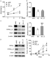
PIPKIγ_i2 deficiency inhibits the in vitro but not in vivo aggressiveness of MDA-MB-231 tumors. (A-C) MDA-MB-231 cells were transfected with siNC, siIγ or siIγ_i2 for 48 hours, and them subjected to MTT assay to determine cell survival/proliferation (A), to Boyden Chamber assay to determine FBS (10%)-induced directional cell migration (B), or immunoblotting to analyze Snail protein levels (C). For Boyden Chamber in vitro cell migration assay (B), results were normalized against the migration index of control cells. Results from three indicated experiments were quantified, statistically analyzed, and plotted. (D and E) MDA-MB-231 cells stably expressing the control (shNC), pan-PIPKIγ (shIγ), or PIPKIγ_i2 (shIγ_i2) shRNAs were established using lentivirus. (D) Depletion of pan-PIPKIγ or PIPKIγ_i2 were confirmed by immunoblotting. Snail protein levels were examined as well. (E) Cells from each of these MDA-MB-231 cell lines (2 × 10⁶ cells/ mouse) were injected into mammary fat pad of in NOD SCID mice (n = 6). Tumor volume was measured with calipers weekly and calculated by using the standard formula to establish the growth curve of in situ tumors. *P < 0.05; **P < 0.01; ***P < 0.001; N.S., no significant difference.

Similar articles

Cited by

References

    1. Bray F, Ferlay J, Soerjomataram I, Siegel RL, Torre LA, Jemal A. Global cancer statistics 2018: GLOBOCAN estimates of incidence and mortality worldwide for 36 cancers in 185 countries. CA Cancer J Clin. 2018;68:394–424. - PubMed
    1. Doughman RL, Firestone AJ, Anderson RA. Phosphatidylinositol phosphate kinases put PI4,5P(2) in its place. J Membr Biol. 2003;194:77–89. - PubMed
    1. van den Bout I, Divecha N. PIP5K-driven PtdIns(4,5)P2 synthesis: regulation and cellular functions. J Cell Sci. 2009;122:3837–3850. - PubMed
    1. Xia Y, Irvine RF, Giudici ML. Phosphatidylinositol 4-phosphate 5-kinase Iγ_v6, a new splice variant found in rodents and humans. Biochem Biophys Res Commun. 2011;411:416–420. - PMC - PubMed
    1. Sun Y, Ling K, Wagoner MP, Anderson RA. Type I gamma phosphatidylinositol phosphate kinase is required for EGF-stimulated directional cell migration. J Cell Biol. 2007;178:297–308. - PMC - PubMed

LinkOut - more resources