

The Use of 3D Printed Vasculature for Simulation-based Medical Education Within Interventional Radiology
- PMID: 31218145
- PMCID: PMC6553672
- DOI: 10.7759/cureus.4381
The Use of 3D Printed Vasculature for Simulation-based Medical Education Within Interventional Radiology
Abstract
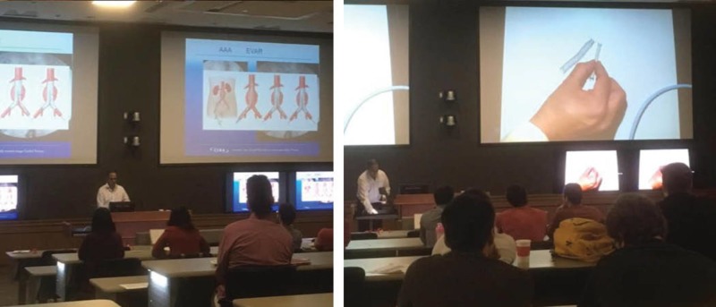
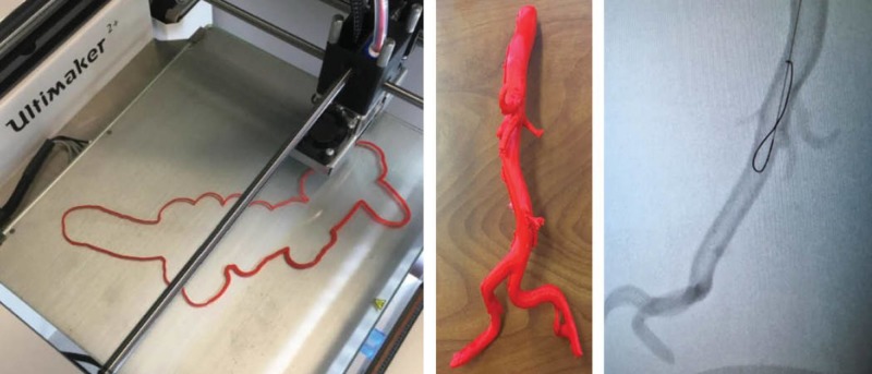
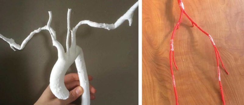
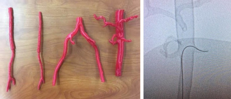
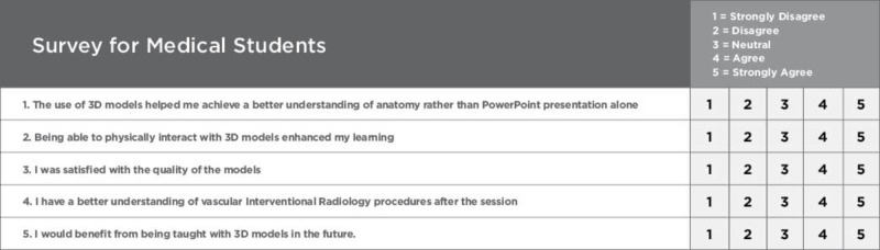
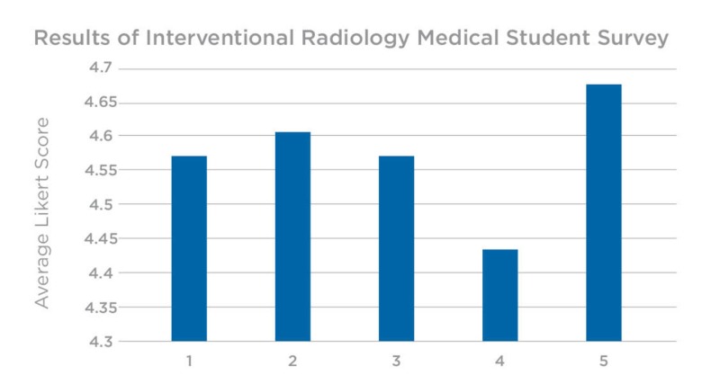
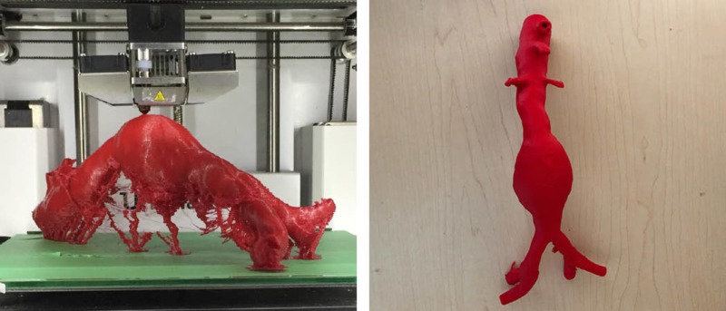
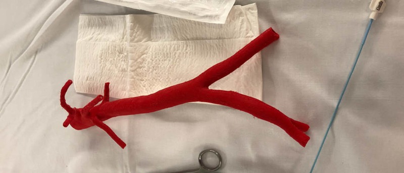
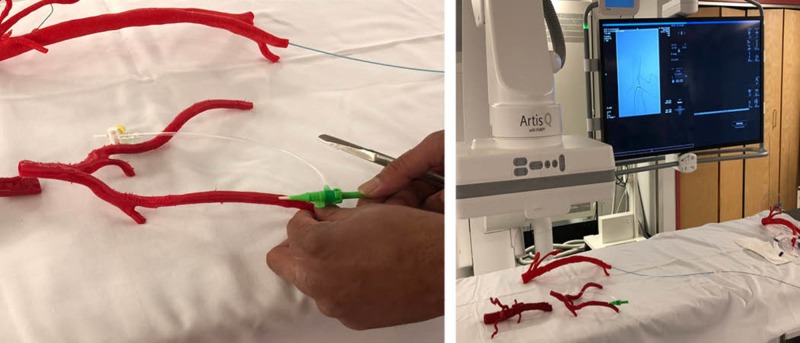
Three-dimensional (3D) printing has become a useful tool within the field of medicine as a way to produce custom anatomical models for teaching, surgical planning, and patient education. This technology is quickly becoming a key component in simulation-based medical education (SBME) to teach hands-on spatial perception and tactile feedback. Within fields such as interventional radiology (IR), this approach to SBME is also thought to be an ideal instructional method, providing an accurate and economical means to study human anatomy and vasculature. Such anatomical details can be extracted from patient-specific and anonymized CT or MRI scans for the purpose of teaching or analyzing patient-specific anatomy. There is evidence that 3D printing in IR can also optimize procedural training, so learners can rehearse procedures under fluoroscopy while receiving immediate supervisory feedback. Such training advancements in IR hold the potential to reduce procedural operating time, thus reducing the amount of time a patient is exposed to radiation and anaesthetia. Using a program evaluation approach, the purpose of this technical report is to describe the development and application of 3D-printed vasculature models within a radiology interest group to determine their efficacy as supplementary learning tools to traditional, lecture-based teaching. The study involved 30 medical students of varying years in their education, involved in the interest group at Memorial University of Newfoundland (MUN). The session was one hour in length and began with a Powerpoint presentation demonstrating the insertion of guide wires and stents using 3D-printed vasculature models. Participants had the opportunity to use the models to attempt several procedures demonstrated during the lecture. These attempts were supervised by an educational expert/facilitator. A survey was completed by all 30 undergraduate medical students and returned to the facilitators, who compiled the quantitative data to evaluate the efficacy of the 3D-printed models as an adjunct to the traditional didactic teaching within IR. The majority of feedback was positive, supporting the use of 3D=printed vasculature as an additional tactile training method for medical students within an IR academic setting. The hands-on experience provides a valuable training approach, with more opportunities for the rehearsal of high-acuity, low-occurrence (HALO) procedures performed in IR.
Keywords: 3d printing; health care; interventional radiology; medical education; point of care; radiology; simulation; simulation based medical education; vasculature.
Conflict of interest statement
The authors have declared that no competing interests exist.
Figures
References
-
- 3D Printing materials and their use in medical education: a review of current technology and trends for the future. [Jul;2018 ];Garcia J, Yang Z, Mongrain R, Leask RL, Lachapelle K. https://stel.bmj.com/content/4/1/27 BMJ Simul Technol Enhanc Learn. 2018 4:27–40. - PMC - PubMed
-
- Three-dimensional printing and medical education: a narrative review of the literature. [Jul;2018 ];Bartellas M. https://uottawa.scholarsportal.info/ojs/index.php/uojm-jmuo/article/view... University of Ottawa Medical Journal. 2016 6:1–38.
-
- Clinical applications of 3D printing: primer for radiologists. [Oct;2018 ];Ballard DH, Trace AP, Ali S, et al. https://www.academicradiology.org/article/S1076-6332(17)30361-6/fulltext. Acad Radiol. 2018 25:52–65. - PMC - PubMed
-
- Search engine for 3D printable models. [Oct;2018 ];https://www.yeggi.com 2018
-
- The role of 3D printing in anatomy education and surgical training: a narrative review. [Jul;2018 ];Li KHC, Kui C, Lee EKM, et al. https://www.mededpublish.org/manuscripts/1010 MedEdPublish. 2017 6:31. - PMC - PubMed
LinkOut - more resources
Full Text Sources